. 2024 Feb 29;25(5):2833.
doi: 10.3390/ijms25052833.

Anabolic Steroids Activate the NF-κB Pathway in Porcine Ovarian Putative Stem Cells Independently of the ZIP-9 Receptor

Affiliations

Anabolic Steroids Activate the NF-κB Pathway in Porcine Ovarian Putative Stem Cells Independently of the ZIP-9 Receptor

Kamil Wartalski et al. Int J Mol Sci. .

Abstract

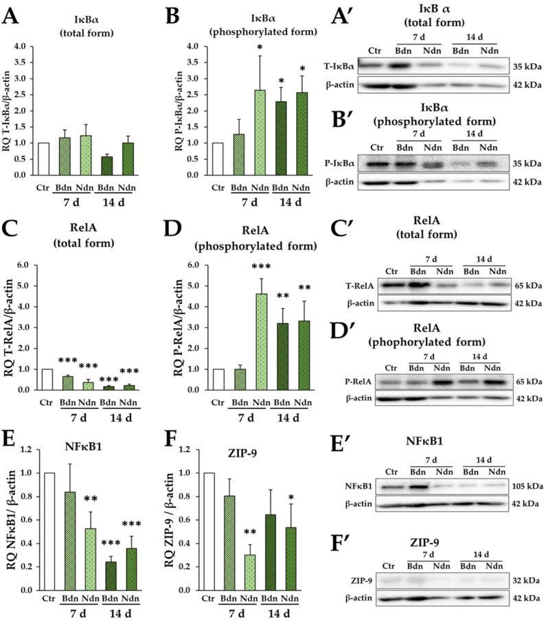
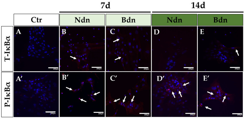
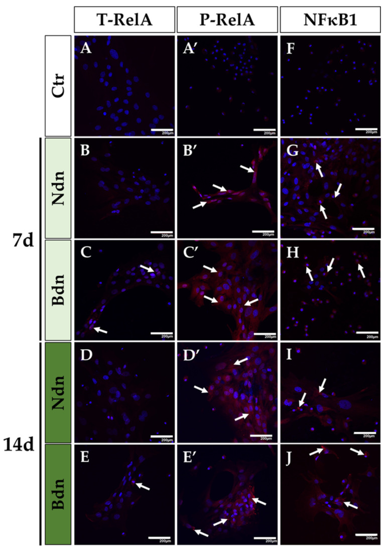
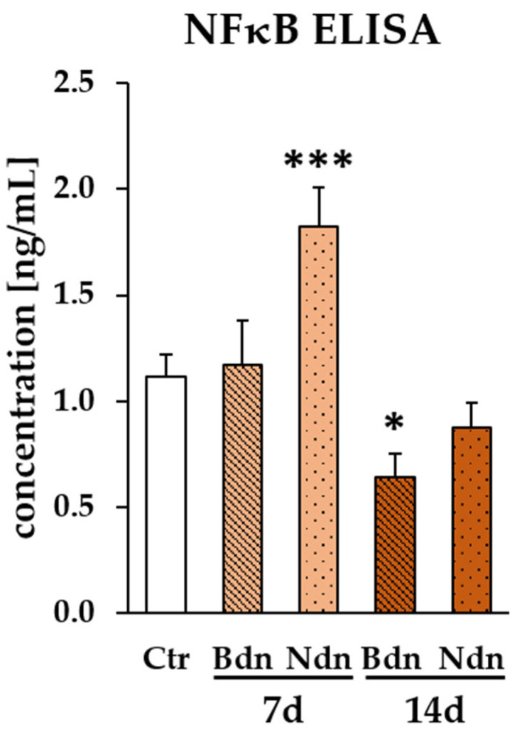
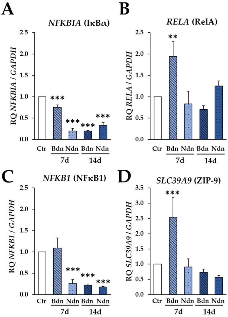
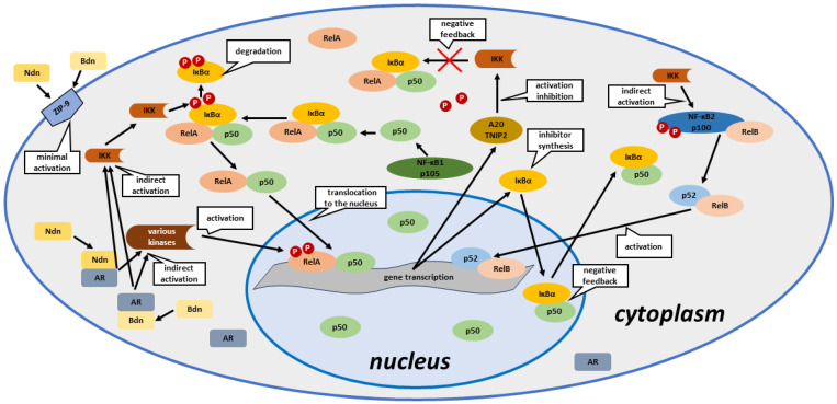
Boldenone (Bdn) and nandrolone (Ndn) are anabolic androgenic steroids (AASs) that, as our previous studies have shown, may increase the risk of neoplastic transformation of porcine ovarian putative stem cells (poPSCs). The NF-κB pathway may be important in the processes of carcinogenesis and tumour progression. Therefore, in this work, we decided to test the hypothesis of whether Bdn and Ndn can activate the NF-κB pathway by acting through the membrane androgen receptor ZIP-9. For this purpose, the expression profiles of both genes involved in the NF-κB pathway and the gene coding for the ZIP-9 receptor were checked. The expression and localization of proteins of this pathway in poPSCs were also examined. Additionally, the expression of the ZIP-9 receptor and the concentration of the NF-κB1 and 2 protein complex were determined. Activation of the NF-κB pathway was primarily confirmed by an increase in the relative abundances of phosphorylated forms of RelA protein and IκBα inhibitor. Reduced quantitative profiles pinpointed not only for genes representing this pathway but also for unphosphorylated proteins, and, simultaneously, decreased concentration of the NF-κB1 and 2 complex may indicate post-activation silencing by negative feedback. However, the remarkably and sustainably diminished expression levels noticed for the SLC39A9 gene and ZIP-9 protein suggest that this receptor does not play an important role in the regulation of the NF-κB pathway.

Keywords: NF-κB signalling pathway; ZIP-9; boldenone; nandrolone; ovary; pig; putative stem cells.


Conflict of interest statement

The authors declare no conflicts of interest. The funders had no role in the design of the study; in the collection, analyses, or interpretation of data; in the writing of the manuscript, or in the decision to publish the results.

Figures

Figure 1
Figure 1
Expression of the NF-κB pathway proteins and the ZIP-9 receptor: total form of IκBα (A,A′), phosphorylated form of IκBα (B,B′), total form of RelA (C,C′), phosphorylated form of RelA (D,D′), NF-kB1 subunit 105 (E,E′) and ZIP-9 (F,F′) at the level of total protein on Days 7 and 14 of culture in the presence of boldenone (Bdn) or nandrolone (Ndn). The graphs show the relative expression of IκBα, RelA, NF-κB1, and ZIP-9 proteins obtained from measurements of the optical density of the bands representing a specific signal. Results represent the mean with n = 3 ± standard deviation (SD). Statistical analysis: homogeneity of variance—Levene’s test, normality of distribution—Shapiro–Wilk test, one-way ANOVA, Dunnett post-hoc test, * p ≤ 0.05; ** p ≤ 0.01; *** p ≤ 0.001.
Figure 2
Figure 2
The presence and specific cytoplasmic localization of T-IκBα (AE) and P-IκBα (A′E′) in poPSCs cultured without the addition of anabolic steroids (A,A′) and in poPSCs cultured in the presence of nandrolone (Ndn; B,B′,D,D′) or boldenone (Bdn; C,C′,E,E′) for 7 and 14 days. Red signals—mediated by DyLight 594 fluorescent dye and derived from T-IκBα and P-IκBα proteins (immunofluorescent signals marked by white arrows), blue signals—dependent on DAPI and originating from the tagged DNA molecules within the cell nuclei; scale bars represent 200 μm.
Figure 3
Figure 3
The presence and specific cytoplasmic localization of T-RelA (AE), P-RelA (A′E′), and NF-κB1 (FJ) in poPSCs cultured without the addition of anabolic steroids (A,A′,F) and in poPSCs cultured for 7 (B,B′,G,C,C′,H) and 14 days (D,D′,I,E,E′,J) in the presence of nandrolone (Ndn; B,B′,G,D,D′,I) or boldenone (Bdn; C,C′,H,E,E′,J). Red signals—mediated by DyLight 594 fluorescent dye and derived from T-RelA, P-RelA and NF-κB1 proteins (immunofluorescent signals marked by white arrows), blue signals—dependent on DAPI and originating from the tagged DNA molecules within the cell nuclei; scale bars represent 200 μm.
Figure 4
Figure 4
Concentrations of NF-κB pathway proteins (NF-κB1 and NF-κB2 complex) [ng/mL] in poPSCs lysates were assessed by ELISA as described in Materials and Methods. The results of each treatment were expressed as the fold change between control and boldenone (Bdn)- or nandrolone (Ndn)-treated cells. Data are expressed as the mean with n = 3 ± standard deviation (SD). Statistical analysis: homogeneity of variance—Levene’s test, normality of distribution—Shapiro–Wilk test, one-way ANOVA, Dunnett post-hoc test, * p < 0.05; *** p < 0.001.
Figure 5
Figure 5
Expression of genes for NF-kB: NFKBIA (A), NFKB1 (C), and RELA (B) and also for ZIP-9 receptor: SLC39A9 (D) at 7th and 14th day of culture in the presence of boldenone (Bdn) and nandrolone (Ndn) versus poPSCs cultured without the addition of steroids at the transcript level as shown by RT-qPCR. The results (2−ΔΔCt) are presented as mean values with n = 3 ± standard deviation (SD). Statistical analysis: homogeneity of variance—Levene’s test, normality of distribution—Shapiro–Wilk test, one-way ANOVA and Dunnett post-hoc test, ** p ≤ 0.01; *** p ≤ 0.001.
Figure 6
Figure 6
Simplified scheme of AAS-triggered activation of the NF-κB pathway and its regulation by negative feedback in poPSCs. Ndn—nandrolone; Bdn—boldenone; ZIP-9—membrane androgen receptor (Zrt- and Irt-like protein 9); IKK—IκB kinase; AR—androgen receptor; IκBα—nuclear factor of κ light polypeptide gene enhancer in B-cells inhibitor, α; RelA—transcription factor p65; RelB—transcription factor RelB; NF-κB1 p105—nuclear factor NF-κB p105 subunit; NF-κB2 p100—nuclear factor NF-κB p100 subunit; p50—NF-κB p50 subunit; p52—NF-κB p52 subunit; A20/TNIP2—A20/tumour necrosis factor (TNF)-interacting protein 2; P—phosphorylation; various kinases—e.g., Akt, PI3K.

References

    1. Sen R., Baltimore D. Inducibility of κ immunoglobulin enhancer-binding protein NF-κB by a posttranslational mechanism. Cell. 1986;47:921–928. doi: 10.1016/0092-8674(86)90807-X. - DOI - PubMed
    1. Nabel G.J., Verma I.M. Proposed NF-κB/IκB family nomenclature. Genes Dev. 1993;7:2063. doi: 10.1101/gad.7.11.2063. - DOI - PubMed
    1. Biancalana M., Natan E., Lenardo M.J., Fersht A.R. NF-κB Rel subunit exchange on a physiological timescale. Protein Sci. 2021;30:1818–1832. doi: 10.1002/pro.4134. - DOI - PMC - PubMed
    1. Hayden M.S., Ghosh S. NF-κB, the first quarter-century: Remarkable progress and outstanding questions. Genes Dev. 2012;26:203–234. doi: 10.1101/gad.183434.111. - DOI - PMC - PubMed
    1. May M.J., Ghosh S. Rel/NF-κB and IκB proteins: An overview. Semin. Cancer Biol. 1997;8:63–73. doi: 10.1006/scbi.1997.0057. - DOI - PubMed

LinkOut - more resources