Review
. 2024 Feb 29;25(5):2838.
doi: 10.3390/ijms25052838.

Mowat-Wilson Syndrome: Case Report and Review of ZEB2 Gene Variant Types, Protein Defects and Molecular Interactions

Affiliations
Review

Mowat-Wilson Syndrome: Case Report and Review of ZEB2 Gene Variant Types, Protein Defects and Molecular Interactions

Caroline St Peter et al. Int J Mol Sci. .

Abstract

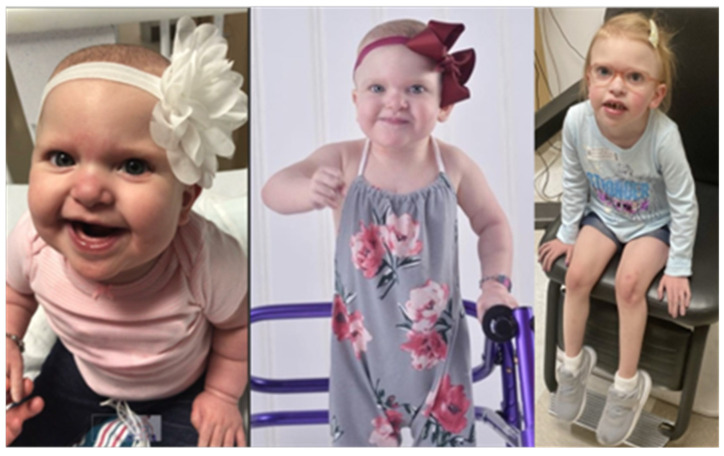
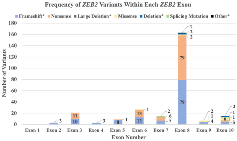
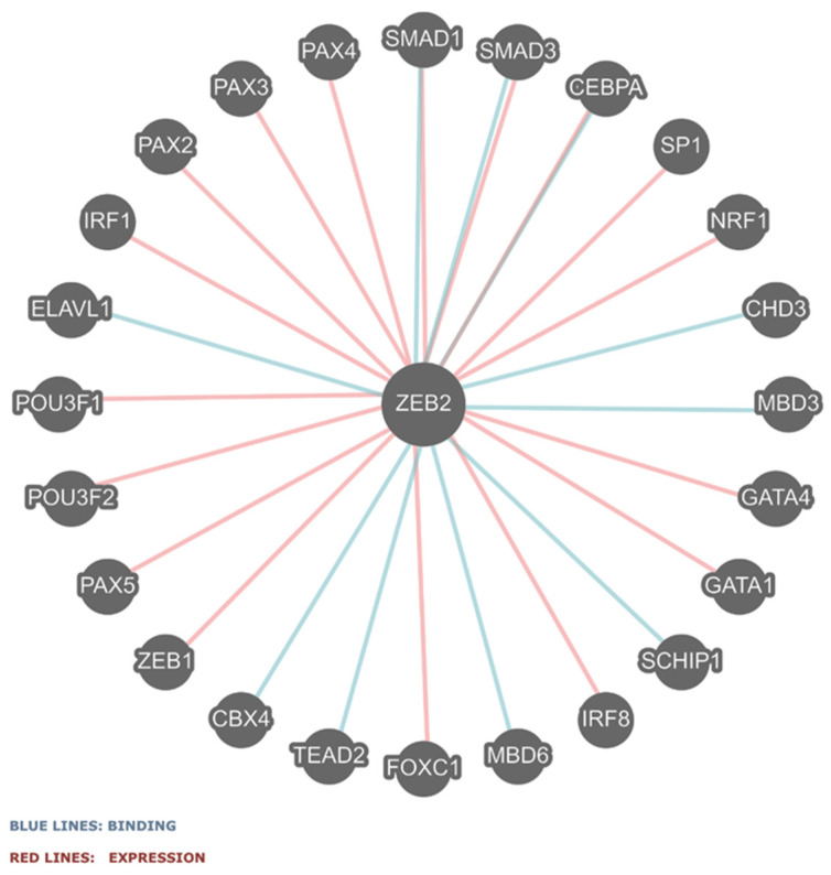
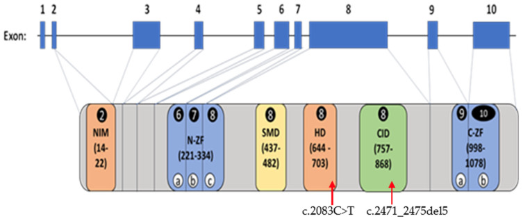
Mowat-Wilson syndrome (MWS) is a rare genetic neurodevelopmental congenital disorder associated with various defects of the zinc finger E-box binding homeobox 2 (ZEB2) gene. The ZEB2 gene is autosomal dominant and encodes six protein domains including the SMAD-binding protein, which functions as a transcriptional corepressor involved in the conversion of neuroepithelial cells in early brain development and as a mediator of trophoblast differentiation. This review summarizes reported ZEB2 gene variants, their types, and frequencies among the 10 exons of ZEB2. Additionally, we summarized their corresponding encoded protein defects including the most common variant, c.2083 C>T in exon 8, which directly impacts the homeodomain (HD) protein domain. This single defect was found in 11% of the 298 reported patients with MWS. This review demonstrates that exon 8 encodes at least three of the six protein domains and accounts for 66% (198/298) of the variants identified. More than 90% of the defects were due to nonsense or frameshift changes. We show examples of protein modeling changes that occurred as a result of ZEB2 gene defects. We also report a novel pathogenic variant in exon 8 in a 5-year-old female proband with MWS. This review further explores other genes predicted to be interacting with the ZEB2 gene and their predicted gene-gene molecular interactions with protein binding effects on embryonic multi-system development such as craniofacial, spine, brain, kidney, cardiovascular, and hematopoiesis.

Keywords: Mowat–Wilson syndrome (MWS); ZEB2 functional molecular interactions; ZEB2 gene variants; ZEB2 protein domains and defects; case report; review.


Conflict of interest statement

The authors declare no conflicts of interest.

Figures

Figure 1
Figure 1
Our proband with typical craniofacial features and phenotype of MWS. She had a pathogenic heterozygous c.2471_2475del5 in exon 8 of the ZEB2 gene. Photos were obtained with consent during infancy, early childhood, and before her death at about five years of age.
Figure 2
Figure 2
Frequency of ZEB2 gene variants identified by variant type in 298 reported patients with MWS. The frequencies of ZEB2 gene variants were identified and grouped. Frameshift* represents a combination of frameshift alone (N = 88), and frameshift plus variants such as frameshift with small deletion (N = 29), frameshift with small insertion (N = 13), or frameshift with small indel (N = 4). Large deletion* represents a combination of large deletions (N = 15) and chromosome deletions (N = 6). Deletion* represents a combination of deletions involving DNA (N = 9) or whole exome based (N = 4). Other* represents a combination of in-frame defects with intragenic deletion (N = 1), partial duplication (N = 1) or insertion deletion (N = 1).
Figure 3
Figure 3
Number of ZEB2 gene variants and types identified in each exon.
Figure 4
Figure 4
ZEB2 gene–gene functional interactions are identified via binding (blue lines) and co-expression (red lines) [http://pathwaycommons.org/pc12/Pathway_751ce7ccec6e191682a33d7252aac8] (accessed on 1 November 2023). These gene interactions relate to TGF receptor pathways in which ZEB1, ZEB2, and SMAD are major players.
Figure 5
Figure 5
Schematic representation of ZEB2 gene structure, exons, and codons listed and incorporated into each of the six protein domains. The protein domains and their corresponding exons are illustrated and modified from Zou et al. [13] such as NIM (nucleosome remodeling and deacetylase-interaction motif), N-ZF (N-terminal zinc finger cluster), SMD (SMAD-binding domain), HD (homeodomain), CID (CtBP-interacting domain), and C-ZF (C-terminal zinc finger cluster) modified from reference [13]. Exons coding the individual protein domains and domain regions are circled. The circled lowercase letters (a/b/c) found in both the N-ZF and C-ZF domains represent the regions coded by different exons (exon numbers circled). The most common gene variant (c.2083C>T) found in our study of 298 patients with MWS and the pathogenetic variant (c.2471_2475del5) found in our 5-year-old proband along with their locations in the HD and CID domains, respectively, are indicated by red arrows.
Figure 6
Figure 6
Predicted AlphaFold2 structure of ZEB2 (www.uniprot.org/uniprotkb/O60315/entry; accessed on 29 September 2023). (A) Full-length structure showing the N-terminal zinc finger domain (tan, L211-C334) and the C-terminal zinc finger domain (green, M998-Y1078) with codon positions at R695 and M824 with their predicted zinc ion binding sites drawn as blue spheres. M824 is a codon that is deleted in our case report of a 5-year-old female with a pathogenic variant c.2472_2475del5 in exon 8, while the R695 protein variant was the most common protein defect found in the 298 reported patients with MWS summarized in Table 1. (B) Same view as panel A but with residues after R695 omitted, resulting in a polypeptide lacking the C-terminal zinc finger domain. (C) N-terminal zinc finger domain shows the predicted zinc binding sites and coordination with ZEB2 residues. (D) C-terminal zinc finger domain shows predicted side chain hydrogen bond interactions (dashed lines) between H1045, Y1055, and S1071 missense ZEB2 changes reported by Ghoumid et al. [12] and other ZEB2 residues (red). Y1055 does not form interactions with the side chain -OH but does form a backbone hydrogen bond interaction with the backbone N-atom of F1064. Other backbone interactions with H1045 and Y1055 are indicated with blue text. (E) Electrostatic surface representation shows packing of Y1055 within a cleft of the ZEB2 C-terminal zinc finger domain.

Similar articles

Cited by

References

    1. Haendel M., Vasilevsky N., Unni D., Bologa C., Harris N., Rehm H., Hamosh A., Baynam G., Groza T., McMurry J., et al. How many rare diseases are there? Nat. Rev. Drug Discov. 2020;19:77–78. doi: 10.1038/d41573-019-00180-y. - DOI - PMC - PubMed
    1. Yang Y., Muzny D.M., Reid J.G., Bainbridge M.N., Willis A., Ward P.A., Braxton A., Beuten J., Xia F., Niu Z., et al. Clinical whole-exome sequencing for the diagnosis of Mendelian Disorders. N. Engl. J. Med. 2013;369:1502–1511. doi: 10.1056/NEJMoa1306555. - DOI - PMC - PubMed
    1. Mowat D.R., Croaker G.D., Cass D.T., Kerr B.A., Chaitow J., Adès L.C., Chia N.L., Wilson M.J. Hirschsprung disease, microcephaly, mental retardation, and characteristic facial features: Delineation of a new syndrome and identification of a locus at chromosome 2q22-q23. J. Med. Genet. 1998;8:617–623. doi: 10.1136/jmg.35.8.617. - DOI - PMC - PubMed
    1. Wakamatsu N., Yamada Y., Yamada K., Ono T., Nomura N., Taniguchi H., Kitoh H., Mutoh N., Yamanaka T., Mushiake K., et al. Mutations in SIP1, encoding Smad interacting protein-1, cause a form of Hirschsprung disease. Nat. Genet. 2001;4:369–370. doi: 10.1038/86860. - DOI - PubMed
    1. Cacheux V., Dastot-Le Moal F., Kääriäinen H., Bondurand N., Rintala R., Boissier B., Wilson M., Mowat D., Goossens M. Loss-of-function mutations in SIP1 Smad interacting protein 1 result in a syndromic Hirschsprung disease. Hum. Mol. Genet. 2001;14:1503–1510. doi: 10.1093/hmg/10.14.1503. - DOI - PubMed

Supplementary concepts