Review
. 2024 Mar 1;25(5):2850.
doi: 10.3390/ijms25052850.

Development of Fluorescence-Based Assays for Key Viral Proteins in the SARS-CoV-2 Infection Process and Lifecycle

Affiliations
Review

Development of Fluorescence-Based Assays for Key Viral Proteins in the SARS-CoV-2 Infection Process and Lifecycle

Mingzhenlong Deng et al. Int J Mol Sci. .

Abstract

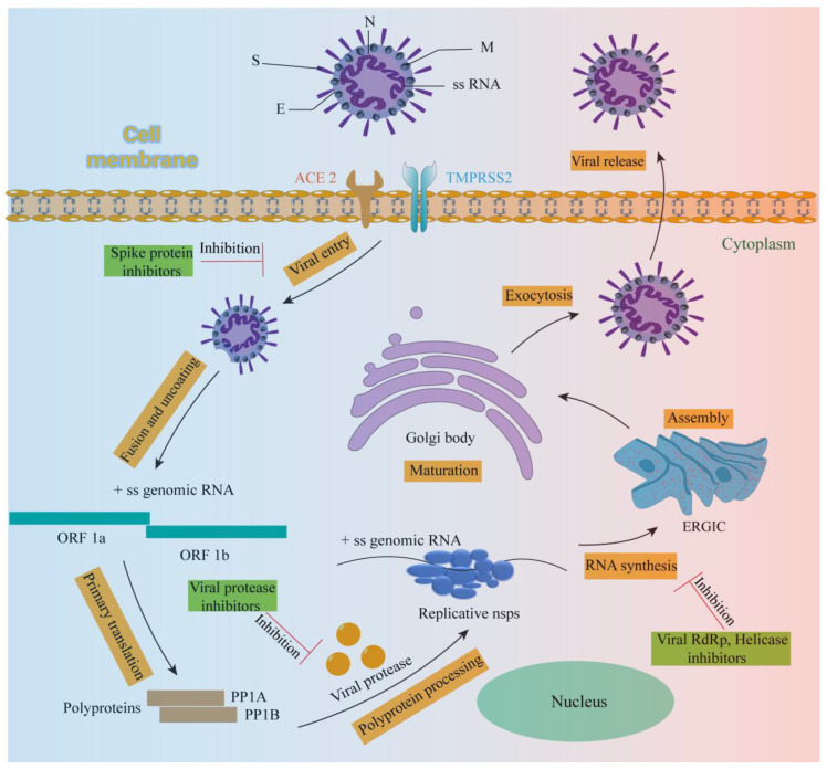
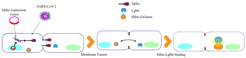
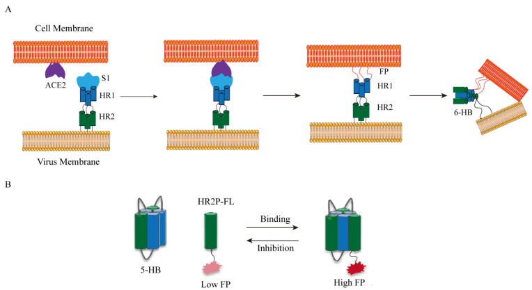
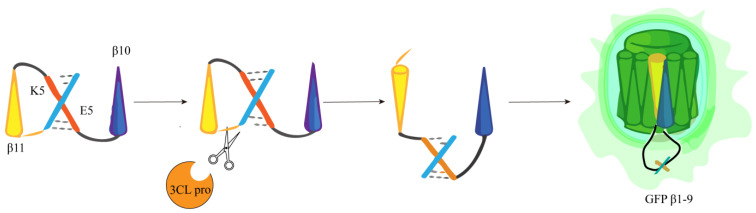
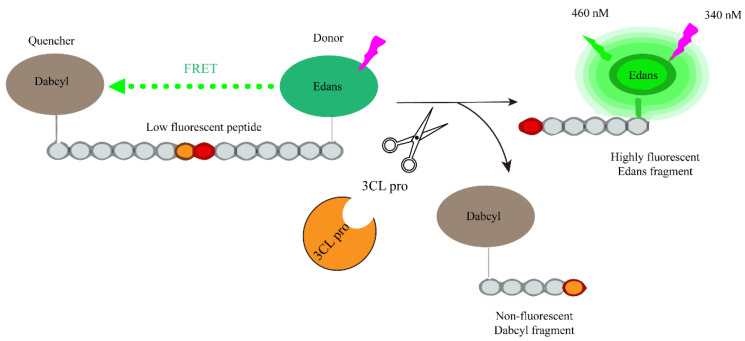
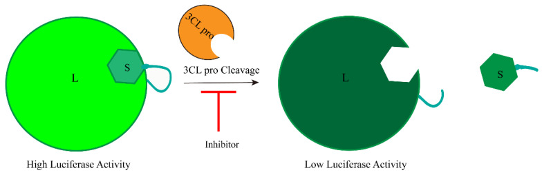
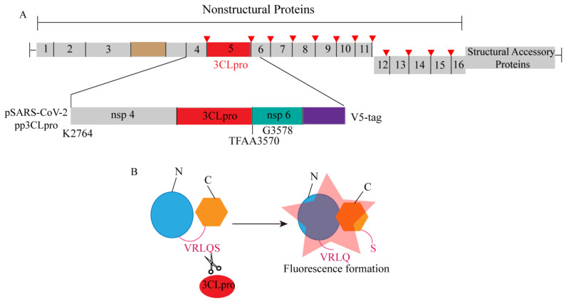
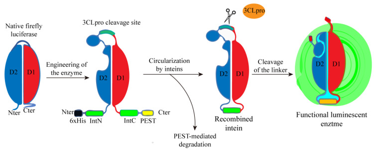
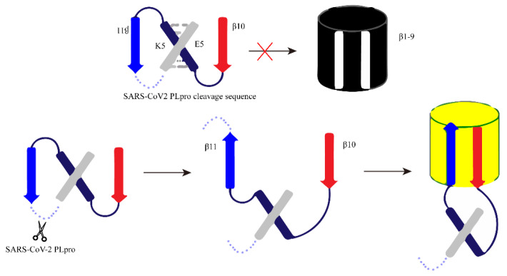
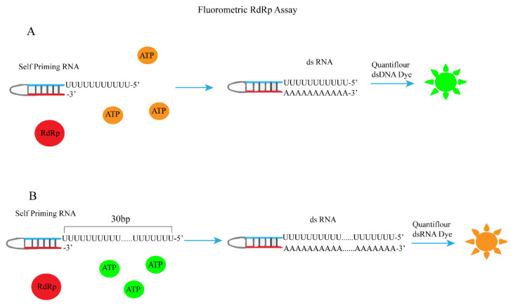
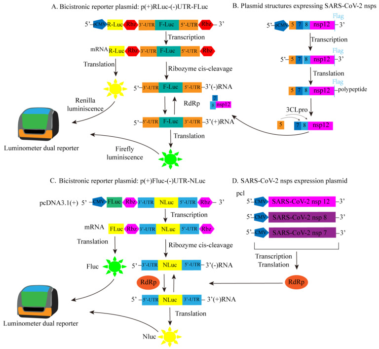
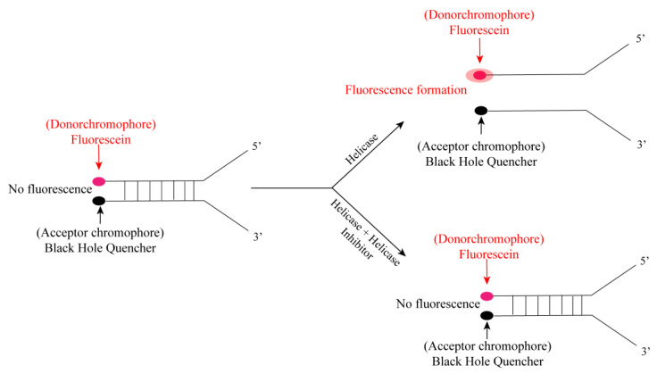
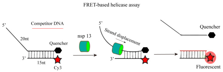
Since the appearance of SARS-CoV-2 in 2019, the ensuing COVID-19 (Corona Virus Disease 2019) pandemic has posed a significant threat to the global public health system, human health, life, and economic well-being. Researchers worldwide have devoted considerable efforts to curb its spread and development. The latest studies have identified five viral proteins, spike protein (Spike), viral main protease (3CLpro), papain-like protease (PLpro), RNA-dependent RNA polymerase (RdRp), and viral helicase (Helicase), which play crucial roles in the invasion of SARS-CoV-2 into the human body and its lifecycle. The development of novel anti-SARS-CoV-2 drugs targeting these five viral proteins holds immense promise. Therefore, the development of efficient, high-throughput screening methodologies specifically designed for these viral proteins is of utmost importance. Currently, a plethora of screening techniques exists, with fluorescence-based assays emerging as predominant contenders. In this review, we elucidate the foundational principles and methodologies underpinning fluorescence-based screening approaches directed at these pivotal viral targets, hoping to guide researchers in the judicious selection and refinement of screening strategies, thereby facilitating the discovery and development of lead compounds for anti-SARS-CoV-2 pharmaceuticals.

Keywords: COVID-19; SARS-CoV-2; antiviral agents; assay development; infection.


Conflict of interest statement

The authors declare no conflicts of interest.

Figures

Figure 1
Figure 1
Virus body and life cycle of SARS-CoV-2.
Figure 2
Figure 2
The scheme elucidates the binding assay between RBD-d2 and SNAP-tagged ACE2 labeled with Lumi4-Tb based on TR-FRET.
Figure 3
Figure 3
Design and characterization of the FRET-based ACE2 biosensor.
Figure 4
Figure 4
Construction of RBD: ACE 2 NanoBiT biosensor. (A) The structure diagram of SRAE 2-BS and the demonstration of its working mechanism. (B) Schematic diagram of Nano-luciferase complementation-based biosensor for the interaction between SARS-CoV-2 Spike S1 protein and ACE2 ectodomain.
Figure 5
Figure 5
(a) PP production and (b) PP entry assay schematics.
Figure 6
Figure 6
A diagram illustrating the structure-based design of the PPI reporter SURF, utilizing split fluorescent proteins, and the initial SURF reporter gene for imaging the interaction between S protein and ACE2 at its peak.
Figure 7
Figure 7
Luciferase quantitative detection of SARS-CoV-2S protein-induced membrane fusion design diagram.
Figure 8
Figure 8
(A) SARS-CoV-2 membrane fusion mechanism. Abbreviations: FP, fusion peptide; HR, heptad repeat. (B) Fluorescence polarization (FP) determination. The FP value of HR 2 P-FL increased after the combination of 5-HB and HR 2 P-FL. Inhibitors could destroy the binding between 5-HB and HR2P-FL, decreasing the FP value.
Figure 9
Figure 9
Diagram of the FlipGFP protease reporter.
Figure 10
Figure 10
Schematic representation of the fluorogenic assay for the enzymatic activity of the SARS-CoV-2 protease.
Figure 11
Figure 11
Development of luciferase complementary reporters based on cell-based inhibition of SARS-CoV-2 3CLpro activity.
Figure 12
Figure 12
A schematic diagram illustrating the development of an enzyme-based biosensor for evaluating the activity of SARS-CoV-2 3CLprotease (3CLpro). (A) The schematic diagram depicts the region of SARS-CoV-2 nonstructural proteins 4 and 5, as well as the amino-terminal region of nsp6, which were cloned into the pcDNA3.1 expression vector with an in-frame V5 epitope tag. (B) The schematic diagram of the pGlo-VRLQS biosensor activated after cleavage by 3CLpro.
Figure 13
Figure 13
Schematic design of a luminescent biosensor for detecting the activity of 3CLpro.
Figure 14
Figure 14
The schematic diagram of the design principle of FlipGFP detection based on cells. Red cross: the configuration is not suitable, and it cannot bind to produce fluorescence.
Figure 15
Figure 15
A schematic diagram of PLpro activity detection based on firefly luciferase (FLuc) reporter was constructed.
Figure 16
Figure 16
The schematic diagram of sandwich-like FP screening method based on fluorescence.
Figure 17
Figure 17
A schematic diagram of a fluorescent protein sensor composed of FRET donor mScarlet and receptor miRFP670 connected by LKGG.
Figure 18
Figure 18
A schematic diagram of RdRp chain displacement determination based on FRET.
Figure 19
Figure 19
A schematic diagram for the determination of RDRP activity using self-initiating RNA, Quan-tiflour dsDNA, or Quan-tiflour dsRNA fluorophores. (A) Schematic of fluorometric RdRp assay with self-priming RNA and Quantiflour dsDNA fluorophore. (B) Schematic of fluorometric RdRp assay with self-priming RNA and Quantiflour dsRNA fluorophore.
Figure 20
Figure 20
The constructed SARS-CoV-2 RdRp bicistron RdRp reporter substructure diagram. (A) Plasmid for dual luciferase reporter: schematic diagram of p(+)RLuc-(−)UTR-NLuc. (B) Express the plasmid structure diagram of SARS-CoV-2 RdRp (nsp12), accessory proteins (nsp7 and nsp8), and viral 3CLpro protease (nsp5). (C) Plasmid for dual luciferase reporter: schematic diagram of p(+)FLuc-(−)UTR-NLuc. (D) Illustration of SARS-CoV-2 nsps expression plasmid.
Figure 21
Figure 21
Schematic diagram of fluorescence determination of helicase activity.
Figure 22
Figure 22
A schematic diagram for the determination of nsp13 helicase activity based on FRET.

Similar articles

Cited by

References

    1. Weiss S.R., Navas-Martin S. Coronavirus pathogenesis and the emerging pathogen severe acute respiratory syndrome coronavirus. Microbiol. Mol. Biol. Rev. 2005;69:635–664. doi: 10.1128/MMBR.69.4.635-664.2005. - DOI - PMC - PubMed
    1. Verstrepen K., Baisier L., De Cauwer H. Neurological manifestations of COVID-19, SARS and MERS. Acta. Neurol. Belg. 2020;120:1051–1060. doi: 10.1007/s13760-020-01412-4. - DOI - PMC - PubMed
    1. Liu J., Xie W., Wang Y., Xiong Y., Chen S., Han J., Wu Q. A comparative overview of COVID-19, MERS and SARS: Review article. Int. J. Surg. 2020;81:1–8. doi: 10.1016/j.ijsu.2020.07.032. - DOI - PMC - PubMed
    1. Jiang H., Yang P., Zhang J. Potential Inhibitors Targeting Papain-Like Protease of SARS-CoV-2: Two Birds with One Stone. Front. Chem. 2022;10:822785. doi: 10.3389/fchem.2022.822785. - DOI - PMC - PubMed
    1. Xie X., Muruato A.E., Zhang X., Lokugamage K.G., Fontes-Garfias C.R., Zou J., Liu J., Ren P., Balakrishnan M., Cihlar T., et al. A nanoluciferase SARS-CoV-2 for rapid neutralization testing and screening of anti-infective drugs for COVID-19. Nat. Commun. 2020;11:5214. doi: 10.1038/s41467-020-19055-7. - DOI - PMC - PubMed