. 2024 Mar 1;25(5):2900.
doi: 10.3390/ijms25052900.

Monocytes as Targets for Immunomodulation by Regional Citrate Anticoagulation

Affiliations

Monocytes as Targets for Immunomodulation by Regional Citrate Anticoagulation

Giovana Seno Di Marco et al. Int J Mol Sci. .

Abstract

Immune alterations in end-stage renal patients receiving hemodialysis are complex and predispose patients to infections. Anticoagulation may also play an immunomodulatory role in addition to the accumulation of uremic toxins and the effects of the dialysis procedure. Accordingly, it has been recently shown that the infection rate increases in patients under regional citrate anticoagulation (RCA) compared with systemic heparin anticoagulation (SHA). We hypothesized that RCA affects the immune status of hemodialysis patients by targeting monocytes. In a cohort of 38 end-stage renal patients undergoing hemodialysis, we demonstrated that whole blood monocytes of patients receiving RCA-but not SHA-failed to upregulate surface activation markers, like human leukocyte antigen class II (HLA-DR), after stressful insults, indicating a state of deactivation during and immediately after dialysis. Additionally, RNA sequencing (RNA-seq) data and gene set enrichment analysis of pre-dialysis monocytes evidenced a great and complex difference between the groups given that, in the RCA group, monocytes displayed a dramatic transcriptional change with increased expression of genes related to the cell cycle regulation, cellular metabolism, and cytokine signaling, compatible with the reprogramming of the immune response. Transcriptomic changes in pre-dialysis monocytes signalize the lasting nature of the RCA-related effects, suggesting that monocytes are affected even beyond the dialysis session. Furthermore, these findings demonstrate that RCA-but not SHA-impairs the response of monocytes to activation stimuli and alters the immune status of these patients with potential clinical implications.

Keywords: anticoagulation; citrate; hemodialysis; heparin; immunomodulation; monocytes.


Conflict of interest statement

The authors declare no conflicts of interest.

Figures

Figure 1
Figure 1
Overview of study workflow. Schematic representation of study design. Citrate, ESRD patients receiving regional citrate anticoagulation (RCA); heparin, ESRD patients receiving systemic heparin anticoagulation (unfractionated heparin); ESRD, end-stage renal disease; iHD, intermittent hemodialysis; RNA-seq, RNA sequencing; PCR, polymerase chain reaction; pre, pre-dialysis, before anticoagulation; post, immediately after dialysis.
Figure 2
Figure 2
Effect of the dialysis procedure and lipopolysaccharide (LPS) stimulation on the expression of surface activation markers on blood monocytes from ESRD patients receiving regional citrate anticoagulation (RCA) or systemic heparin anticoagulation (SHA). (a) Whole blood was collected from patients before (pre) and after (post) intermittent hemodialysis. The graphs show the mean fluorescence intensity (MFI) of the specific markers measured by multicolor flow cytometry. (b) Changes in monocyte surface marker expression in post-dialysis blood relative to pre-dialysis. (c) Whole blood was incubated in the presence or not of 2 ng/mL LPS for 4 h before flow cytometry analysis. The graphs show the changes in the expression in LPS-treated relative to non-treated monocytes. Data obtained from healthy subjects were used as reference (control) only. A Wilcoxon matched-pairs signed rank test ((a), pre vs. post) or a Mann–Whitney U test ((b,c), RCA vs. SHA) was applied. ESRD, end-stage renal disease; HLA-DR, human leucocyte antigen–DR.
Figure 3
Figure 3
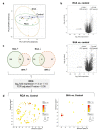
Transcriptomic profiling of pre-dialysis monocytes isolated from ESRD patients from ESRD patients receiving regional citrate anticoagulation (RCA) or systemic heparin anticoagulation (SHA). Blood samples were collected before dialysis, and isolated monocytes were left to rest overnight before RNA isolation. (a) Principal component (PC) analysis of bulk RNA-seq data of cells isolated from patients receiving RCA (n = 7) or SHA (n = 7) and healthy control subjects (n = 6). Each dot represents one subject, and the variance percentage is shown in brackets. (b) Volcano plot representation of differential expression analysis calculated by DESeq2 package. Each dot represents a single gene. Significantly regulated genes (log2-fold expression > 2 or <−2, and p-value < 0.05) are highlighted in gray. (c) Venn diagram showing the number of shared differentially regulated genes in patient monocytes relative to control monocytes (left, upregulated genes; right, downregulated genes). Only significant genes with a log2-fold expression > 1.5 or <−1.5 and false discovery rate (FDR)-adjusted p-value < 0.05 were included. (d) REVIGO scatterplots showing clusters representative of enriched gene ontology biological process terms. The color indicates the log10(p-value), and the size indicates the number of annotations for the term in the gene ontology annotation database. ESRD, end-stage renal disease.
Figure 4
Figure 4
Top enriched gene ontology hits for biological process 2023 and KEGG 2021 human pathways for the differentially expressed genes (log2-fold expression > 1.5 and adjusted p-value < 0.05) in patient monocytes relative to control. (a) Venn diagram showing the number of enriched GOBP terms in patient monocytes relative to control. Bar charts showing (b) the GOBP terms shared between RCA and SHA groups, (c) representatives of terms exclusively enriched in the RCA group (see also Table S1), and (d) enriched KEGG pathways. The terms are displayed based on the −log10(adjusted p-value) according to the data obtained from ENRICHR analysis. The dashed box indicates the single enriched KEGG pathway shared between RCA and SHA groups. RCA, regional citrate anticoagulation; SHA, systemic heparin anticoagulation.
Figure 5
Figure 5
Gene expression analysis and plasma changes in ESRD patients receiving regional citrate anticoagulation (RCA) and systemic heparin anticoagulation (SHA). (a) Blood samples were collected before dialysis, and isolated monocytes were left to rest overnight before RNA isolation. Graphs show real-time PCR data of cells isolated from patients receiving RCA or SHA relative to healthy control subjects (n = 6). (b) Phosphate levels in plasma samples before (pre) and after (post) intermittent hemodialysis. Dashed lines indicate the lower and upper limits of the phosphate reference range. One-way analysis of variance was applied. (c) THP-1 monocytes were cultured in a medium containing 1 and 5 mM phosphate for three days, and gene expression was analyzed by real-time PCR. Graphs show expression fold change relative to high Pi. In (a,b), results are log-transformed and given as mean ± SEM. The Mann–Whitney U test was applied. ESRD, end-stage renal disease; HLA-DR, human leucocyte antigen–DR; MCP-1, monocyte chemoattractant protein-1; PCR, polymerase chain reaction; Pi, phosphate; TLR4, toll-like receptor 4.

Similar articles

References

    1. Kato S., Chmielewski M., Honda H., Pecoits-Filho R., Matsuo S., Yuzawa Y., Tranaeus A., Stenvinkel P., Lindholm B. Aspects of immune dysfunction in end-stage renal disease. Clin. J. Am. Soc. Nephrol. 2008;3:1526–1533. doi: 10.2215/CJN.00950208. - DOI - PMC - PubMed
    1. Rossaint J., Oehmichen J., Van Aken H., Reuter S., Pavenstädt H.J., Meersch M., Unruh M., Zarbock A. FGF23 signaling impairs neutrophil recruitment and host defense during CKD. J. Clin. Investig. 2016;126:962–974. doi: 10.1172/JCI83470. - DOI - PMC - PubMed
    1. Girndt M., Trojanowicz B., Ulrich C. Monocytes in Uremia. Toxins. 2020;12:340. doi: 10.3390/toxins12050340. - DOI - PMC - PubMed
    1. Cohen G. Immune Dysfunction in Uremia 2020. Toxins. 2020;12:439. doi: 10.3390/toxins12070439. - DOI - PMC - PubMed
    1. Girndt M., Sester U., Sester M., Kaul H., Kohler H. Impaired cellular immune function in patients with end-stage renal failure. Nephrol. Dial. Transpl. 1999;14:2807–2810. doi: 10.1093/ndt/14.12.2807. - DOI - PubMed