

Investigating the Role of Brain Natriuretic Peptide (BNP) and N-Terminal-proBNP in Thrombosis and Acute Ischemic Stroke Etiology
- PMID: 38474245
- PMCID: PMC10931830
- DOI: 10.3390/ijms25052999
Investigating the Role of Brain Natriuretic Peptide (BNP) and N-Terminal-proBNP in Thrombosis and Acute Ischemic Stroke Etiology
Abstract
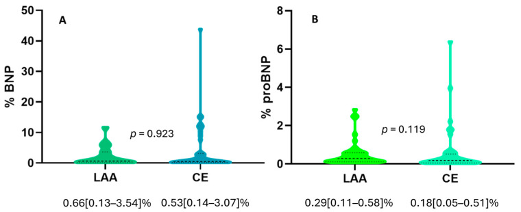
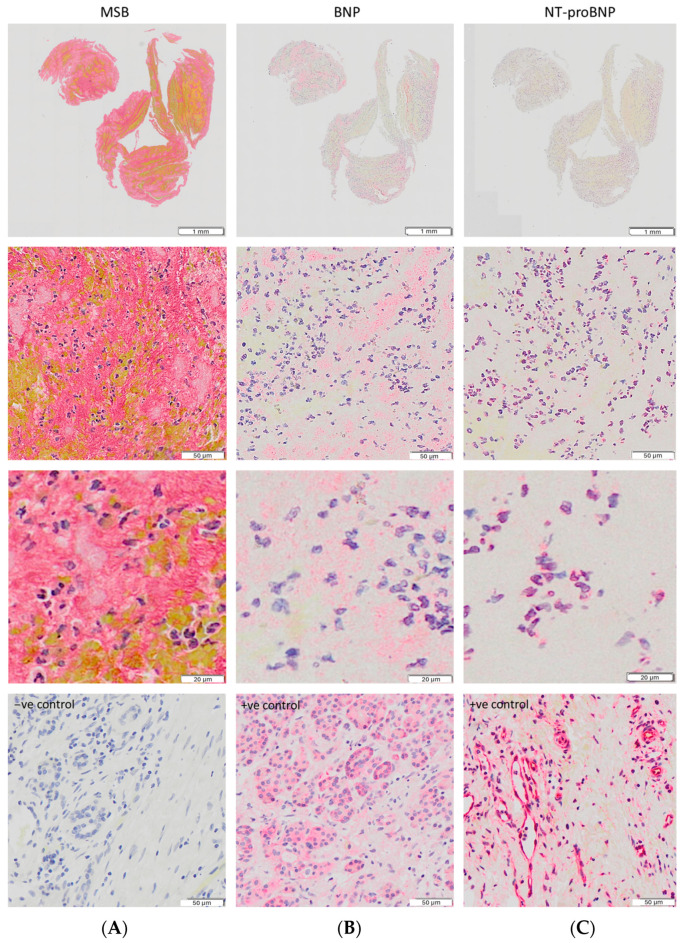
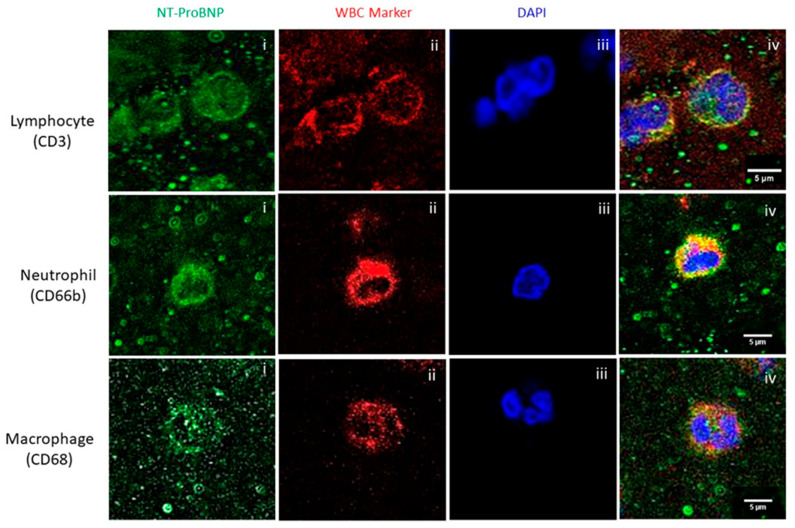
The need for biomarkers for acute ischemic stroke (AIS) to understand the mechanisms implicated in pathological clot formation is critical. The levels of the brain natriuretic peptides known as brain natriuretic peptide (BNP) and NT-proBNP have been shown to be increased in patients suffering from heart failure and other heart conditions. We measured their expression in AIS clots of cardioembolic (CE) and large artery atherosclerosis (LAA) etiology, evaluating their location inside the clots, aiming to uncover their possible role in thrombosis. We analyzed 80 thrombi from 80 AIS patients in the RESTORE registry of AIS clots, 40 of which were of CE and 40 of LAA etiology. The localization of BNP and NT-BNP, quantified using immunohistochemistry and immunofluorescence, in AIS-associated white blood cell subtypes was also investigated. We found a statistically significant positive correlation between BNP and NT-proBNP expression levels (Spearman's rho = 0.668 p < 0.0001 *). We did not observe any statistically significant difference between LAA and CE clots in BNP expression (0.66 [0.13-3.54]% vs. 0.53 [0.14-3.07]%, p = 0.923) or in NT-proBNP expression (0.29 [0.11-0.58]% vs. 0.18 [0.05-0.51]%, p = 0.119), although there was a trend of higher NT-proBNP expression in the LAA clots. It was noticeable that BNP was distributed throughout the thrombus and especially within platelet-rich regions. However, NT-proBNP colocalized with neutrophils, macrophages, and T-lymphocytes, suggesting its association with the thrombo-inflammatory process.
Keywords: BNP; NT-proBNP; acute ischemic stroke; brain natriuretic peptide; stroke biomarkers; stroke etiology; thrombus.
Conflict of interest statement
Authors M.G. and R.M. were employed by the company Cerenovus. The remaining authors declare that the research was conducted in the absence of any commercial or financial relationships that could be construed as a potential conflict of interest.
Figures
References
-
- Douglas A., Fitzgerald S., Mereuta O.M., Rossi R., O’Leary S., Pandit A., McCarthy R., Gilvarry M., Holmegaard L., Abrahamsson M., et al. Platelet-rich emboli are associated with von Willebrand factor levels and have poorer revascularization outcomes. J. Neurointerv. Surg. 2020;12:557–562. doi: 10.1136/neurintsurg-2019-015410. - DOI - PubMed
-
- Rossi R., Molina S., Mereuta O.M., Douglas A., Fitzgerald S., Tierney C., Pandit A., Brennan P., Power S., O’Hare A., et al. Does prior administration of rtPA influence acute ischemic stroke clot composition? Findings from the analysis of clots retrieved with mechanical thrombectomy from the RESTORE registry. J. Neurol. 2022;269:1913–1920. doi: 10.1007/s00415-021-10758-5. - DOI - PMC - PubMed
-
- Fitzgerald S., Rossi R., Mereuta O.M., Jabrah D., Okolo A., Douglas A., Gil S.M., Pandit A., McCarthy R., Gilvarry M., et al. Per-pass analysis of acute ischemic stroke clots: Impact of stroke etiology on extracted clot area and histological composition. J. Neurointerv. Surg. 2021;13:1111–1116. doi: 10.1136/neurintsurg-2020-016966. - DOI - PMC - PubMed
MeSH terms
Substances
Grants and funding
LinkOut - more resources
Full Text Sources
Medical
Research Materials
