. 2024 Feb 23;13(5):382.
doi: 10.3390/cells13050382.

DPPA2/4 Promote the Pluripotency and Proliferation of Bovine Extended Pluripotent Stem Cells by Upregulating the PI3K/AKT/GSK3β/β-Catenin Signaling Pathway

Affiliations

DPPA2/4 Promote the Pluripotency and Proliferation of Bovine Extended Pluripotent Stem Cells by Upregulating the PI3K/AKT/GSK3β/β-Catenin Signaling Pathway

Shu Fang et al. Cells. .

Abstract

Developmental pluripotency-associated 2 (DPPA2) and DPPA4 are crucial transcription factors involved in maintaining pluripotency in humans and mice. However, the role of DPPA2/4 in bovine extended pluripotent stem cells (bEPSCs) has not been investigated. In this study, a subset of bEPSC-related differentially expressed genes (DEGs), including DPPA2 and DPPA4, was identified based on multiomics data (ATAC-seq and RNA-seq). Subsequent investigations revealed that double overexpression of DPPA2/4 facilitates the reprogramming of bovine fetal fibroblasts (BFFs) into bEPSCs, whereas knockout of DPPA2/4 in BFFs leads to inefficient reprogramming. DPPA2/4 overexpression and knockdown experiments revealed that the pluripotency and proliferation capability of bEPSCs were maintained by promoting the transition from the G1 phase to the S phase of the cell cycle. By activating the PI3K/AKT/GSK3β/β-catenin pathway in bEPSCs, DPPA2/4 can increase the nuclear accumulation of β-catenin, which further upregulates lymphoid enhancer binding factor 1 (LEF1) transcription factor activity. Moreover, DPPA2/4 can also regulate the expression of LEF1 by directly binding to its promoter region. Overall, our results demonstrate that DPPA2/4 promote the reprogramming of BFFs into bEPSCs while also maintaining the pluripotency and proliferation capability of bEPSCs by regulating the PI3K/AKT/GSK3β/β-catenin pathway and subsequently activating LEF1. These findings expand our understanding of the gene regulatory network involved in bEPSC pluripotency.

Keywords: DPPA2; DPPA4; LEF1; PI3K/AKT pathway; bEPSCs; pluripotency; proliferation; reprogramming.


Conflict of interest statement

The authors declare no conflicts of interest.

Figures

Figure 1
Figure 1
Identification of hub genes involved in EPSC reprogramming. (A) Principal component analysis (PCA) of the RNA-seq data between FFs and EPSCs. (B) Heatmap showing the correlation of the samples. (C) The differential gene expression analysis of human, mouse, and bovine. Significantly differentially expressed peaks are denoted by red and blue dots. (D,E) Venn diagrams showing the overlap of differentially upregulated (D) and downregulated (E) genes. (F) The Venn diagram shows genes with open chromatin in human EPSCs and human FFs. (G) The number of overlapping genes between up-regulated genes (RNA-seq) and genes with open chromatin in EPSCs only (ATAC-seq). (H) Eight hub genes were selected by the cytoHubba plugin in Cytoscape. (I) GO-molecular function analysis revealed that hub gene FGF4, DPPA2, DPPA4, and NANOG were significantly associated with chromatin binding (p value < 0.05). GO biological process analysis revealed that these hub genes were enriched mainly in system development and stem cell population maintenance.
Figure 2
Figure 2
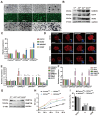
Reprogramming of BFFs into bEPSCs requires DPPA2 and DPPA4. (A) Experimental strategy for generating bovine EPSCs from BFFs. (B) AP staining of reprogrammed BFFs from the control, DPPA2/4-double overexpressing, and DPPA2/4-double knockout groups. (C) Karyotype analysis of bEPSCsC and bEPSCsA2/4. (D) AP staining for bEPSCsC and bEPSCsA2/4. GFP (bEPSCsA2/4) in the upper panel represents cell colonies successfully transfected by PB-CAG-bovine DPPA2/4, while GFP (bEPSCsC) in the lower panel represents the control group cell colonies successfully transfected by PB-CAG-GFP. (E) qRT-PCR analysis of key endogenous pluripotency genes in bEPSCsA2/4 and bEPSCsC. (F) Immunostaining of OCT4, SOX2, NANOG, CDX2, SSEA1, and SSEA4 in bEPSCsC and bEPSCsA2/4 cells. Scale bar, 50 μm. GFP in the left panel represents the control group cell clones successfully transfected with PB-CAG-GFP, whereas the GFP in the right panel represents cell clones successfully transfected with PB-CAG-bovine DPPA2/4. (G) Protein levels of OCT4 and SOX2 in bEPSCsC and bEPSCsA2/4; GAPDH served as a loading control. The data are presented as the means ± SDs; n = 3 independent experiments (* p < 0.05; ** p < 0.01; *** p < 0.001).
Figure 3
Figure 3
bEPSCsA2/4 cells exhibited greater differentiation ability than control bEPSCsC cells. (A) A schematic illustration of the generation of EBs from bEPSCsC and bEPSCsA2/4 and the morphologies of the EBs. Scale bar, 100 μm. (B) qRT-PCR analysis of EBs at day 20. (C) Immunostaining of AFP, GFAP, and SMA in the EBs derived from bEPSCsC and bEPSCsA2/4 cells at day 20. Scale bar, 100 μm. (D) Teratoma derived from bEPSCsC and bEPSCsA2/4 (passage 35). HE staining analysis revealed the presence of the three germ layers (mesoderm, ectoderm, and endoderm). Scale bar, 50 μm. The data are presented as the means ± SDs; n = 3 independent experiments (*** p < 0.001).
Figure 4
Figure 4
DPPA2/4 knockdown affects the pluripotency and early differentiation of bEPSCs. (A) Morphology and AP staining of ControlKD, DPPA2KD, DPPA4KD, and DPPA2/4KD bEPSCs. Scale bar, 50 μm. (B) Protein levels of OCT4, SOX2, and NANOG in ControlKD, DPPA2KD, DPPA4KD, and DPPA2/4KD bEPSCs. (C) qRT-PCR analysis of OCT4, SOX2, NANOG, and CDX2. (D) Immunofluorescence staining for SOX2 and NANOG in ControlKD, DPPA2KD, DPPA4KD, and DPPA2/4KD bEPSCs. Scale bars, 50 µm. (E) qRT-PCR analysis of T, GFAP, GBX2, ASCC1, FGF5, GATA4, HNF4A, and PAX6. (F) Western blotting analysis of DNMT3A and DNMT3B protein expression. (G) Proliferation of ControlKD, DPPA2KD, DPPA4KD, and DPPA2/4KD bEPSCs. (H) Cell cycle analysis of ControlKD, DPPA2KD, DPPA4KD, and DPPA2/4KD bEPSCs. (I) qRT-PCR analysis of cell cycle regulatory genes. (J) qRT-PCR was used to detect changes in the expression levels of pluripotent and differentiation marker genes at 1–3 days. (K) EB morphology of ControlKD, DPPA2KD, DPPA4KD, and DPPA2/4KD bEPSCs (n = 3). Scale bars, 100 µm. The data are presented as the means ± SDs; n = 3 independent experiments (* p < 0.05; ** p < 0.01; *** p < 0.001).
Figure 5
Figure 5
Overexpression of DPPA2 and DPPA4 increases the pluripotency of bEPSCs and promotes their proliferation. (A) AP staining of ControlOE, DPPA2OE, DPPA4OE, and DPPA2/4OE bEPSC colonies cultured for 3 days. Scale bars, 100 µm. GFP indicates the successfully transfected cells. The black arrow indicates the "dome" form of bEPSCs. (B) Protein levels of OCT4, SOX2, and NANOG in different treatment groups of bEPSCs. (C) qRT-PCR analysis of pluripotency marker genes and trophectoderm marker genes. (D) Immunofluorescence staining for OCT4, SOX2, and NANOG in ControlOE, DPPA2OE, DPPA4OE, and DPPA2/4OE bEPSCs. Scale bars, 50 µm. (E,F) qRT-PCR analysis of naïve and primed marker genes. (G) Western blotting analysis of DNMT3A and DNMT3B protein expression in different treatment groups of bEPSCs. (H) The proliferation rates of ControlOE, DPPA2OE, DPPA4OE, and DPPA2/4OE bEPSCs. (I) Cell cycle analysis of ControlOE, DPPA2OE, DPPA4OE, and DPPA2/4OE bEPSCs. The data are presented as the means ± SDs; n = 3 independent experiments (* p < 0.05; ** p < 0.01; *** p < 0.001).
Figure 6
Figure 6
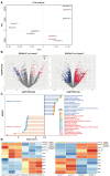
RNA-seq analysis revealed that DPPA2/4 affect the PI3K/AKT signaling pathway in bEPSCs. (A) PCA of DPPA2/4-knockdown and DPPA2/4-overexpressing RNA-seq datasets. (B) Volcano plot of the differentially expressed genes. (C) KEGG enrichment analysis of the differentially expressed genes. (D) Heatmap of PI3K/AKT pathway-related genes in ControlKD, DPPA2/4KD, ControlOE, and DPPA2/4OE bEPSCs.
Figure 7
Figure 7
Induction of LEF1 by DPPA2/4 activates the PI3K/AKT/GSK3β/β-catenin pathway and maintains cell proliferation and pluripotency. (A) Western blotting analysis of p-PI3K, PI3K, p-AKT, AKT, p-GSK3β, GSK3β, β-catenin, and LEF1 protein levels in DPPA2/4 knockdown and overexpression bEPSCs. (B) β-catenin localization was detected by immunofluorescence staining with an anti-β-catenin antibody in ControlOE, DPPA2/4OE, ControlKD, and DPPA2/4KD bEPSCs. Scale bars, 50 µm. (C) Western blotting analysis of p-PI3K, PI3K, p-AKT, AKT, p-GSK3β, GSK3β, p-β-catenin, and β-catenin protein expression in bEPSCs following treatment with the PI3K inhibitor LY294002. (D) qRT-PCR analysis of LEF1 expression in different cell lines. (E) Western blotting analysis of OCT4, SOX2, NANOG, DPPA2, and DPPA4 protein expression in different treatment groups of bEPSCs. (F) Immunofluorescence images were obtained by the EdU incorporation (red, Alexa Fluor 555) assay. Scale bars, 20 µm. (G) Cell cycle analysis of LEF1-knockdown bEPSCs. The percentages of cells in different phases are indicated. (H) Prediction results of the binding of DPPA2/4 to the site upstream of the TSS of LEF1. (I) ChIP–qPCR was used to detect DPPA2/4 binding to the LEF1 promoter region in bEPSCs. The data are presented as the means ± SDs; n = 3 independent experiments ** p < 0.01; *** p < 0.001.). (J) The hypothesized model for the signaling pathways involved in DPPA2/4-induced bEPSC proliferation and pluripotency.

Similar articles

Cited by

References

    1. Yang Y., Liu B., Xu J., Wang J., Wu J., Shi C., Xu Y., Dong J., Wang C., Lai W., et al. Derivation of Pluripotent Stem Cells with In Vivo Embryonic and Extraembryonic Potency. Cell. 2017;169:243–257.e225. doi: 10.1016/j.cell.2017.02.005. - DOI - PMC - PubMed
    1. Yang J., Ryan D.J., Wang W., Tsang J.C., Lan G., Masaki H., Gao X., Antunes L., Yu Y., Zhu Z., et al. Establishment of mouse expanded potential stem cells. Nature. 2017;550:393–397. doi: 10.1038/nature24052. - DOI - PMC - PubMed
    1. Gao X., Nowak-Imialek M., Chen X., Chen D., Herrmann D., Ruan D., Chen A.C.H., Eckersley-Maslin M.A., Ahmad S., Lee Y.L., et al. Establishment of porcine and human expanded potential stem cells. Nat. Cell Biol. 2019;21:687–699. doi: 10.1038/s41556-019-0333-2. - DOI - PMC - PubMed
    1. Yang J., Ryan D.J., Lan G., Zou X., Liu P. In vitro establishment of expanded-potential stem cells from mouse pre-implantation embryos or embryonic stem cells. Nat. Protoc. 2019;14:350–378. doi: 10.1038/s41596-018-0096-4. - DOI - PubMed
    1. Zhao L., Gao X., Zheng Y., Wang Z., Zhao G., Ren J., Zhang J., Wu J., Wu B., Chen Y., et al. Establishment of bovine expanded potential stem cells. Proc. Natl. Acad. Sci. USA. 2021;118:e2018505118. doi: 10.1073/pnas.2018505118. - DOI - PMC - PubMed

Publication types

MeSH terms

LinkOut - more resources