Review
. 2024 Feb 27;13(5):418.
doi: 10.3390/cells13050418.

Hedgehog on the Move: Glypican-Regulated Transport and Gradient Formation in Drosophila

Affiliations
Review

Hedgehog on the Move: Glypican-Regulated Transport and Gradient Formation in Drosophila

Carlos Jiménez-Jiménez et al. Cells. .

Abstract

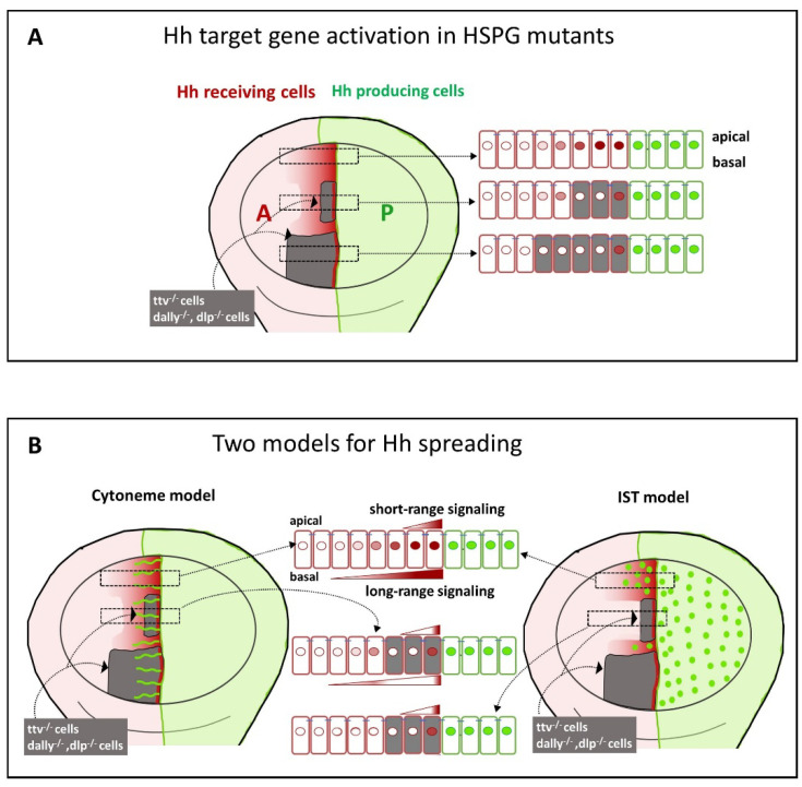
Glypicans (Glps) are a family of heparan sulphate proteoglycans that are attached to the outer plasma membrane leaflet of the producing cell by a glycosylphosphatidylinositol anchor. Glps are involved in the regulation of many signalling pathways, including those that regulate the activities of Wnts, Hedgehog (Hh), Fibroblast Growth Factors (FGFs), and Bone Morphogenetic Proteins (BMPs), among others. In the Hh-signalling pathway, Glps have been shown to be essential for ligand transport and the formation of Hh gradients over long distances, for the maintenance of Hh levels in the extracellular matrix, and for unimpaired ligand reception in distant recipient cells. Recently, two mechanistic models have been proposed to explain how Hh can form the signalling gradient and how Glps may contribute to it. In this review, we describe the structure, biochemistry, and metabolism of Glps and their interactions with different components of the Hh-signalling pathway that are important for the release, transport, and reception of Hh.

Keywords: Dally; Dally like; Hedgehog; glypicans; heparan sulphate proteoglycans.


Conflict of interest statement

The authors declare no conflicts of interest.

Figures

Figure 1
Figure 1
Overview of Glp structure and predicted cell surface distribution. (A) Representation of Glp1 lacking the most C-terminal disordered domain (Glp1DC) [24]. The disordered C-terminal Glp domain (aa Asn474-Ser530) contains the attachment sites of three closely spaced HS chains located close to the folded core (linked to serine residues Ser486, Ser488, and Ser490) and connects the Glp core domain to the GPI anchor. The HS structures were resolved separately [25] and manually added to the schematic. Three different lobes can be assigned to the Glp1DC structure: The cysteine-rich N-lobe, the central or M-lobe, and the C-lobe (also called the protease lobe because it has been described in many Glp family members to be susceptible to processing by furin proteases [26]). The structure of Glp1 is very similar to that of Drosophila melanogaster Dlp, despite only 25% sequence similarity. Note that the GPI membrane anchor and the unstructured flexible C-terminal domain give the core a large degree of freedom to tilt, move laterally, and rotate relative to the membrane (arrows). Shown are protein data bank (pdb) structures 3irl (HS) and 4ad7 (Glp). The structures are not to scale. (B) HS biosynthesis starts with a xylose residue (star) linked to a serine of the proteoglycan protein, followed by two galactose (circles) and a glucuronic acid residue (diamond). The subsequent addition of an N-acetylglucosamine residue (square) to the tetrasaccharide linker region initiates the biosynthesis of HS chains by the HS copolymerase complex. The growing chain (eventually consisting of 50–150 sugar residues) is simultaneously modified by N- and 2O-, 3O-, and 6O-sulphotransferases and an epimerase that generates iduronic acid residues (inverted diamond) from glucuronic acid residues. In vertebrates, high sulphated domains (NS domains) are separated by low-sulphated domains called NA domains (top). In contrast, Drosophila HS consists of a continuous sulphated domain (bottom) [27]. (C) A bird’s eye view of modelled multiple highly dynamic (brown arrows) interaction sites of Glp HS chains with neighbouring Glp core proteins, other HS chains and lipid head groups at the cell surface [28].
Figure 2
Figure 2
Wing imaginal disc as a model to study the function of Glps in Hh signalling. (A) The imaginal wing disc consists of a sheet of epithelial cells that form a sac-like fold of epithelium in fly larvae, called the wing pouch (marked in blue in the schematic representation of the polarised epithelium of the wing disc in a transverse section shown on the left), overlain by a fluid-filled closed compartment called the peripodial space (shown in white). Hh is produced throughout the posterior (P) compartment of the epithelial layer of the disc (green) and moves into the anterior (A) compartment to bind the Ptc receptor on the same epithelial layer (red). During this movement, Hh is thought to form a gradient of decreasing concentration with increasing distance from its source. Note that Hh movement must be confined to the epithelial layer to prevent morphogen loss into the overlying peripodial space, effectively ruling out free Hh diffusion as the underlying transport mode. Instead, Hh movement is thought to be confined to the epithelial layer by Glps. (B) Expression patterns of the Glps Dally and Dlp in the wing imaginal disc. Note that Dally shows reduced expression levels in the central area of the A-P boundary. Dlp shows higher expression levels in the A compartment adjacent to the P compartment and a marked decrease along the dorsoventral (D-V) axis.
Figure 3
Figure 3
Schematic of a possible mechanism of presentation and reception of Hh. (A) Model showing direct recycling of Hh and Ptc to the basal surface of the Drosophila wing disc epithelium. Hh (green) and Ptc (red) undergo vesicular trafficking from the apical to the basal surface of their respective expressing cells by endocytosis and multivesicular body (MVB) formation (arrows indicate the direction of vesicular trafficking). At the basal surface, thin extensions of the plasma membrane called cytonemes mediate the transfer of Hh from Hh-producing cells in the posterior (P) compartment to Ptc-expressing, Hh-receiving cells in the anterior (A) compartment [12,80,81]. Reception occurs at specific contact points on cytonemes, similar to synaptic boutons [85,86]. (B) Schematic of the synaptic process involved in Hh signalling. Hh is released to be received by Ptc at contact sites on cytonemes. Hh release from producer cells may involve Dispatched (Disp), Ihog, the Dally-like proteoglycan (Dlp), and the diffusible protein Shf. In receiving cells, Dlp and Ihog proteins are involved in the co-reception of Hh.
Figure 4
Figure 4
Effects of HS or Glp mutagenesis on Hh signalling in the wing disc and proposed mechanisms to explain these effects. (A) The wing imaginal disc, which later develops into the Drosophila wing, is divided into two compartments: the posterior (P) compartment, where Hh is produced (green), and the anterior (A) compartment, where Hh signals and induces the expression of target genes (red dots). Mutations in Glps, the HS biosynthetic enzymes of the EXT family, or in genes that determine the degree of N- and O-sulfation (such as the Drosophila gene sulphateless, clonal tissue is shown in grey) disrupt the formation of the Hh-signalling gradient in the A compartment (shown in red) [82,87,89]. Under these conditions, only clone cells directly adjacent to the P compartment are able to activate Hh signalling targets [17,70,87,89]. However, when the mutant clones are narrow, it has been shown that the low-threshold Hh-signalling targets can be activated beyond the anterior clone boundary, but not in all published experiments. (B) Two models have been proposed to explain how Hh spreads to the A compartment: the cytoneme model and the intersegmental transfer (IST) model. The cytoneme model suggests that cytonemes can traverse mutant territories and deliver Hh to distant cells as long as the mutant territories are small [17,85]. The IST model postulates direct morphogen transfer (green dots) from one Glp–HS chain to the next in the gradient field. In this model, lack of HSPG expression in small or large clones disrupts the formation of the Hh-signalling gradient in the A compartment (red) but still allows target activation in cells immediately adjacent to posterior Hh-producing cells.
Figure 5
Figure 5
Proposed Hh relay by the IST or “monkey bar” mechanism. In the monkey bar (or intersegmental transfer (IST)) mechanism, two HS-binding domains on the Hh protein interact simultaneously with one or two HS chains. In the first situation, the protein can move along the sugar chain (sliding) and in the second situation, it can switch directly between them without a diffusible intermediate (intersegmental transfer). The process is initiated by flexible N-terminal tail interactions with the new acceptor chain, followed by Hh switching through an intermediate in which the second binding site is retained on the original HS chain. This “monkey bar” mechanism avoids intermittent steps of free protein diffusion and protein loss from the HS chain. We note that the same mechanism could potentially be used by Hhs associated with lipid carriers such as lipoprotein particles [10] and exosomes [13], or after their association in micelles [9]. Shown are the pdb structures 3irl (HS), 4ad7 (Glp), and 3m1n (Shh). The structures are not to scale. Brown arrows indicate the dynamic movement of Glp HSPGs on the cell surface and black arrows indicate electrostatically restricted Hh movement on and between the HS chains.
Figure 6
Figure 6
Possible roles of HSPGs in the two models of Hh signaling. (A) During Hh trafficking in wild-type epithelia, Hh protein is exchanged between Hh-producing and Hh-receiving cells in a process that requires Glp HSPGs. One possibility is that Glp-associated Hh moves directly on the apical cell surface via the HS chains of the Glps to form a signalling gradient (short-range but possibly also long-range). In the cytoneme-signalling model in wild-type epithelia, Hh is exchanged at basal cytoneme contact sites in a synapse-like process. In this exchange process, Glps play a key role in facilitating both cytoneme contact and Hh reception. (B) Hh transport in Glp-HSPG-deficient clones (ttv-/-, or dally-/- dlp-/-), signalling occurs only in the first row of Hh-receiving cells of the A compartment. This is presumably because Ptc can contact Hh exposed by the Hh-producing cells, with the HSPGs in the P compartment possibly rescuing the first row of cells touching the A/P boundary in a non-autonomous manner. In the clone interior, however, Hh-loaded cytonemes protruding from Hh-producing cells are unable to contact the Glp-deficient Hh-receiving cells, preventing the activation of Hh-signalling targets. However, Hh-loaded cytonemes can pass through these Glp-deficient areas and contact wild-type cells anterior to the clone, allowing activation of low Hh-signalling targets far from the A/P compartment boundary. Alternatively, the IST model proposes that the lack of HS acceptors for mobile Hh in the HSPG-deficient clone inhibits Hh transport at the cell surface and traps Hh at the clone boundary.

Similar articles

Cited by

References

    1. Rogers K., Schier A.F. Morphogen Gradients: From Generation to Interpretation. Annu. Rev. Cell Dev. Biol. 2011;27:377–407. doi: 10.1146/annurev-cellbio-092910-154148. - DOI - PubMed
    1. Fico A., Maina F., Dono R. Fine-tuning of cell signaling by glypicans. Cell. Mol. Life Sci. 2011;68:923–929. doi: 10.1007/s00018-007-7471-6. - DOI - PMC - PubMed
    1. Bilioni A., Sánchez-Hernández D., Callejo A., Gradilla A.-C., Ibáñez C., Mollica E., Rodríguez-Navas M.C., Simon E., Guerrero I. Balancing Hedgehog, a retention and release equilibrium given by Dally, Ihog, Boi and shifted/DmWif. Dev. Biol. 2013;376:198–212. doi: 10.1016/j.ydbio.2012.12.013. - DOI - PubMed
    1. Norman M., Vuilleumier R., Springhorn A., Gawlik J., Pyrowolakis G. Pentagone internalises glypicans to fine-tune multiple signalling pathways. eLife. 2016;5:e13301. doi: 10.7554/eLife.13301. - DOI - PMC - PubMed
    1. Kolluri A., Ho M. The Role of Glypican-3 in Regulating Wnt, YAP, and Hedgehog in Liver Cancer. Front. Oncol. 2019;9:708. doi: 10.3389/fonc.2019.00708. - DOI - PMC - PubMed

Publication types

LinkOut - more resources