. 2024 Mar 5;13(5):455.
doi: 10.3390/cells13050455.

Nicotinamide Riboside Augments Human Macrophage Migration via SIRT3-Mediated Prostaglandin E2 Signaling

Affiliations

Nicotinamide Riboside Augments Human Macrophage Migration via SIRT3-Mediated Prostaglandin E2 Signaling

Jing Wu et al. Cells. .

Abstract

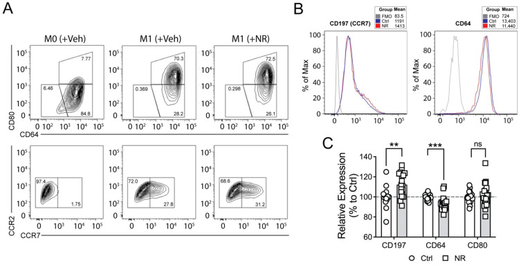
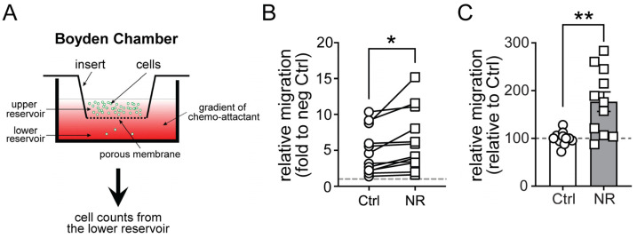
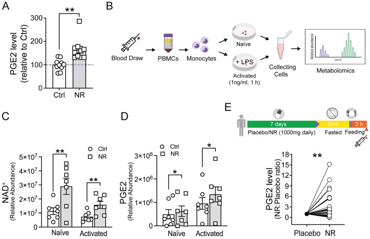
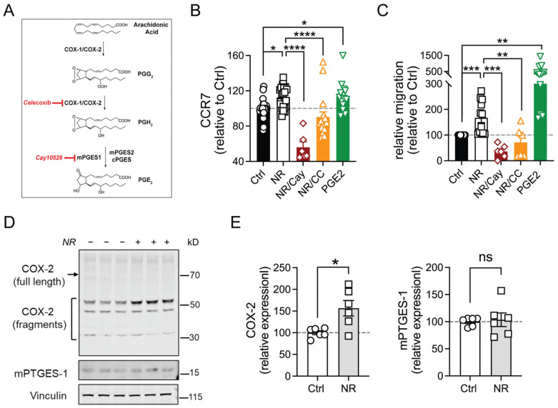
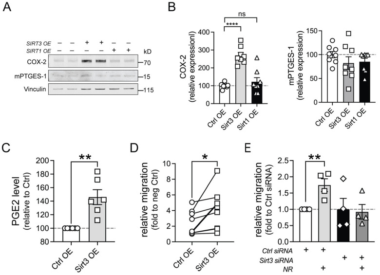
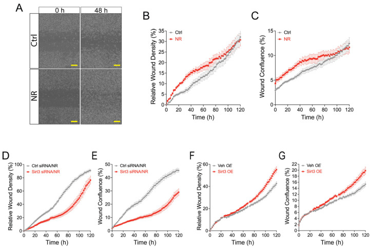
NAD+ boosting via nicotinamide riboside (NR) confers anti-inflammatory effects. However, its underlying mechanisms and therapeutic potential remain incompletely defined. Here, we showed that NR increased the expression of CC-chemokine receptor 7 (CCR7) in human M1 macrophages by flow cytometric analysis of cell surface receptors. Consequently, chemokine ligand 19 (CCL19, ligand for CCR7)-induced macrophage migration was enhanced following NR administration. Metabolomics analysis revealed that prostaglandin E2 (PGE2) was increased by NR in human monocytes and in human serum following in vivo NR supplementation. Furthermore, NR-mediated upregulation of macrophage migration through CCL19/CCR7 was dependent on PGE2 synthesis. We also demonstrated that NR upregulated PGE2 synthesis through SIRT3-dependent post-transcriptional regulation of cyclooxygenase 2 (COX-2). The NR/SIRT3/migration axis was further validated using the scratch-test model where NR and SIRT3 promoted more robust migration across a uniformly disrupted macrophage monolayer. Thus, NR-mediated metabolic regulation of macrophage migration and wound healing may have therapeutic potential for the topical management of chronic wound healing.

Keywords: NAD+ boosting; SIRT3; chemotaxis; macrophage migration; nicotinamide riboside; prostaglandin E2.


Conflict of interest statement

Dr. Sack’s laboratory studies mechanisms of action of nicotinamide riboside (NR) on immunomodulation in human inflammatory disease. His laboratory receives no direct financial support or compensation but does acquire NR and matching placebo from Chromadex Inc. through Cooperative and Development Research Agreements (CRADA) Material Transfer Agreements.

Figures

Figure 1
Figure 1
NR administration differentially regulates the expression of M1 macrophage markers in human MDMs. (A) Representative flow cytometry contour plots showing CD64/CD80 expression (top panel) and CCR2/CCR7 expression (bottom panel) in human M0 or M1 MDMs treated with vehicle or NR for 48 h. The gates represent surface marker expression as a percentage of living cells. The population of CCR7+ macrophages was increased following NR administration. CCR7 was not detected in M0 macrophages. CCR2 was low in both M0 and M1 macrophages. (B) Representative histograms showing cell surface expression of CCR7 (CD197) and CD64. Data shown are representative of 11–12 independent experiments. (C) Quantification of relative surface expression of CCR7 (CD197), CD64, and CD80 in human M1 MDM treated with vehicle (Ctrl) or NR. NR increased the surface expression of CCR7 (CD197), decreased CD64, and had no effect on CD80. Data are represented as mean ± SEM. ** p < 0.01; *** p < 0.001; ns, not significant. Unpaired two-tailed Student’s t-test.
Figure 2
Figure 2
NR administration increases CCR7 (CD197)-mediated migration in cultured human M1 MDMs. (A) Diagram showing the migration assay using the Boyden chamber apparatus followed by cell counting from the lower reservoir. (B) Pairwise comparison of relative migration in vehicle (Ctrl)- or NR-treated human M1 MDMs in response to CCL19. Each line represents one experiment. Human MDMs were derived from a total of 8 healthy subjects. (C) Quantification of relative migration in vehicle (Ctrl)- or NR-treated human M1 MDMs in response to CCL19. NR application increased CCL19/CCR7-mediated migration by ~70%. Data are represented as mean ± SEM. * p < 0.05; ** p < 0.01. Unpaired two-tailed Student’s t-test.
Figure 3
Figure 3
NR administration increases the PGE2 level in cultured human monocytes, MDMs, and human serum. (A) The PGE2 level in human primary M1 MDMs treated with vehicle or NR for 48 h (n = 10) was measured by the Parameter PGE2 Immunoassay (R&D systems). Human MDMs were derived from 3 healthy subjects. (B) Scheme showing unstimulated (naïve) and LPS-stimulated (activated) human monocytes treated with vehicle or NR for 24 h and collected for metabolomic analysis. (C,D) Relative abundance of NAD+ and PGE2 in vehicle control and NR-supplemented groups determined by metabolomics analysis (n = 10 healthy subjects). (E) (Inset) Design of the clinical protocol. The horizontal bar depicts volunteers consuming NR or placebo for 7 days followed by 24 h of fasting and 3 h of refeeding. The syringe symbol depicts the blood draw time point for serum collection and metabolomics. The PGE2 level in human serum after in vivo NR administration compared to placebo control was measured in a cohort of 36 healthy subjects. Data were analyzed using an unpaired two-tailed Student’s t-test (A,C,D) or paired two-tailed t-test (E). All data are represented as mean ± SEM. * p < 0.05; ** p < 0.01.
Figure 4
Figure 4
NR-mediated increases in CCR7 expression and CCL19-induced migration are attenuated by PGE2 synthesis blockers. (A) Diagram showing the PGE2 synthesis pathway and inhibitors used in this study. (B) Relative surface expression of CCR7 was measured by flow cytometry in human M1 MDMs treated with vehicle, NR (±inhibitor: Celecoxib (CC) or Cay10526 (Cay)), or PGE2 during the 48 h polarization. (C) Relative migration of human M1 MDMs treated with vehicle, NR (±inhibitor), or PGE2 in response to CCL19. (D) Representative immunoblot of PGE2 synthesis enzymes from human M1 MDMs treated with vehicle or NR for 48 h during the polarization. (E) Quantification of the COX-2 or mPTGES-1 level in vehicle (Ctrl)- or NR-incubated M1 MDMs, normalized to the vinculin level (n = 6 replicates, two experiments). Data were analyzed using a one-way ANOVA followed by Dunnett’s multiple comparisons test (B,C) or unpaired two-tailed Student’s t-test (E). All data are represented as mean ± SEM. * p < 0.05; ** p < 0.01; *** p < 0.001; **** p < 0.0001; ns, not significant.
Figure 5
Figure 5
SIRT3 is required for PGE2 synthesis and CCL19-induced migration elicited by NR administration. (A) Representative immunoblot of PGE2 synthesis enzymes from LPS-activated human monocytes transfected with an empty vector or SIRT1- or SIRT3-expression plasmids. (B) Quantification of the COX-2 or mPTGES-1 level in empty vector (Ctrl OE)-, SIRT1-, or SIRT3-transfected monocytes (n = 8 replicates from three experiments). (C) PGE2 levels from human M1 MDMs transfected with an empty vector (Ctrl OE) or SIRT3-expression plasmid (SIRT3 OE) measured using the Parameter PGE2 Immunoassay. Human M1 MDMs were derived from 6 healthy subjects. (D) Relative migration of human M1 MDMs transfected with an empty vector (Ctrl OE) or SIRT3-expression plasmid (SIRT3 OE) in response to CCL19. Human M1 MDMs were derived from 8 healthy subjects. (E) Relative migration of vehicle- or NR-treated human M1 MDMs transfected with either control siRNA or SIRT3 siRNA in response to CCL19. Human M1 MDMs were derived from 4 healthy subjects. Data were analyzed using a one-way ANOVA followed by Dunnett’s multiple comparisons test (B,E) or an unpaired two-tailed Student’s t-test (C,D). All data are represented as mean ± SEM. * p < 0.05; ** p < 0.01; **** p < 0.0001; ns, not significant.
Figure 6
Figure 6
NR facilitates collective cell migration in a SIRT3-dependent manner in human M1 MDMs during wounding. (A) Human MDMs were subjected to standardized wounding and wound closure was monitored by an IncuCyte Live-Cell Analysis System. The images show closure of the wound for up to 48 h post-wounding in control vs. NR-treated MDMs. The scale bars denote 200 μM. (B,C) Relative wound density (RWD) and wound confluence (WC) showing wound closure of human MDMs treated with vehicle (control) or NR over time. (D,E) RWD and WC showing temporal wound closure of NR-treated human MDMs transfected with control siRNA or SIRT3 siRNA. (F,G) RWD and WC showing temporal wound closure of human MDMs transfected with empty vector (Veh OE) or SIRT3 (SIRT3 OE).

Similar articles

References

    1. Youm Y.H., Nguyen K.Y., Grant R.W., Goldberg E.L., Bodogai M., Kim D., D’Agostino D., Planavsky N., Lupfer C., Kanneganti T.D., et al. The ketone metabolite beta-hydroxybutyrate blocks NLRP3 inflammasome-mediated inflammatory disease. Nat. Med. 2015;21:263–269. doi: 10.1038/nm.3804. - DOI - PMC - PubMed
    1. Johnson J.B., Summer W., Cutler R.G., Martin B., Hyun D.-H., Dixit V.D., Pearson M., Nassar M., Tellejohan R., Maudsley S., et al. Alternate day calorie restriction improves clinical findings and reduces markers of oxidative stress and inflammation in overweight adults with moderate asthma. Free Radic. Biol. Med. 2007;42:665–674. doi: 10.1016/j.freeradbiomed.2006.12.005. - DOI - PMC - PubMed
    1. Fraser D.A., Thoen J., Djoseland O., Forre O., Kjeldsen-Kragh J. Serum levels of interleukin-6 and dehydroepiandrosterone sulphate in response to either fasting or a ketogenic diet in rheumatoid arthritis patients. Clin. Exp. Rheumatol. 2000;18:357–362. - PubMed
    1. Fann D.Y.-W., Santro T., Manzanero S., Widiapradja A., Cheng Y.-L., Lee S.-Y., Chunduri P., Jo D.-G., Stranahan A.M., Mattson M.P., et al. Intermittent fasting attenuates inflammasome activity in ischemic stroke. Exp. Neurol. 2014;257:114–119. doi: 10.1016/j.expneurol.2014.04.017. - DOI - PubMed
    1. Choi I.Y., Piccio L., Childress P., Bollman B., Ghosh A., Brandhorst S., Suarez J., Michalsen A., Cross A.H., Morgan T.E., et al. A Diet Mimicking Fasting Promotes Regeneration and Reduces Autoimmunity and Multiple Sclerosis Symptoms. Cell Rep. 2016;15:2136–2146. doi: 10.1016/j.celrep.2016.05.009. - DOI - PMC - PubMed

Publication types