. 2024 Mar 5;13(5):458.
doi: 10.3390/cells13050458.

Chlorine-Induced Toxicity on Murine Cornea: Exploring the Potential Therapeutic Role of Antioxidants

Affiliations

Chlorine-Induced Toxicity on Murine Cornea: Exploring the Potential Therapeutic Role of Antioxidants

Seungwon An et al. Cells. .

Abstract

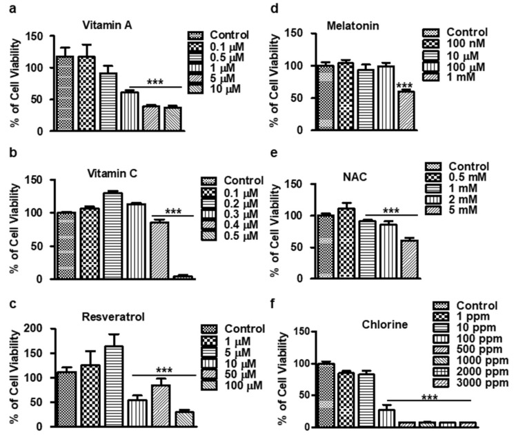
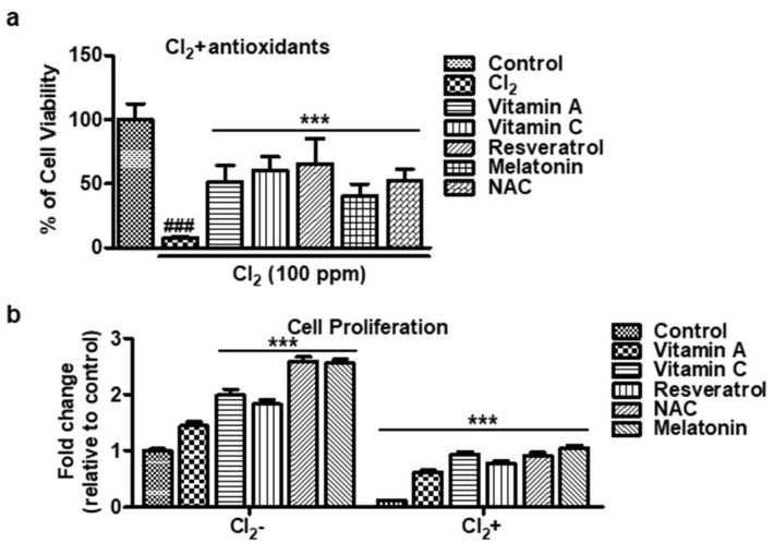
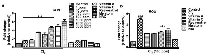
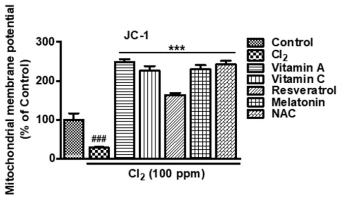
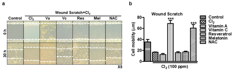
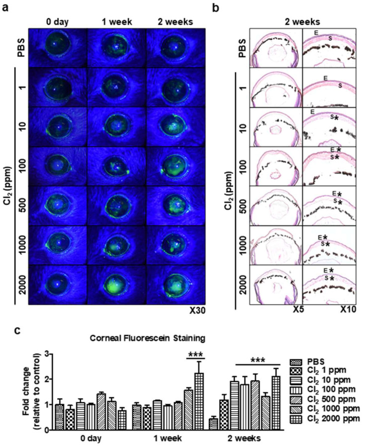
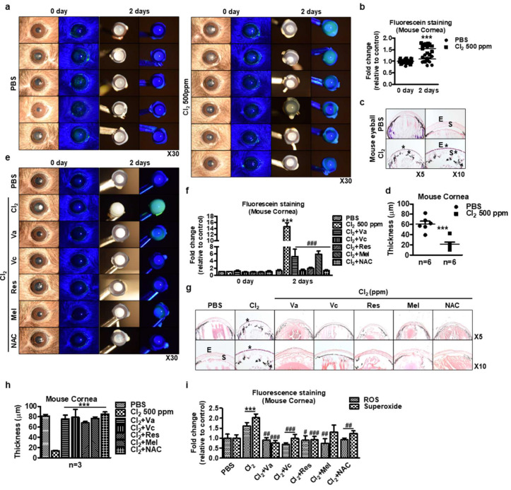
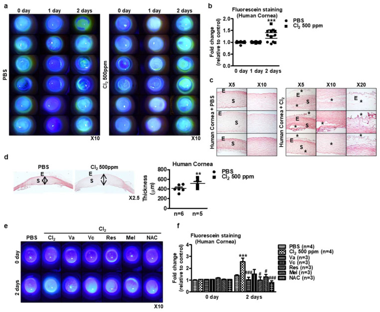
Chlorine (Cl2) exposure poses a significant risk to ocular health, with the cornea being particularly susceptible to its corrosive effects. Antioxidants, known for their ability to neutralize reactive oxygen species (ROS) and alleviate oxidative stress, were explored as potential therapeutic agents to counteract chlorine-induced damage. In vitro experiments using human corneal epithelial cells showed decreased cell viability by chlorine-induced ROS production, which was reversed by antioxidant incubation. The mitochondrial membrane potential decreased due to both low and high doses of Cl2 exposure; however, it was recovered through antioxidants. The wound scratch assay showed that antioxidants mitigated impaired wound healing after Cl2 exposure. In vivo and ex vivo, after Cl2 exposure, increased corneal fluorescein staining indicates damaged corneal epithelial and stromal layers of mice cornea. Likewise, Cl2 exposure in human ex vivo corneas led to corneal injury characterized by epithelial fluorescein staining and epithelial erosion. However, antioxidants protected Cl2-induced damage. These results highlight the effects of Cl2 on corneal cells using in vitro, ex vivo, and in vivo models while also underscoring the potential of antioxidants, such as vitamin A, vitamin C, resveratrol, and melatonin, as protective agents against acute chlorine toxicity-induced corneal injury. Further investigation is needed to confirm the antioxidants' capacity to alleviate oxidative stress and enhance the corneal healing process.

Keywords: JC-1; ROS; antioxidants; chlorine; wound healing.


Conflict of interest statement

The authors declare no conflicts of interest.

Figures

Figure 1
Figure 1
Cytotoxicity assay of antioxidants in the HCECs. (af) The cells were treated for 24 h with varying concentrations. The cell viability is reported as the percentage of the control group (100%). All data are presented as the mean ± SEM (n = 6). A significant difference *** p < 0.001 using one-way ANOVA analysis witsh Tukey’s post hoc analysis was observed in the percentage of cell viability vs. the control group (untreated). NAC: N-Acetyl Cysteine.
Figure 2
Figure 2
Cell proliferation of antioxidants in Cl2-treated HCECs. (a,b) The HCECs are exposed to 100 ppm Cl2 for 30 min and are followed by treatment with antioxidants for 24 h. The results indicate the percentage of cell proliferation vs. the control cells (untreated). Values are the mean ± SEM (n = 6). The data were analyzed by one-way ANOVA analysis with Tukey’s post hoc analysis. A significant difference, ### p < 0.001 was observed in the percentage of cell viability vs. untreated cells and Cl2-treated cells. A significant difference, *** p < 0.001 was observed in the percentage of cell viability vs. antioxidant-treated cells. Vitamin A: 100 μM, vitamin C: 300 μM, resveratrol: 5 μM, melatonin: 100 μM, NAC: 500 μM.
Figure 3
Figure 3
Effect of antioxidants on Cl2-induced ROS production in HCECs. (a,b) The HCECs were pretreated with Cl2 for 30 min followed by 24 h of antioxidant treatment. The results indicate the fold change of ROS level vs. the control cells (untreated). Values are the mean ± SEM (n = 6). A significant difference, *** p < 0.001 was observed in the fold change of ROS vs. untreated cells and Cl2-treated cells. ### p < 0.001 was observed in the fold change of ROS vs. untreated cells and antioxidant-treated cells. Vitamin A: 100 μM, vitamin C: 300 μM, resveratrol: 5 μM, melatonin: 100 μM, NAC: 500 μM.
Figure 4
Figure 4
Mitochondrial membrane potential in HCECs. Cells are exposed to 100 ppm Cl2 for 30 min prior to treating antioxidants. The results indicate the percentage of mitochondrial membrane potential vs. the control cells (untreated). Values are the mean ± SEM (n = 6). A significant difference, ### p < 0.001 was observed in the percentage of cell viability vs. untreated cells. A significant difference, *** p < 0.001 was observed in the percentage of cell viability vs. Cl2-treated cells. Vitamin A: 100 μM, vitamin C: 300 μM, resveratrol: 5 μM, melatonin: 100 μM, NAC: 500 μM.
Figure 5
Figure 5
Wound-healing assay to detect cell migration of HCECs. (a,b) Wound scratch in HCECs exposed to 100 ppm Cl2 for 30 min prior to treating antioxidants. (a) Representative images showing scratch wound assay in HCLE cells. White dot: wound area. (b) Graph showing wound healing rate for different conditions in epithelial scratch wounds (n = 5/group) at 30 h. *** p < 0.001 was observed in the cell mobility (μm) vs. untreated cells and Cl2-treated cells. Va: vitamin A, Vc: vitamin C, Res: resveratrol, Mel: melatonin, vitamin A: 100 μM, vitamin C: 300 μM, resveratrol: 5 μM, melatonin: 100 μM, NAC: 500 μM.
Figure 6
Figure 6
In vivo evaluation of chlorine’s impact on mice eyes using corneal fluorescein staining. Mice corneas were applied to various doses of Cl2 (1, 10, 100, 500, 1000, and 2000 ppm; 10 µL, 30 s) once a day for 2 weeks. (a) Representative images of murine corneas showing fluorescein staining with Cl2 treatment. (b) H&E staining of various doses of Cl2-treated murine corneas. E: epithelium, S: stroma. *: damaged area. (c) Graph showing the intensity fold change of corneal fluorescein staining after application of Cl2 treatment (n = 4/group) for 2 weeks. Values are the mean ± SEM (n = 4). The results indicate that corneal fluorescein staining was greatly increased in a dose-dependent manner compared to the control group (PBS-treated). A significant difference, *** p < 0.001 was observed in the fold change of fluorescein staining vs. the control groups (PBS-treated on 1 week or 2 weeks).
Figure 7
Figure 7
Ex vivo evaluation of chlorine’s effects on mice eyeballs. (ac) Mouse eyeballs were exposed to 500 ppm Cl2 for 2 h. Subsequently, the eyeballs were washed two times and then incubated in 1x PBS for 2 days. (a) Representative images of murine whole eyeballs showing fluorescein staining with or without Cl2 treatment. (b) Graph showing the intensity fold change of corneal fluorescein staining after application of Cl2 treatment (n = 12/group) for 2 days. Values are the mean ± SEM (n = 11). The results indicate that corneas with 500 ppm Cl2 became opaque and hazy with more fluorescein staining of the cornea and conjunctiva than the PBS-treated group. (c) H&E staining on murine whole eyeballs after application of PBS or chlorine 500 ppm for 2 days. E: epithelium, S: stroma. *: damaged area. (d) Murine cornea thickness after 500 ppm Cl2 exposure. *** p < 0.001 was observed in the cornea thickness vs. the control group (PBS treatment). Values are the mean ± SEM (PBS: n = 11, Cl2: n = 12). (e) Representative images of murine whole eyeballs showed fluorescein staining. (f) Graph showing the intensity fold change of corneal fluorescein staining after application of antioxidants (n = 4/group) for 2 days. *** p < 0.001 was observed in the corneal fluorescein staining vs. the control group (PBS-treated on day 2). ### p < 0.001 was observed in the corneal fluorescein staining vs. Cl2-treated group on day 2. (g) H&E staining on murine whole eyeballs with antioxidants after application of Cl2 500 ppm for 2 days. E: epithelium, S: stroma. *: damaged area. Vitamin A (Va): 100 μM, vitamin C (Vc): 300 μM, resveratrol (Res): 5 μM, melatonin (Mel): 100 μM, and NAC: 500 μM for 2 days. (h) Murine cornea thickness. *** p < 0.001 was observed in the cornea thickness vs. the control group (PBS-treated). (i) Relative fold changes of immunofluorescence intensity from Figure S1c. *** p < 0.001 was observed in the ROS and superoxide groups vs. the control group (PBS-treated). # p < 0.05, ## p < 0.01, ### p < 0.001 were observed in the ROS and superoxide groups vs. the Cl2 treatment group.
Figure 8
Figure 8
Ex vivo evaluation of Cl2 effects on human corneas. (ad) Human corneas were exposed to 500 ppm Cl2 for 3 days. (a) Representative images of human corneas showing fluorescein staining with or without Cl2 treatment. (b) Graph showing the intensity fold change of human cornea fluorescein staining after Cl2 exposure (n = 12/group) for 3 days. *** p < 0.001 was observed in the fold change of fluorescein staining vs. the control group (PBS). The results indicate that corneas exposure to 500 ppm Cl2 showed higher levels of fluorescein staining on both the cornea and conjunctival compared to the PBS-exposed group. (c) H&E staining of Cl2-treated human corneas. Black star: damaged area, E: epithelium, S: stroma. (d) Human cornea thickness after 500 ppm Cl2 exposure. ** p < 0.05 was observed in the cornea thickness vs. the control group (PBS). Values are the mean ± SEM (PBS: n = 12, Cl2: n = 12). (e) Representative images of human corneas showed fluorescein staining by Cl2 treatment and followed by antioxidants (Va, Vc, Res, Mel, and NAC). (f) Graph showing the intensity fold change of corneal fluorescein staining after application of antioxidants (n = 4/group) for 2 days. *** p < 0.001 was observed in the corneal fluorescein staining vs. the control group (PBS treatment on day 2). # p < 0.05, ### p < 0.001 were observed in the corneal fluorescein staining vs. Cl2-treated group on day 2.

Similar articles

Cited by

References

    1. Sridhar M.S. Anatomy of cornea and ocular surface. Indian J. Ophthalmol. 2018;66:190–194. doi: 10.4103/ijo.IJO_646_17. - DOI - PMC - PubMed
    1. Wilson S.E. Corneal wound healing. Exp. Eye Res. 2020;197:108089. doi: 10.1016/j.exer.2020.108089. - DOI - PMC - PubMed
    1. Andley U.P., Rhim J.S., Chylack L.T., Jr., Fleming T.P. Propagation and immortalization of human lens epithelial cells in culture. Investig. Ophthalmol. Vis. Sci. 1994;35:3094–3102. - PubMed
    1. Balasubramanian D. Ultraviolet radiation and cataract. J. Ocul. Pharmacol. Ther. 2000;16:285–297. doi: 10.1089/jop.2000.16.285. - DOI - PubMed
    1. Zigman S. Lens UVA photobiology. J. Ocul. Pharmacol. Ther. 2000;16:161–165. doi: 10.1089/jop.2000.16.161. - DOI - PubMed

Publication types