. 2024 Feb 22;29(5):953.
doi: 10.3390/molecules29050953.

Neuroprotective Effects of Polysaccharides and Gallic Acid from Amauroderma rugosum against 6-OHDA-Induced Toxicity in SH-SY5Y Cells

Affiliations

Neuroprotective Effects of Polysaccharides and Gallic Acid from Amauroderma rugosum against 6-OHDA-Induced Toxicity in SH-SY5Y Cells

Panthakarn Rangsinth et al. Molecules. .

Abstract

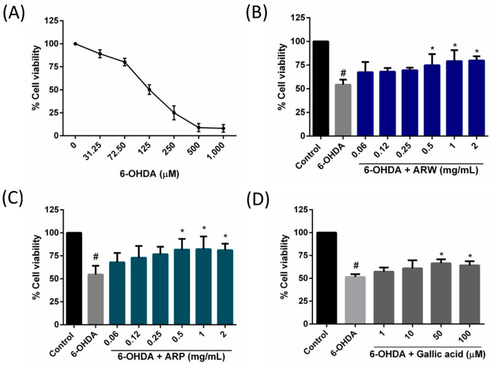
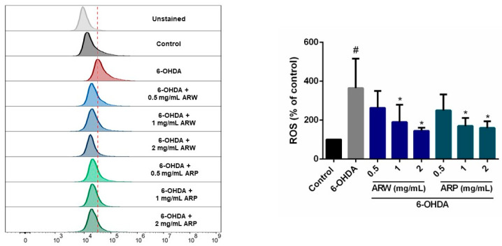
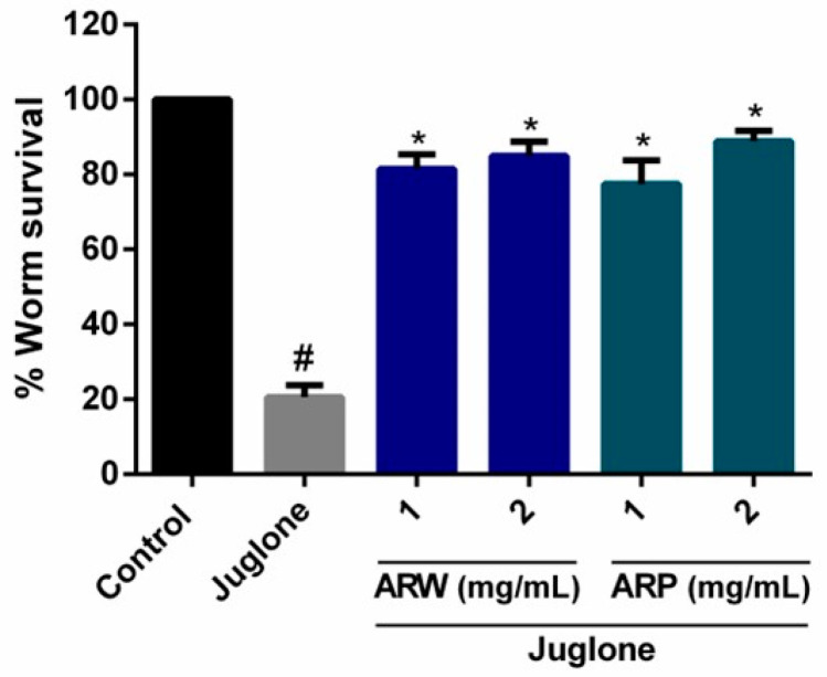
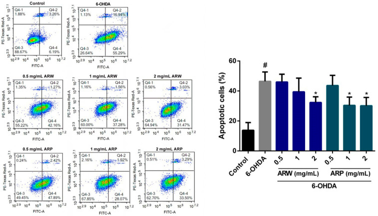
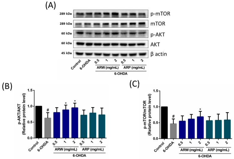
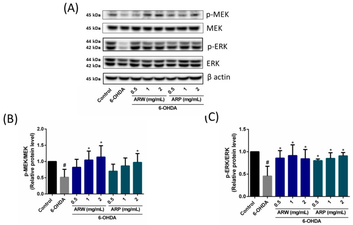
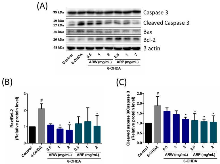
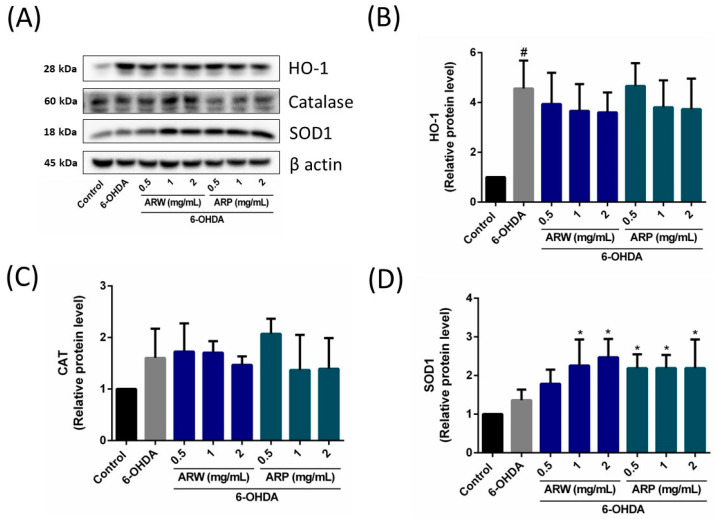
The pharmacological activity and medicinal significance of Amauroderma rugosum (AR) have rarely been documented. We examined the antioxidant and neuroprotective effects of AR on 6-hydroxydopamine (6-OHDA)-induced neurotoxicity in an SH-SY5Y human neuroblastoma cell model of Parkinson's disease (PD) and explored the active ingredients responsible for these effects. The results showed that the AR aqueous extract could scavenge reactive oxygen species and reduce SH-SY5Y cell death induced by 6-OHDA. In addition, the AR aqueous extract increased the survival of Caenorhabditis elegans upon juglone-induced toxicity. Among the constituents of AR, only polysaccharides and gallic acid exhibited antioxidant and neuroprotective effects. The AR aqueous extract reduced apoptosis and increased the expression of phospho-Akt, phospho-mTOR, phospho-MEK, phospho-ERK, and superoxide dismutase-1 in 6-OHDA-treated SH-SY5Y cells. The polysaccharide-rich AR extract was slightly more potent than the aqueous AR extract; however, it did not affect the expression of phospho-Akt or phospho-mTOR. In conclusion, the AR aqueous extract possessed antioxidant and neuroprotective properties against 6-OHDA-induced toxicity in SH-SY5Y cells. The mechanism of action involves the upregulation of the Akt/mTOR and MEK/ERK-dependent pathways. These findings indicate the potential utility of AR and its active ingredients in preventing or treating neurodegenerative disorders associated with oxidative stress such as PD.

Keywords: Amauroderma rugosum; antioxidant; gallic acid; neuroprotective; polysaccharides.


Conflict of interest statement

Author Timothy Man-Yau Cheung was employed by the company Tian Ran Healthcare Limited. The remaining authors declare that the research was conducted in the absence of any commercial or financial relationships that could be construed as a potential conflict of interest.

Figures

Figure 1
Figure 1
Effects of ARW, ARP, and gallic acid on protecting SH-SY5Y cells against 6-OHDA-induced cytotoxicity. (A) SH-SY5Y cells were treated with various concentrations of 6-OHDA for 24 h to evaluate their cytotoxicity. SH-SY5Y cell viability was then measured using the MTT assay. After obtaining the concentration-dependent response curve of 6-OHDA, the cells were pre-incubated with different concentrations of (B) ARW, (C) ARP, and (D) gallic acid for 2 h, followed by treatment with 125 µM 6-OHDA for 24 h. Untreated cells served as controls. Data are presented as the percentage relative to the control group values (mean ± SD of three independent experiments). # p < 0.05 indicates a statistically significant difference compared to the control group. * p < 0.05 indicates a statistically significant difference compared to the 6-OHDA-treated group.
Figure 2
Figure 2
Effects of ARW and ARP on intracellular reactive oxygen species levels in SH-SY5Y cells. SH-SY5Y cells were pre-treated with different concentrations of ARW, ARP, and gallic acid for 2 h and then treated with or without 125 μM of 6-OHDA for 24 h. Untreated cells served as the control. SH-SY5Y cells were subjected to CM-H2DCFDA staining and the fluorescence signals were quantified using flow cytometry analysis. Data are presented as a percentage of the control group values (mean ± SD of three independent experiments). # p < 0.05 indicates a statistically significant difference compared to the control group. * p < 0.05 indicates a statistically significant difference compared to the 6-OHDA-treated group.
Figure 3
Figure 3
Effects of ARW and ARP on Caenorhabditis elegans survival. Oxidative stress in C. elegans was induced by treatment with 80 µM juglone for 24 h. The survival rate of the worms was calculated. The experiment was conducted using a minimum of 100 worms per group. All values are reported as the mean ± SEM. # p < 0.05 indicates a statistically significant difference compared to the control group. * p < 0.05 indicates a statistically significant difference compared to the 6-OHDA-treated group.
Figure 4
Figure 4
Effects of ARW and ARP treatment on 6-OHDA-induced apoptosis in SH-SY5Y cells. SH-SY5Y cells were pretreated with different concentrations of ARW and ARP for 2 h and then treated with 125 μM of 6-OHDA for 24 h. Untreated cells served as the control. The cells were stained with annexin V-FITC and PI, and the number of apoptotic cells was quantified using flow cytometry. Data are presented as a percentage relative to the control group values (mean ± SD of three independent experiments). # p < 0.05 indicates a statistically significant difference compared to the control group. * p < 0.05 indicates a statistically significant difference compared to the 6-OHDA-treated group.
Figure 5
Figure 5
Effects of ARW and ARP treatment on the total and phosphorylated protein levels of Akt and mTOR in the SH-SY5Y cells. SH-SY5Y cells were treated with different concentrations of ARW and ARP for 2 h and then treated with 125 μM of 6-OHDA for 24 h. Untreated cells served as the control. (A) Protein expression levels of Akt, p-Akt, mTOR, and p-mTOR in the SH-SY5Y cells were determined using Western blotting analysis. β-Actin was used as a reference. (B,C) The amounts of the different proteins after being normalized to that of β-actin. Data are presented as the mean ± SD of three independent experiments. # p < 0.05 indicates a statistically significant difference compared to the control group. * p < 0.05 indicates a statistically significant difference compared to the 6-OHDA-treated group.
Figure 6
Figure 6
Effects of ARW and ARP treatment on the total and phosphorylated protein levels of MEK and ERK in the SH-SY5Y cells. SH-SY5Y cells were treated with different concentrations of ARW and ARP for 2 h and then treated with 125 μM of 6-OHDA for 24 h. Untreated cells served as the control. (A) Protein expression levels of MEK, p-MEK, ERK, and p-ERK in the SH-SY5Y cells were determined using Western blotting analysis. β-Actin was used as a reference. (B,C) The amounts of the different proteins after being normalized to that of β-actin. Data are presented as the mean ± SD of three independent experiments. # p < 0.05 indicates a statistically significant difference compared to the control group. * p < 0.05 indicates a statistically significant difference compared to the 6-OHDA-treated group.
Figure 7
Figure 7
Effects of ARW and ARP treatment on the protein levels of Bax, Bcl-2, caspase 3, and cleaved caspase 3 in SH-SY5Y cells. SH-SY5Y cells were treated with different concentrations of ARW and ARP for 2 h and then treated with 125 μM of 6-OHDA for 24 h. Untreated cells served as the control. (A) Protein expression levels of Bax, Bcl-2, caspase 3, and cleaved caspase 3 in SH-SY5Y cells were determined using western blotting analysis. β-Actin was used as a reference. (B,C) The amounts of the different proteins after being normalized to that of β-actin. Data are presented as the mean ± SD of three independent experiments. # p < 0.05 indicates a statistically significant difference compared to the control group. * p < 0.05 indicates a statistically significant difference compared to the 6-OHDA-treated group.
Figure 8
Figure 8
Effects of ARW and ARP treatment on the protein levels of heme-oxygenase (HO)-1, catalase (CAT), and superoxide dismutase (SOD)-1 in SH-SY5Y cells. SH-SY5Y cells were treated with different concentrations of ARW and ARP for 2 h and then treated with 125 μM of 6-OHDA for 24 h. Cells without any treatment served as the control. (A) Protein expression levels of HO-1, CAT, and SOD-1 in SH-SY5Y cells were determined using western blotting analysis. β-Actin was used as a reference. (BD) The amounts of the different proteins were normalized to that of β-actin. Data are presented as the mean ± SD of three independent experiments. # p < 0.05 indicates a statistically significant difference compared to the control group. * p < 0.05 indicates a statistically significant difference compared to the 6-OHDA-treated group.

Similar articles

Cited by

References

    1. Hirsch L., Jette N., Frolkis A., Steeves T., Pringsheim T. The incidence of Parkinson’s disease: A systematic review and meta-analysis. Neuroepidemiology. 2016;46:292–300. doi: 10.1159/000445751. - DOI - PubMed
    1. Elkouzi A., Vedam-Mai V., Eisinger R.S., Okun M.S. Emerging therapies in Parkinson disease—Repurposed drugs and new approaches. Nat. Rev. Neurol. 2019;15:204–223. doi: 10.1038/s41582-019-0155-7. - DOI - PMC - PubMed
    1. Guo J.D., Zhao X., Li Y., Li G.R., Liu X.L. Damage to dopaminergic neurons by oxidative stress in Parkinson’s disease (Review) Int. J. Mol. Med. 2018;41:1817–1825. doi: 10.3892/ijmm.2018.3406. - DOI - PubMed
    1. Mittal P., Dhankhar S., Chauhan S., Garg N., Bhattacharya T., Ali M., Chaudhary A.A., Rudayni H.A., Al-Zharani M., Ahmad W., et al. A review on natural antioxidants for their role in the treatment of Parkinson’s disease. Pharmaceuticals. 2023;16:908. doi: 10.3390/ph16070908. - DOI - PMC - PubMed
    1. Li J., Li R., Wu X., Hoo R.L., Lee S.M., Cheung T.M., Ho B.S., Leung G.P. Amauroderma rugosum protects PC12 Cells against 6-OHDA-induced neurotoxicity through antioxidant and antiapoptotic effects. Oxid. Med. Cell. Longev. 2021;2021:6683270. doi: 10.1155/2021/6683270. - DOI - PMC - PubMed

MeSH terms

Supplementary concepts