. 2024 Feb 23;16(5):623.
doi: 10.3390/nu16050623.

Sulforaphane Inhibits Adhesion and Migration of Cisplatin- and Gemcitabine-Resistant Bladder Cancer Cells In Vitro

Affiliations

Sulforaphane Inhibits Adhesion and Migration of Cisplatin- and Gemcitabine-Resistant Bladder Cancer Cells In Vitro

Hui Xie et al. Nutrients. .

Abstract

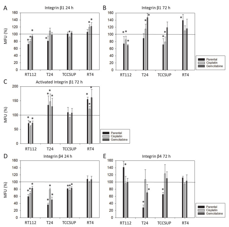
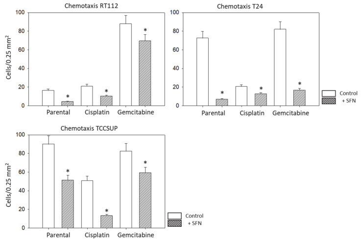
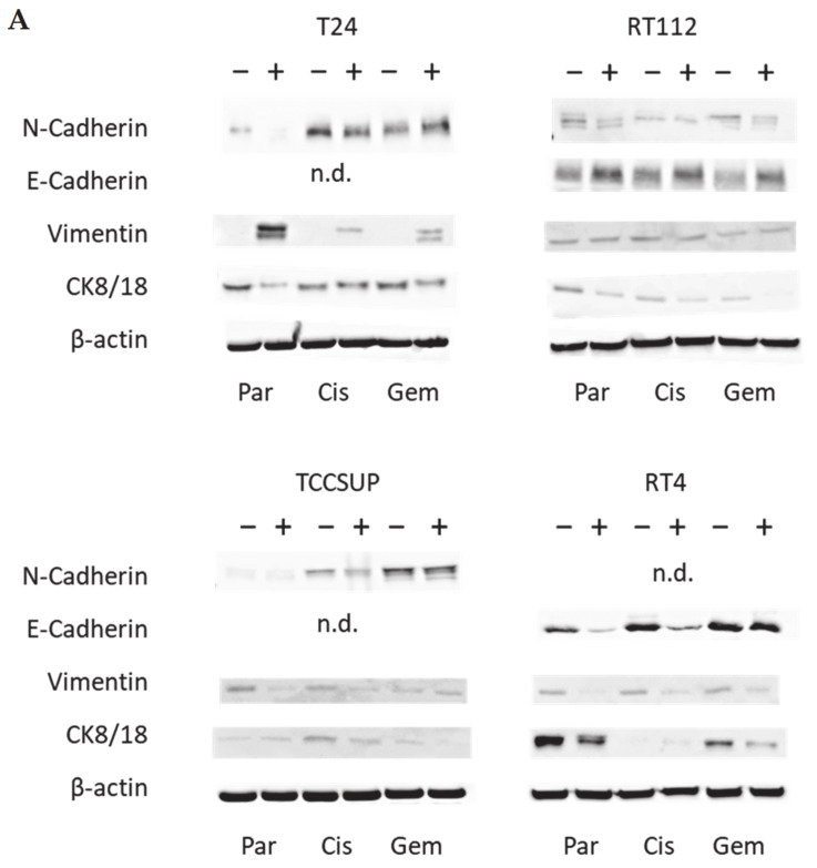
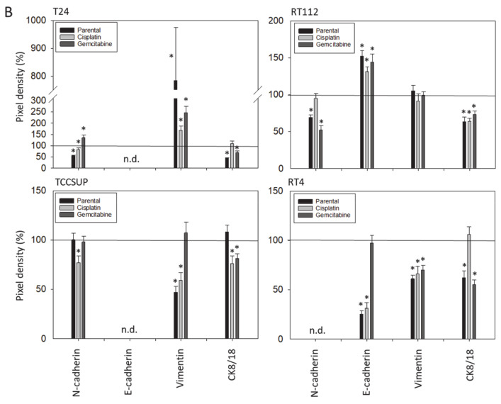
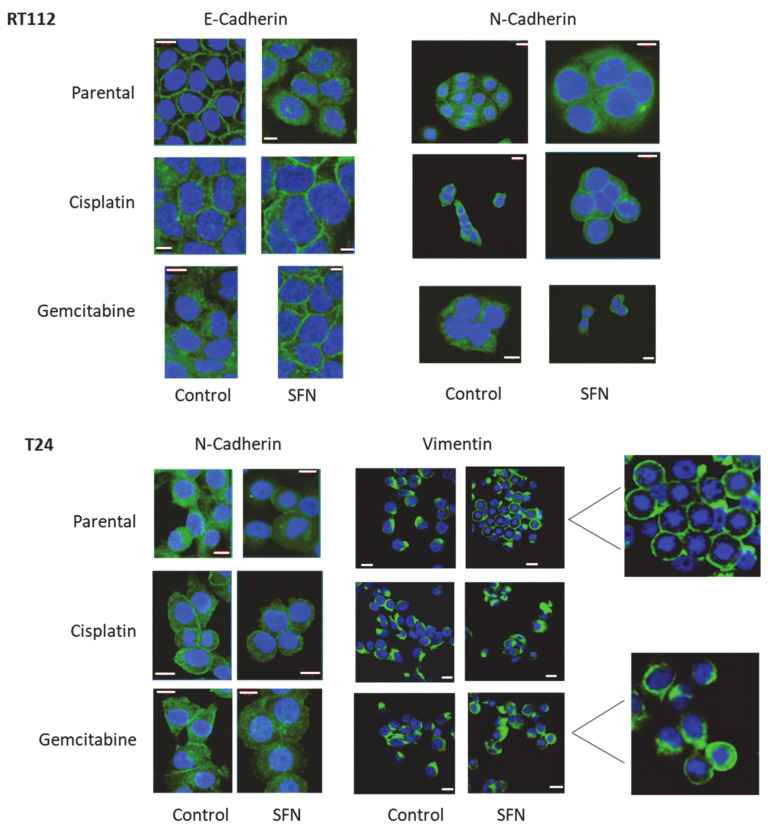
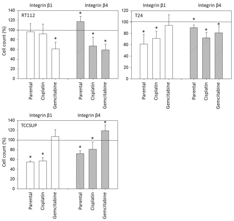
Only 20% of patients with muscle-invasive bladder carcinoma respond to cisplatin-based chemotherapy. Since the natural phytochemical sulforaphane (SFN) exhibits antitumor properties, its influence on the adhesive and migratory properties of cisplatin- and gemcitabine-sensitive and cisplatin- and gemcitabine-resistant RT4, RT112, T24, and TCCSUP bladder cancer cells was evaluated. Mechanisms behind the SFN influence were explored by assessing levels of the integrin adhesion receptors β1 (total and activated) and β4 and their functional relevance. To evaluate cell differentiation processes, E- and N-cadherin, vimentin and cytokeratin (CK) 8/18 expression were examined. SFN down-regulated bladder cancer cell adhesion with cell line and resistance-specific differences. Different responses to SFN were reflected in integrin expression that depended on the cell line and presence of resistance. Chemotactic movement of RT112, T24, and TCCSUP (RT4 did not migrate) was markedly blocked by SFN in both chemo-sensitive and chemo-resistant cells. Integrin-blocking studies indicated β1 and β4 as chemotaxis regulators. N-cadherin was diminished by SFN, particularly in sensitive and resistant T24 and RT112 cells, whereas E-cadherin was increased in RT112 cells (not detectable in RT4 and TCCSup cells). Alterations in vimentin and CK8/18 were also apparent, though not the same in all cell lines. SFN exposure resulted in translocation of E-cadherin (RT112), N-cadherin (RT112, T24), and vimentin (T24). SFN down-regulated adhesion and migration in chemo-sensitive and chemo-resistant bladder cancer cells by acting on integrin β1 and β4 expression and inducing the mesenchymal-epithelial translocation of cadherins and vimentin. SFN does, therefore, possess potential to improve bladder cancer therapy.

Keywords: bladder cancer; cadherins; chemotaxis; drug-resistance; integrins; sulforaphane.


Conflict of interest statement

The authors declare no conflicts of interest. The funders had no role in the design of the study; in the collection, analyses, or interpretation of data; in the writing of the manuscript; or in the decision to publish the results.

Figures

Figure 1
Figure 1
Integrin subtype expression on parental (sensitive to chemotherapy) and cisplatin- or gemcitabine-resistant RT112, RT4, T24, and TCCSUP cells. Single representative of three separate experiments. Solid line shows specific fluorescence; dashed line shows isotype IgG1.
Figure 2
Figure 2
Integrin β1 (total and activated) and β4 expression on parental (sensitive to chemotherapy), cisplatin-resistant, gemcitabine-resistant RT112, T24, TCCSUP, and RT4 cells following SFN exposure (20 µM) for 24 and 72 h ((A): total β1 24 h, (B): total β1 72 h, (C): activated β1 72 h, (D): total β4 24 h, (E): total β4 72 h). Values are means relative to controls not treated with SFN (100%, marked by a horizontal line). MFUs = mean fluorescence units. Error bars indicate SD. * = significant difference to controls not treated with SFN; n = 3.
Figure 3
Figure 3
Influence of SFN (20 µM) on adhesion of parental (sensitive to chemotherapy), cisplatin-resistant, gemcitabine-resistant RT112, T24, TCCSUP, and RT4 cells to a collagen or fibronectin matrix. Counts from five fields of 0.25 mm2 (means ± SD, n = 4). All values are related to cells treated with SFN and expressed as a percentage thereof. Untreated cells served as controls and were set to 100% (marked by horizontal line). * indicates significant difference to controls not treated with SFN.
Figure 4
Figure 4
Influence of SFN (20 µM) on chemotaxis of parental (sensitive to chemotherapy), cisplatin-resistant, gemcitabine-resistant RT112, T24 and TCCSUP cells towards an FBS gradient. Counts from 5 separate 0.25 mm2 fields (means ± SD, n = 4); * indicates significant difference to controls not treated with SFN.
Figure 5
Figure 5
(A) Western blots of differentiation-related proteins in parental (sensitive to chemotherapy), cisplatin-resistant, gemcitabine-resistant RT112, T24, TCCSUP, and RT4 cells following SFN (20 µM) exposure for 24 h. All bands are representative of n = 3. β-actin was used to control protein loading and is representatively shown once. In total, 50 µg was used per sample. − indicates untreated controls, + indicates SFN treated cells. n.d. = non-detectable. (B) pixel density. All values are expressed as a percentage, related to control cells (set to 100% and indicated by a horizontal line), not treated with SFN. * indicates significant difference to controls.
Figure 5
Figure 5
(A) Western blots of differentiation-related proteins in parental (sensitive to chemotherapy), cisplatin-resistant, gemcitabine-resistant RT112, T24, TCCSUP, and RT4 cells following SFN (20 µM) exposure for 24 h. All bands are representative of n = 3. β-actin was used to control protein loading and is representatively shown once. In total, 50 µg was used per sample. − indicates untreated controls, + indicates SFN treated cells. n.d. = non-detectable. (B) pixel density. All values are expressed as a percentage, related to control cells (set to 100% and indicated by a horizontal line), not treated with SFN. * indicates significant difference to controls.
Figure 6
Figure 6
Distribution of E-cadherin and N-cadherin in parental (sensitive to therapy), cisplatin-resistant, gemcitabine-resistant RT112 cells and of N-cadherin and vimentin in T24 cells after 24 h SFN exposure (20 µM). Pictures taken by fluorescence microscopy (×630 magnification, oil immersion objective). White scale bar = 20 µm. Blue shows cell nuclei, green shows cadherin or vimentin staining.
Figure 7
Figure 7
Influence of integrin β1 and β4 on the chemotactic movement of parental (sensitive to chemotherapy), cisplatin-resistant, gemcitabine-resistant RT112, T24, and TCCSUP cells towards an FBS gradient. Cell number expressed relative to unblocked controls (100%). Error bars indicate standard deviation; * = p ≤ 0.05; n = 3.

Similar articles

Cited by

References

    1. Ferlay J., Colombet M., Soerjomataram I., Parkin D.M., Piñeros M., Znaor A., Bray F. Cancer statistics for the year 2020: An overview. Int. J. Cancer. 2021;149:778–789. doi: 10.1002/ijc.33588. - DOI - PubMed
    1. Wang M., Zhang Z., Li Z., Zhu Y., Xu C. E3 ubiquitin ligases and deubiquitinases in bladder cancer tumorigenesis and implications for immunotherapies. Front. Immunol. 2023;14:1226057. doi: 10.3389/fimmu.2023.1226057. - DOI - PMC - PubMed
    1. Bonucci M., Geraci A., Pero D., Villivà C., Cordella D., Condello M., Meschini S., Del Campo L., Tomassi F., Porcu A., et al. Complementary and Integrative Approaches to Cancer: A Pilot Survey of Attitudes and Habits among Cancer Patients in Italy. Evid. Based Complement. Altern. Med. 2022;2022:2923967. doi: 10.1155/2022/2923967. - DOI - PMC - PubMed
    1. Kanimozhi T., Hindu K., Maheshvari Y., Khushnidha Y.G., Kumaravel M., Srinivas K.S., Manickavasagam M., Mangathayaru K. Herbal supplement usage among cancer patients: A questionnaire-based survey. J. Cancer Res. Ther. 2021;17:136–141. - PubMed
    1. Kennelley G.E., Amaye-Obu T., Foster B.A., Tang L., Paragh G., Huss W.J. Mechanistic review of sulforaphane as a chemoprotective agent in bladder cancer. Am. J. Clin. Exp. Urol. 2023;11:103–120. - PMC - PubMed