Review
. 2024 Feb 26;24(5):1510.
doi: 10.3390/s24051510.

Recent Advances in Biosensors for Diagnosis of Autoimmune Diseases

Affiliations
Review

Recent Advances in Biosensors for Diagnosis of Autoimmune Diseases

Ahlem Teniou et al. Sensors (Basel). .

Abstract

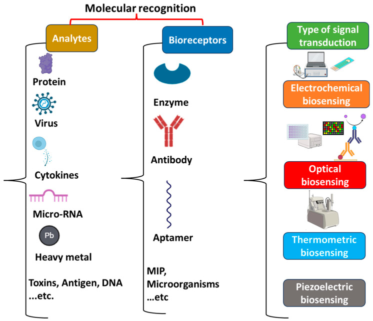
Over the last decade, autoimmune diseases (ADs) have undergone a significant increase because of genetic and/or environmental factors; therefore, their simple and fast diagnosis is of high importance. The conventional diagnostic techniques for ADs require tedious sample preparation, sophisticated instruments, a dedicated laboratory, and qualified personnel. For these reasons, biosensors could represent a useful alternative to these methods. Biosensors are considered to be promising tools that can be used in clinical analysis for an early diagnosis due to their high sensitivity, simplicity, low cost, possible miniaturization (POCT), and potential ability for real-time analysis. In this review, recently developed biosensors for the detection of autoimmune disease biomarkers are discussed. In the first part, we focus on the main AD biomarkers and the current methods of their detection. Then, we discuss the principles and different types of biosensors. Finally, we overview the characteristics of biosensors based on different bioreceptors reported in the literature.

Keywords: autoimmune disease; biomarkers; biosensors; diagnosis.


Conflict of interest statement

The authors declare no conflicts of interest.

Figures

Figure 1
Figure 1
Schematic illustration of the main components of a biosensor.
Figure 2
Figure 2
Schematic representations of the following: (A): A portable device exploiting the integration of screen-printed electrode-based immunosensors and remote-controlled IoT-WiFi for IgA and IgG anti-tissue transglutaminase detection. Reproduced with the permission from Elsevier (Amsterdam, The Netherlands) [173]. Copyright © 2018 Elsevier B.V. All rights reserved. (B): Electrochemical immunosensor based on nanoelectrode ensemble (NEE) technology for the detection of the IgG-TGA (PC = polycarbonate; tTG = tissue transglutaminase; anti-tTG = antibody for tissue transglutaminase; HRP = horseradish peroxidase; H2Q = hydroquinone; BQ = benzoquinone) [112]. (C): Novel NEE biosensor for the detection of IgA-TGA (FA2+ = (ferrocenylmethyl)trimethylammonium, Gox = glucose oxidase). Reproduced with permission from Elsevier, [174]. Copyright © 2021 Elsevier B.V. All rights reserved. (D): Electrochemical dual immunosensor for the simultaneous detection of IgA and IgG types of AGA and TGA antibodies using a carbon–metal hybrid system as the transducer surface (tTG = tissue transglutaminase; AP = alkaline phosphatase; 3-IP: 3-indoxyl phosphate; Ag+ silver ions). Reproduced with permission from Elsevier, [175]. Copyright © 2012 Elsevier B.V. All rights reserved.
Figure 3
Figure 3
(A): Label-free electrochemical immunosensing platform for anti-MBP detection based on the alginate-titanium dioxide (TiO2) nanocomposite-film-modified platinum (Pt) electrode. Reproduced with permission from Elsevier [181]. Copyright © 2013 Elsevier B.V. All rights reserved. (B): Schematic representation of the different steps involved in the construction of the magnetic MBs-based immunosensor for anti-MBP detection involving MBP immobilization onto cMBs and specific conjugation with the target antibody followed by HRP-anti-hIgG conjugation and amperometric detection in the presence of the H2O2/HQ system (cMB: carboxylated magnetic microparticle; HRP = horseradish peroxidase; HQ = hydroquinone; H2O2 = hydrogen peroxide; hIgG = human immunoglobulin G). Reproduced with permission from Elsevier, [182]. Copyright © 2022 Elsevier B.V. All rights reserved. (C): Illustrative scheme of a lateral flow aptasensor for osteopontin (OPN) detection. The OPN-aptamer complex-containing samples were subjected to the LFB sample pad flow and reacted with AuNPs-SA on the conjugate pad; they were then subsequently captured by the anti-OPN antibody at the test line. Then, the excess biotinylated OPN aptamers were captured by the partially complementary ssDNA probes immobilized on the control line. Reproduced with permission from Elsevier [183]. Copyright © 2019 Elsevier B.V. All rights reserved.
Figure 4
Figure 4
(A): Schematic representation of the fabrication of anti-cyclic citrullinated peptide antibodies based on polyaniline (PANI)/MoS2-modified screen-printed electrodes with PANI-Au nanomaterial-based signal amplification for the sensitive detection of the anti-CCP (MoS2 = molybdenum disulfide). Reproduced with permission from Elsevier [194]. Copyright © 2021 Elsevier B.V. All rights reserved. (B): (a) Photograph of the fabricated IDWμE with dimensions of 3.5 × 14 mm. Both low-magnification (200×) and high-magnification (400×) microscopic images of the IDWμE array show a finger width and spacing of 7 μm. (b) Schematic representation of SAM functionalization on IDWμE and the crosslinking of IgG-Fc fragments onto the functionalized electrode array (IDWμE = interfingered wave microelectrode array). Reproduced with permission from Elsevier [195]. Copyright © 2019 Elsevier B.V. All rights reserved.
Figure 5
Figure 5
(A): Schematic illustration of the different steps involved in the preparation of the dual immunosensor using MWCNTs/MoS2(-HRP) -dAb nanocarriers for the determination of the BAFF and APRIL biomarkers by amperometric transduction (MWCNTs = multiwalled carbon nanotubes, MoS2 = molybdenum disulfide, HRP = horseradish peroxidase, dAb = detector antibody) [210]. (B): Schematic display of the different steps involved in the preparation of the developed dual immunoplatform for the (a) BAFF and (b) APRIL biomarkers as well as the reactions implied in the amperometric transduction (BAFF = B-cell activation factor, APRIL = a proliferation-induced ligand). Reproduced with permission from Elsevier, [211]. Copyright © 2022 Elsevier B.V. All rights reserved.

Similar articles

Cited by

References

    1. Davidson A., Diamond B. Autoimmune diseases. N. Engl. J. Med. 2001;345:340–350. doi: 10.1056/NEJM200108023450506. - DOI - PubMed
    1. Chwastiak L., Ehde D.M., Gibbons L.E., Sullivan M., Bowen J.D., Kraft G.H. Depressive symptoms and severity of illness in multiple sclerosis: Epidemiologic study of a large community sample. Am. J. Psychiatry. 2002;159:1862–1868. doi: 10.1176/appi.ajp.159.11.1862. - DOI - PubMed
    1. Alegria G.C., Guellec D., Mariette X., Gottenberg J.E., Dernis E., Dubost J.J., Trouvin A.P., Hachulla E., Larroche C., Le Guern V., et al. Epidemiology of neurological manifestations in Sjögren’s syndrome: Data from the French ASSESS Cohort. RMD Open. 2016;2:e000179. doi: 10.1136/rmdopen-2015-000179. - DOI - PMC - PubMed
    1. Lublin F.D., Reingold S.C., Cohen J.A., Cutter G.R., Sørensen P.S., Thompson A.J., Wolinsky J.S., Balcer L.J., Banwell B., Barkhof F. Defining the clinical course of multiple sclerosis: The 2013 revisions. Neurology. 2014;83:278–286. doi: 10.1212/WNL.0000000000000560. - DOI - PMC - PubMed
    1. Rook G.A. Hygiene hypothesis and autoimmune diseases. Clin. Rev. Allergy Immunol. 2012;42:5–15. doi: 10.1007/s12016-011-8285-8. - DOI - PubMed