

Pleural epithelioid hemangioendothelioma in a 39-Year-old female: a case report
- PMID: 38475834
- PMCID: PMC10935829
- DOI: 10.1186/s13019-024-02602-4
Pleural epithelioid hemangioendothelioma in a 39-Year-old female: a case report
Abstract
Background: Epithelioid hemangioendothelioma (EHE) is a rare malignancy of vascular origin which can be primarily be seen in various tissues. EHE originating from the pleura is an even more uncommon subtype which may mimic mesothelioma and pleural carcinomatosis. The prognosis of pleural EHE is poor and there is no consensus on the optimal therapeutic approach.
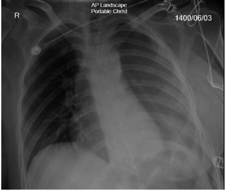
Case presentation: A 39-year-old middle-eastern female presented with progressive dyspnea and left shoulder discomfort. Chest computed tomography scan revealed a left side pleural effusion and pleural thickening. Pleuroscopy was done and biopsies were taken which were positive for CD31, CD34, CK, factor 8-R-antigen, and vimentin. Patient was diagnosed with pleural epithelioid hemangioendothelioma (PEHE) and chemotherapy was started and underwent extrapleural pneumonectomy 7 months later. Unfortunately, the patient passed away 10 months after diagnosis due to disease complications.
Conclusions: Once PEHE is suspected in histology it can be confirmed with immunohistochemistry. Chemotherapy, surgery or a combination of both is currently used as the treatment but the standard treatment remains a question.
Keywords: Epithelioid hemangioendothelioma; Pleural tumors; Thoracic cancer; Vascular tumors.
© 2024. The Author(s).
Conflict of interest statement
The authors declare no competing interests.
Figures


Similar articles
-
[Pleural epithelioid hemangioendothelioma: a case report and review of the literature].Zhonghua Jie He He Hu Xi Za Zhi. 2015 Mar;38(3):174-8. Zhonghua Jie He He Hu Xi Za Zhi. 2015. PMID: 26269304 Review. Chinese.
-
Difficulty of treatment for pleural epithelioid hemangioendothelioma: a report of a case.Gen Thorac Cardiovasc Surg. 2020 Feb;68(2):190-193. doi: 10.1007/s11748-019-01135-1. Epub 2019 May 2. Gen Thorac Cardiovasc Surg. 2020. PMID: 31049818
-
Malignant pleuropulmonary epithelioid hemangioendothelioma - unusual presentation of an aggressive angiogenic neoplasm.Pathol Res Pract. 2014 Sep;210(9):613-8. doi: 10.1016/j.prp.2014.04.011. Epub 2014 May 27. Pathol Res Pract. 2014. PMID: 24939148
-
Pleural Epithelioid Hemangioendothelioma: A Case Report and Literature Review.J Natl Med Assoc. 2016 May;108(2):124-9. doi: 10.1016/j.jnma.2016.05.001. J Natl Med Assoc. 2016. PMID: 27372473 Review.
-
Primary pleural epithelioid hemangioendothelioma compressing the myocardium.J Card Surg. 2013 May;28(3):266-8. doi: 10.1111/jocs.12094. Epub 2013 Apr 1. J Card Surg. 2013. PMID: 23551388
Cited by
-
Dual immunotherapy alternating with anti-PD-1 antibody plus liposomal doxorubicin show good efficacy in prostate epithelioid hemangioendothelioma: a case report.Front Immunol. 2024 Jun 14;15:1384111. doi: 10.3389/fimmu.2024.1384111. eCollection 2024. Front Immunol. 2024. PMID: 38947327 Free PMC article.
References
-
- Dail DH, Liebow AA, Gmelich JT, Friedman PJ, Miyai K, Myer W, Patterson SD, Hammar SP. Intravascular, bronchiolar, and alveolar tumor of the lung (IVBAT): an analysis of twenty cases of a peculiar sclerosing endothelial tumor. Cancer. 1983;51(3):452–64. doi: 10.1002/1097-0142(19830201)51:3<452::AID-CNCR2820510317>3.0.CO;2-M. - DOI - PubMed
-
- Bocchino M, Barra E, Lassandro F, Ranieri F, Muto R, Rea G. Primary pleural haemangioendothelioma in an Italian female patient: a case report and review of the literature. Monaldi Arch Chest Dis 2016, 73(3). - PubMed
Publication types
MeSH terms
LinkOut - more resources
Full Text Sources
Research Materials
