

Menstrual blood-derived mesenchymal stem cells combined with collagen I gel as a regenerative therapeutic strategy for degenerated disc after discectomy in rats
- PMID: 38475906
- PMCID: PMC10935903
- DOI: 10.1186/s13287-024-03680-w
Menstrual blood-derived mesenchymal stem cells combined with collagen I gel as a regenerative therapeutic strategy for degenerated disc after discectomy in rats
Abstract
Background: Annulus fibrosis (AF) defects have been identified as the primary cause of disc herniation relapse and subsequent disc degeneration following discectomy. Stem cell-based tissue engineering offers a promising approach for structural repair. Menstrual blood-derived mesenchymal stem cells (MenSCs), a type of adult stem cell, have gained attention as an appealing source for clinical applications due to their potential for structure regeneration, with ease of acquisition and regardless of ethical issues.
Methods: The differential potential of MenSCs cocultured with AF cells was examined by the expression of collagen I, SCX, and CD146 using immunofluorescence. Western blot and ELISA were used to examine the expression of TGF-β and IGF-I in coculture system. An AF defect animal model was established in tail disc of Sprague-Dawley rats (males, 8 weeks old). An injectable gel containing MenSCs (about 1*106/ml) was fabricated and transplanted into the AF defects immediately after the animal model establishment, to evaluate its repairment properties. Disc degeneration was assessed via magnetic resonance (MR) imaging and histological staining. Immunohistochemical analysis was performed to assess the expression of aggrecan, MMP13, TGF-β and IGF-I in discs with different treatments. Apoptosis in the discs was evaluated using TUNEL, caspase3, and caspase 8 immunofluorescence staining.
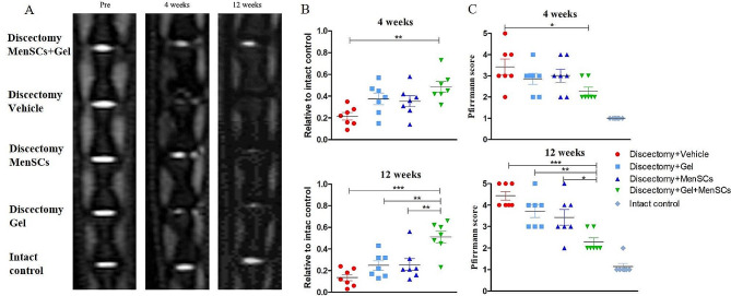
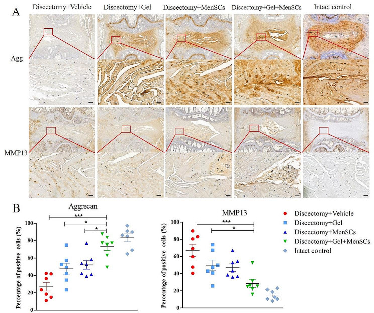
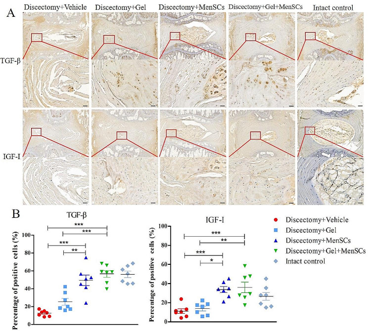
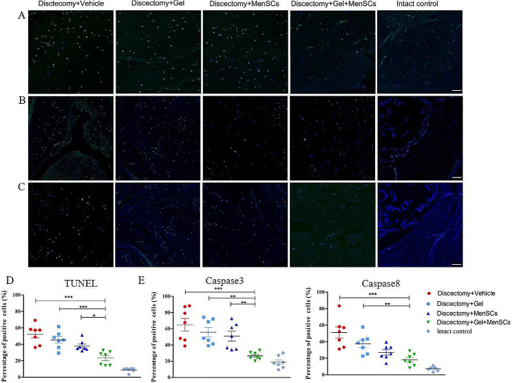
Results: Coculturing MenSCs with AF cells demonstrated ability to express collagen I and biomarkers of AF cells. Moreover, the coculture system presented upregulation of the growth factors TGF-β and IGF-I. After 12 weeks, discs treated with MenSCs gel exhibited significantly lower Pffirrmann scores (2.29 ± 0.18), compared to discs treated with MenSCs (3.43 ± 0.37, p < 0.05) or gel (3.71 ± 0.29, p < 0.01) alone. There is significant higher MR index in disc treated with MenSCs gel than that treated with MenSCs (0.51 ± 0.05 vs. 0.24 ± 0.04, p < 0.01) or gel (0.51 ± 0.05 vs. 0.26 ± 0.06, p < 0.01) alone. Additionally, MenSCs gel demonstrated preservation of the structure of degenerated discs, as indicated by histological scoring (5.43 ± 0.43 vs. 9.71 ± 1.04 in MenSCs group and 10.86 ± 0.63 in gel group, both p < 0.01), increased aggrecan expression, and decreased MMP13 expression in vivo. Furthermore, the percentage of TUNEL and caspase 3-positive cells in the disc treated with MenSCs Gel was significantly lower than those treated with gel alone and MenSCs alone. The expression of TGF-β and IGF-I was higher in discs treated with MenSCs gel or MenSCs alone than in those treated with gel alone.
Conclusion: MenSCs embedded in collagen I gel has the potential to preserve the disc structure and prevent disc degeneration after discectomy, which was probably attributed to the paracrine of growth factors of MenSCs.
Keywords: Annulus fibrosis defects; Disc degeneration; Disc repairment; Discectomy; Growth factors; Menstrual blood-derived mesenchymal stem cells; Paracrine; Tissue engineering.
© 2024. The Author(s).
Conflict of interest statement
The authors declare that they have no competing interests.
Figures


Similar articles
-
Therapeutic effects of PDGF-AB/BB against cellular senescence in human intervertebral disc.Elife. 2025 Jul 16;13:RP103073. doi: 10.7554/eLife.103073. Elife. 2025. PMID: 40668091 Free PMC article.
-
Human amnion mesenchymal stem cells promote endometrial repair via paracrine, preferentially than transdifferentiation.Cell Commun Signal. 2024 May 31;22(1):301. doi: 10.1186/s12964-024-01656-0. Cell Commun Signal. 2024. PMID: 38822356 Free PMC article.
-
Can treatment with human mesenchymal stem cells rescue the degenerative disc phenotype? An in vitro pilot study of induced cytokine expression.Spine J. 2025 Aug;25(8):1830-1840. doi: 10.1016/j.spinee.2025.03.026. Epub 2025 Mar 26. Spine J. 2025. PMID: 40154630
-
Progesterone or progestogen-releasing intrauterine systems for heavy menstrual bleeding.Cochrane Database Syst Rev. 2005 Oct 19;(4):CD002126. doi: 10.1002/14651858.CD002126.pub2. Cochrane Database Syst Rev. 2005. Update in: Cochrane Database Syst Rev. 2015 Apr 30;(4):CD002126. doi: 10.1002/14651858.CD002126.pub3. PMID: 16235297 Updated.
-
Intravenous magnesium sulphate and sotalol for prevention of atrial fibrillation after coronary artery bypass surgery: a systematic review and economic evaluation.Health Technol Assess. 2008 Jun;12(28):iii-iv, ix-95. doi: 10.3310/hta12280. Health Technol Assess. 2008. PMID: 18547499
Cited by
-
Risk factors and treatment strategies for adjacent segment disease following spinal fusion (Review).Mol Med Rep. 2025 Feb;31(2):33. doi: 10.3892/mmr.2024.13398. Epub 2024 Nov 22. Mol Med Rep. 2025. PMID: 39575466 Free PMC article. Review.
-
Recent Advances in the Development and Application of Cell-Loaded Collagen Scaffolds.Int J Mol Sci. 2025 Apr 24;26(9):4009. doi: 10.3390/ijms26094009. Int J Mol Sci. 2025. PMID: 40362249 Free PMC article. Review.
References
-
- Ukeba D, Sudo H, Tsujimoto T, Ura K, Yamada K, Iwasaki N. Bone marrow mesenchymal stem cells combined with ultra-purified alginate gel as a regenerative therapeutic strategy after discectomy for degenerated intervertebral discs. EBioMedicine. 2020;53:102698. doi: 10.1016/j.ebiom.2020.102698. - DOI - PMC - PubMed
Publication types
MeSH terms
Substances
Grants and funding
- 82102599/National Natural Science Foundation of China
- LGF19H060012/Public Welfare Program of Science and Technology Department of Zhejiang Province
- LTGY24H060003/Public Welfare Program of Science and Technology Department of Zhejiang Province
- 2020RC058/Medicine and health science and technology plan in Zhejiang province
- 2022KY493/Medicine and health science and technology plan in Zhejiang province
LinkOut - more resources
Full Text Sources
Research Materials
