. 2024 May;115(5):1492-1504.
doi: 10.1111/cas.16138. Epub 2024 Mar 13.

LncRNA CTBP1-AS inhibits TP63-mediated activation of S100A14 during prostate cancer progression

Affiliations

LncRNA CTBP1-AS inhibits TP63-mediated activation of S100A14 during prostate cancer progression

Guangzheng Wu et al. Cancer Sci. 2024 May.

Abstract

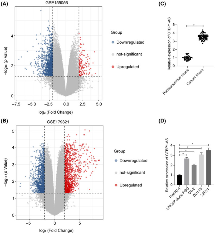
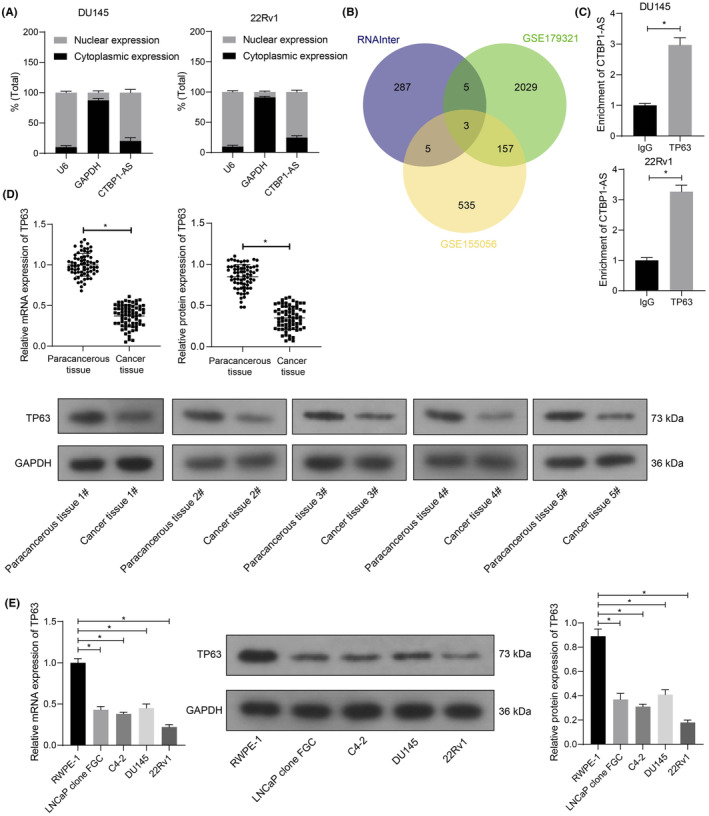
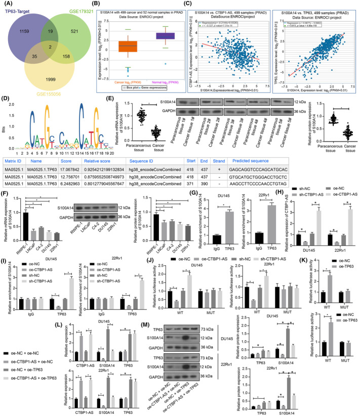
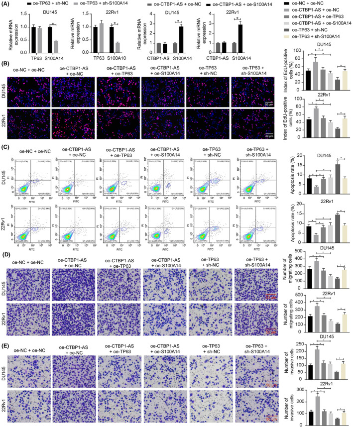
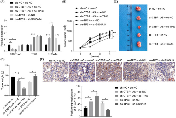
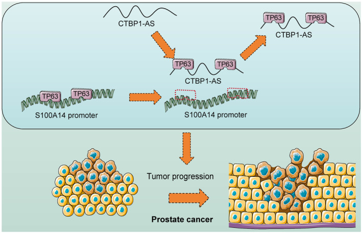
Long noncoding RNAs (lncRNAs) have emerged as important molecules and potential new targets for human cancers. This study investigates the function of lncRNA CTBP1 antisense RNA (CTBP1-AS) in prostate cancer (PCa) and explores the entailed molecular mechanism. Aberrantly expressed genes potentially correlated with PCa progression were probed using integrated bioinformatics analyses. A cohort of 68 patients with PCa was included, and their tumor and para-cancerous tissues were collected. CTBP1-AS was highly expressed in PCa tissues and cells and associated with poor patient prognosis. By contrast, tumor protein p63 (TP63) and S100 calcium binding protein A14 (S100A14) were poorly expressed in the PCa tissues and cells. CTBP1-AS did not affect TP63 expression; however it blocked the TP63-mediated transcriptional activation of S100A14, thereby reducing its expression. CTBP1-AS silencing suppressed proliferation, apoptosis resistance, migration, invasion, and tumorigenicity of PCa cell lines, while its overexpression led to inverse results. The malignant phenotype of cells was further weakened by TP63 overexpression but restored following artificial S100A14 silencing. In conclusion, this study demonstrates that CTBP1-AS plays an oncogenic role in PCa by blocking TP63-mediated transcriptional activation of S100A14. This may provide insight into the management of PCa.

Keywords: CTBP1‐AS; S100A14; TP63; prostate cancer; transcription.


Conflict of interest statement

The authors declare no conflict of interest.

Figures

FIGURE 1
FIGURE 1
CTBP1‐AS is significantly highly expressed in PCa tissues and cells. (A, B) Volcano plots for differentially expressed genes between PCa and normal tissues (adj. p‐value <0.05 and |Log FC| > 2 as the thresholds) in the GSE155056 and GSE179321 datasets; (C) expression of CTBP1‐AS in the tumor and the paracancerous tissues from clinical PCa patients (N = 68) determined using RT‐qPCR; (D) expression of CTBP1‐AS in the procured PCa cell lines (LNCaP clone FGC, C4‐2, DU145 and 22Rv1) and normal RWPE‐1 cells determined using RT‐qPCR. For cellular experiments, three biological replicates were performed. Differences were compared by paired t‐test (C) or one‐way ANOVA (D). *p < 0.05.
FIGURE 2
FIGURE 2
CTBP1‐AS interacts with TP63, a molecule poorly expressed in PCa. (A) Subcellular localization of CTBP1‐AS in DU145 and 22Rv1 cells analyzed by nucleus/cytoplasm separation assay; (B) bioinformatics analyses of the candidate interactive proteins of CTBP1‐AS in PCa predicted using the RNAInter system and their intersections with the differentially expressed genes in the GSE179321 and GSE155056 datasets; (C) direct binding between CTBP1‐AS and the TP63 protein in DU145 and 22Rv1 cells examined by RIP assay; (D) TP63 mRNA and protein levels in the clinically collected PCa cancer and paracancerous tissues (N = 68) examined by RT‐qPCR and WB assays, respectively; (E) TP63 mRNA and protein levels in the procured PCa cell lines (LNCaP clone FGC, C4‐2, DU145, and 22Rv1) and normal RWPE‐1 cells determined by RT‐qPCR and WB assays, respectively. For cellular experiments, three biological replicates were performed. Differences were analyzed by unpaired t‐test (C), paired t‐test (D), or one‐way ANOVA (E). *p < 0.05.
FIGURE 3
FIGURE 3
CTBP1‐AS interacts with TP63 to suppress S100A14 transcription. (A) Downstream targets of TP63 predicted in the hTFtarget system and their intersections with differentially expressed genes in the GSE179321 and GSE155056 datasets; (B) expression pattern of S100A14 in PRAD and normal samples in the StarBase system; (C) correlations of S100A14 with CTBP1‐AS and TP63 in PRAD in the StarBase system; (D) putative binding sites between TP63 and S100A14 promoter obtained from the Jaspar system; (E) S100A14 mRNA and protein levels in the clinically collected PCa cancer and paracancerous tissues (N = 68) examined by RT‐qPCR and WB assays, respectively; (F) S100A14 mRNA and protein levels in the procured PCa cell lines (LNCaP clone FGC, C4‐2, DU145 and 22Rv1) and normal RWPE‐1 cells determined by RT‐qPCR and WB assays, respectively; (G) binding relationship between TP63 and S100A14 promoter in DU145 and 22Rv1 cells determined by the ChIP‐qPCR assay; (H) expression of CTBP1‐AS in DU145 and 22Rv1 cells after sh‐CTBP1‐AS or oe‐CTBP1‐AS transfection determined by RT‐qPCR; (I) binding relationship between TP63 and S100A14 promoter in DU145 and 22Rv1 cells with artificial CTBP1‐AS upregulation or silencing determined by ChIP‐qPCR assay; (J) transcriptional activity of the WT or MUT S100A14 promoter luciferase reporter vectors in DU145 and 22Rv1 cells with artificial CTBP1‐AS upregulation or silencing examined by dual luciferase reporter gene assay; (K) transcriptional activity of the WT or MUT S100A14 promoter luciferase reporter vectors in DU145 and 22Rv1 cells with artificial TP63 upregulation examined by dual luciferase reporter gene assay; (L) expression of CTBP1‐AS, TP63 mRNA, and S100A14 mRNA in DU145 and 22Rv1 cells after different transfection combinations determined by RT‐qPCR; (M) protein levels of TP63 and S100A14 in DU145 and 22Rv1 cells after different transfection combinations determined by WB analysis. For cellular experiments, three biological replicates were performed. Differences were analyzed by paired t‐test (E), unpaired t‐test (G), one‐way ANOVA (F) or two‐way ANOVA (H–M). *p < 0.05.
FIGURE 4
FIGURE 4
The CTBP1‐AS/TP63/S100A14 axis affects the malignant properties of PCa cells in vitro. (A) Expression of TP63 and S100A14 in DU145 and 22Rv1 cells after several combinations of oe‐NC, oe‐TP63, sh‐NC, and sh‐S100A14 administration determined by RT‐qPCR; (B) proliferation of DU145 and 22Rv1 cells determined by EdU labeling assay; (C) apoptosis of DU145 and 22Rv1 cells determined by flow cytometry; (D, E) migration (D) and invasion (E) of DU145 and 22Rv1 cells determined by Transwell assays. Three biological replicates were performed. Differences were compared using one‐way ANOVA (B–E) or two‐way ANOVA (A). *p < 0.05.
FIGURE 5
FIGURE 5
The CTBP1‐AS/TP63/S100A14 axis affects the tumorigenicity of PCa cells in nude mice. (A) Expression of CTBP1‐AS, TP63, and S100A14 in 22Rv1 cells after several combinations of sh‐NC, oe‐NC, sh‐CTBP1‐AS, oe‐TP63, and sh‐S100A14 administration determined by RT‐qPCR; (B) volume of the xenograft tumors formed by the 22Rv1 cells; (C) representative images of the xenograft tumors on week 4 after animal euthanasia; (D) weight of the collected xenograft tumors; (E) expression of S100A14 in the xenograft tumor tissues analyzed by IHC. In each group, n = 8. Differences were compared by one‐way ANOVA (D, E) or two‐way ANOVA (A, B). *p < 0.05.
FIGURE 6
FIGURE 6
CTBP1‐AS interacts with TP63 and negates the transcription activation of S100A14, resulting in increased proliferation and dissemination of PCa cells.

Similar articles

Cited by

References

    1. Sung H, Ferlay J, Siegel RL, et al. Global cancer statistics 2020: GLOBOCAN estimates of incidence and mortality worldwide for 36 cancers in 185 countries. CA Cancer J Clin. 2021;71:209‐249. - PubMed
    1. Conteduca V, Oromendia C, Eng KW, et al. Clinical features of neuroendocrine prostate cancer. Eur J Cancer. 2019;121:7‐18. - PMC - PubMed
    1. Chin YF, Lynn N. Systematic review of focal and salvage cryotherapy for prostate cancer. Cureus. 2022;14:e26400. - PMC - PubMed
    1. Xin S, Sun X, Jin L, et al. The prognostic signature and therapeutic value of phagocytic regulatory factors in prostate adenocarcinoma (PRAD). Front Genet. 2022;13:877278. - PMC - PubMed
    1. Akova Olken E, Aszodi A, Taipaleenmaki H, et al. SFRP2 overexpression induces an osteoblast‐like phenotype in prostate cancer cells. Cells. 2022;11:4081. - PMC - PubMed

MeSH terms

Grants and funding