. 2024 Jan:148:101765.
doi: 10.1016/j.progpolymsci.2023.101765. Epub 2023 Dec 9.

Structural Determinants of Stimuli-Responsiveness in Amphiphilic Macromolecular Nano-assemblies

Affiliations

Structural Determinants of Stimuli-Responsiveness in Amphiphilic Macromolecular Nano-assemblies

Hongxu Liu et al. Prog Polym Sci. 2024 Jan.

Abstract

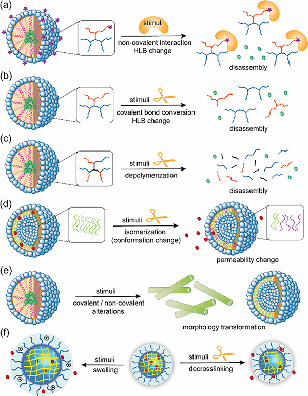
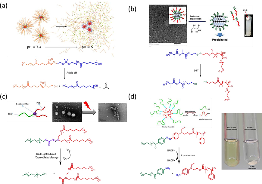
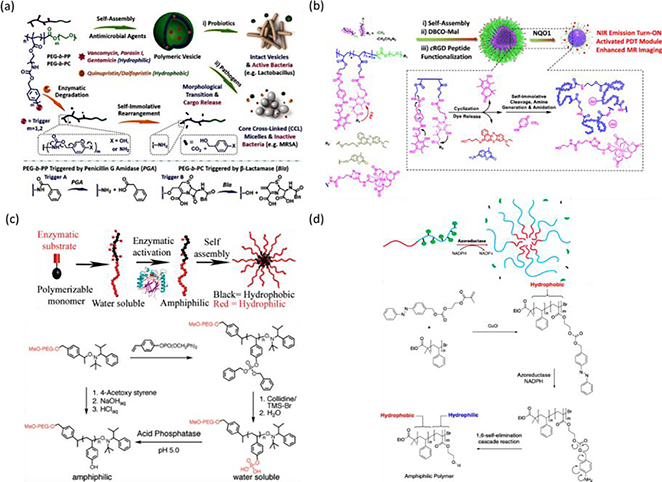
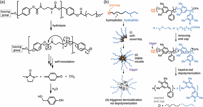
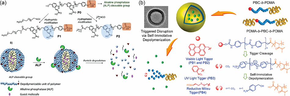
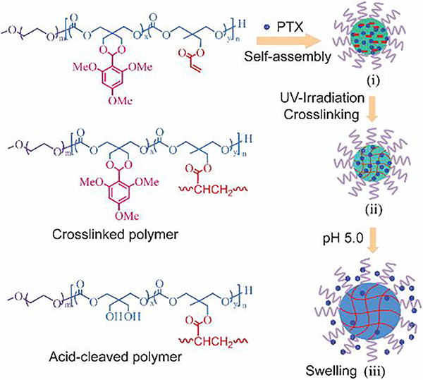
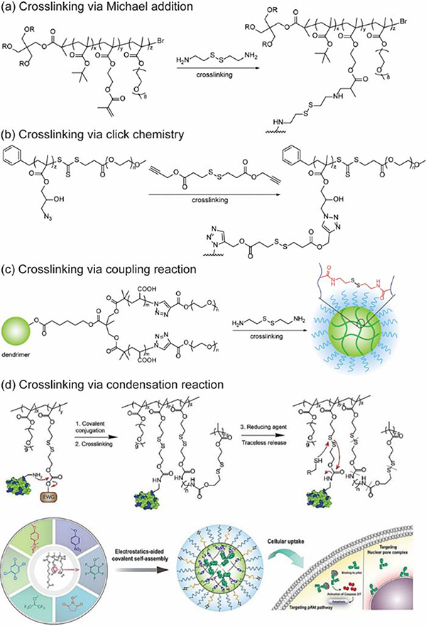
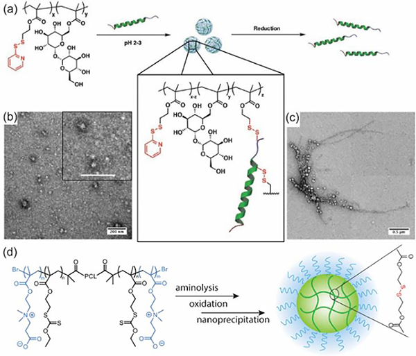
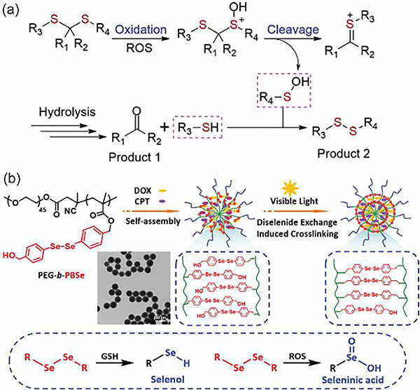
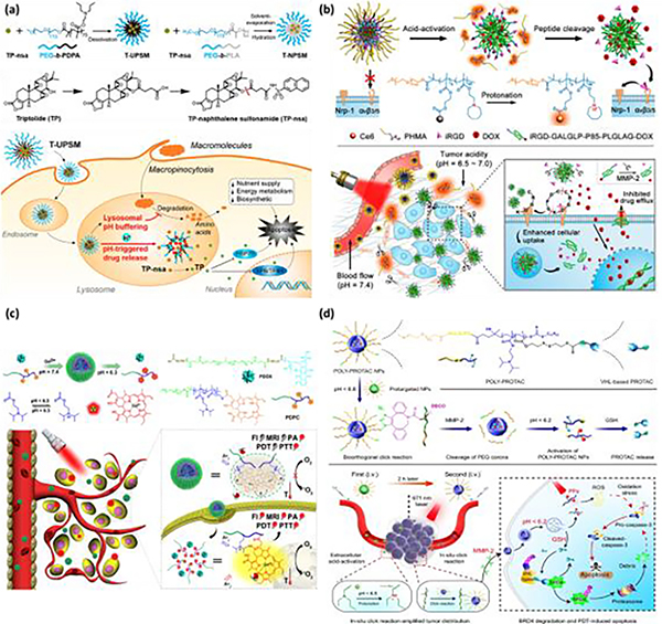
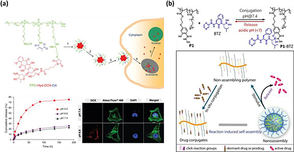
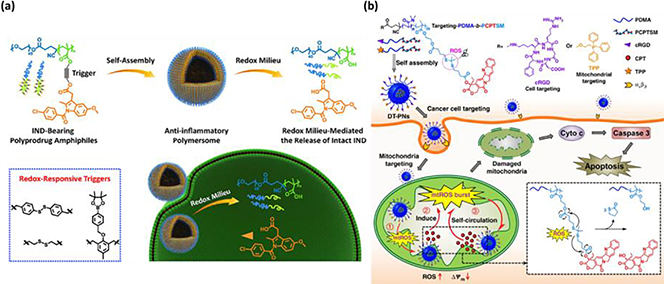
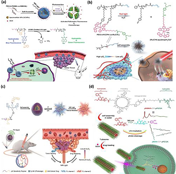
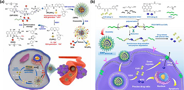
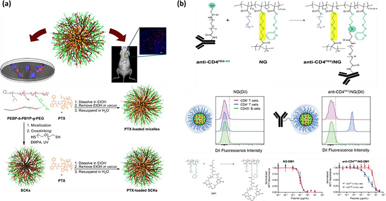
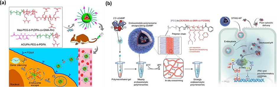
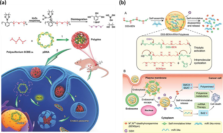
Stimuli-responsive nano-assemblies from amphiphilic macromolecules could undergo controlled structural transformations and generate diverse macroscopic phenomenon under stimuli. Due to the controllable responsiveness, they have been applied for broad material and biomedical applications, such as biologics delivery, sensing, imaging, and catalysis. Understanding the mechanisms of the assembly-disassembly processes and structural determinants behind the responsive properties is fundamentally important for designing the next generation of nano-assemblies with programmable responsiveness. In this review, we focus on structural determinants of assemblies from amphiphilic macromolecules and their macromolecular level alterations under stimuli, such as the disruption of hydrophilic-lipophilic balance (HLB), depolymerization, decrosslinking, and changes of molecular packing in assemblies, which eventually lead to a series of macroscopic phenomenon for practical purposes. Applications of stimuli-responsive nano-assemblies in delivery, sensing and imaging were also summarized based on their structural features. We expect this review could provide readers an overview of the structural considerations in the design and applications of nanoassemblies and incentivize more explorations in stimuli-responsive soft matters.

Keywords: Amphiphilic macromolecules; Biomedical applications; Disassembly; Nanoassemblies; Self-assembly; Stimuli-responsive; Structural determinants.


Conflict of interest statement

Declaration of Competing Interest The authors declare that they have no known competing financial interests or personal relationships that could have appeared to influence the work reported in this paper.

Figures

Fig. 1.
Fig. 1.
Different strategies for stimuli-triggered alterations in nano-assemblies: (a) Disassembly via HLB change by the disruption of non-covalent interactions, (b) disassembly via HLB change by the disruption of covalent bond, (c) disassembly via depolymerization, (d) permeability change via conformation/configuration alterations, (e) stimuli-triggered morphology transformation, (f) stimuli-triggered swelling or decrosslinking of crosslinked nano-assemblies.
Fig. 2.
Fig. 2.
Esterification equilibrium between boronic acids and diols.
Fig. 3.
Fig. 3.
(a) The copolymerization of methacrylic acid and benzyl methacrylate by RAFT aqueous emulsion polymerization (top panel), and the schizophrenic micellization behavior of PDEA88-P(MAA-stat-BzMA)y diblock copolymers in acidic and basic aqueous solution (bottom panel) [134], Copyright 2017. Adapted with permission from the American Chemical Society. (b) Mechanisms of homo Förster resonance energy transfer and photoinduced electron transfer behind tunable, ultrasensitive pH-responsive nanoparticles, where the neutralized PR segments that could self-assemble into the micelle cores at pH > pKa led to quenching of fluorophores while formation of unimers at pH < pKa lighted on fluorescence (top panel). Structures of the PEO-b-(PR-r-TMR) copolymers (PEO = poly(ethylene oxide), PR = ionizable block, TMR = tetramethyl rhodamine, and R = dialkyl and cyclic substituents) (bottom panel) [138], Copyright 2011. Adapted with permission from John Wiley & Sons Inc.
Fig. 4.
Fig. 4.
(a) General structure of dye-conjugated PEG-b-PNIPAM (top panel) and fluorescence transition from polymeric ratiometric fluorescent thermometers upon heating and cooling (bottom panel) [145], Copyright 2015. Adapted with permission from the American Chemical Society. (b) Structure of PEG-b-P(NAGA-co-AN) (top panel), and UCST-type structural transformation that induced payload release (bottom panel) [154], Copyright 2018. Adapted with permission from John Wiley & Sons Inc.
Fig. 5.
Fig. 5.
Cleavage at hydrophobic and hydrophilic junction led to the disruption of nanoparticles in a form of precipitation. (a) Chemical structure of the acid-labile block copolymer MPEO44-b-PCL17 (top panel) and the schematic illustration of assembly/disassembly transition of nanoparticles at pH ~5.0 (bottom panel) [169], Copyright 2021. Adapted with permission from the authors. (b) Illustration of particle disruption of PCL-SS-POEOMA micelles in response to DTT (top panel) and chemical structure of the reduction-responsive block copolymer PCL-SS-POEOMA (bottom panel) [170], Copyright 2015. Adapted with permission from the American Chemical Society. (c) Schematic illustration of amphiphilic miktoarm PEG-b-PCL2 copolymer with a junction of 1O2-Labile β-aminoacrylate (top panel), and their self-assembly and particle disruption 1O2-mediated dissociation upon red-laser (bottom panel) [171], Copyright 2018. Adapted with permission from the American Chemical Society. (d) Schematic representation (top panel) and a digital picture (right panel) of the micellar assembly from PEG-N=N-PS block copolymer and triggered disruption into PEG and PS homopolymers by the enzyme azoreductase in the presence of NADPH. Chemical structure of the acid-labile block copolymer PEG-N=N-PS (bottom panel) [116], Copyright 2018. Adapted with permission from the American Chemical Society.
Fig. 6.
Fig. 6.
(a) Enzyme-responsive polymeric vesicles for bacterial strain-selective delivery of antibiotics and structural transformation in response to enzyme (top panel). Illustration of bond cleavage of built-in triggers in response to penicillin G amidase, and β-lactamase (bottom panel) [203], Copyright 2016. Adapted with permission from John Wiley & Sons Inc. (b) Self-assembly of amphiphilic BCPs containing quinone trimethyl lock-capped self-immolative side linkages and triggered particle transformation under exposure to NQO1 [204], Copyright 2020. Adapted with permission from the American Chemical Society. (c) The illustration of enzyme-induced self-assembly from hydrophilic block copolymer (top panel) and synthetic approach for the preparation of amphiphilic diblock copolymers (bottom panel) [117], Copyright 2009. Adapted with permission from the American Chemical Society. (d) Schematic illustration of enzyme-induced self-assembly from hydrophobic block copolymers (top panel). Synthesis of hydrophobic precursor of block copolymer, and hydrophobic-to-amphiphilic transition behind the enzyme-induced particle formation (bottom panel) [205], Copyright 2014. Adapted with permission from the American Chemical Society.
Fig. 7.
Fig. 7.
(a) Self-Immolation of polymers via an alternating alternating electronic cascade 1,6-elimination and cyclization mechanism [214]. (b) Poly(benzylether)-based self-immolative polymer for the design of stimuli-responsive assemblies [215], Copyright 2022. Adapted with permission from the American Chemical Society.
Fig. 8.
Fig. 8.
(a) ALP-responsive self-immolative nano-assemblies [130], Copyright 2020. Adapted with permission from the Royal Society of Chemistry. (b) Self-Immolative Polymersomes from poly(benzylcabomate)-PDMA block copolymers [54], Copyright 2014. Adapted with permission from the American Chemical Society.
Fig. 9.
Fig. 9.
Depolymerization of (a) polyglyoxylates [226], (b) poly(phthalaldehyde) [227], and (c) poly(O-vinyl carbamate-alt-sulfones) [228].
Fig. 10.
Fig. 10.
Synthesis of polydisulfide-based random and block copolymers via (a) thiol-initiated ring-opening polymerization [245], (b) thermal polymerization [248], and (c) thiol-disulfide exchange and RAFT polymerization [249].
Fig. 11.
Fig. 11.
(a) Synthesis of poly (CHTA-co-HD)-PEG. (b) Structures of OxaPt(IV) and 56MESS. (c) Formation of NP-OxaPt(IV), and (d) NP-56MESS via nanoprecipitation [252], Copyright 2021. Adapted with permission from the Nature Publishing Group.
Fig. 12.
Fig. 12.
(a) Examples of degradable polymers linked by diselenide [256], thioacetal [258] and palladium [111]. (b) Mechanisms for CARTs degradation [260].
Fig. 13.
Fig. 13.
Schematic illustration of the cytosolic protein delivery using ROS-degradable polymer with built-in phenylboronic acid in the backbone and the degradation mechanisms [268], Copyright 2022. Reproduced with permission from John Wiley & Sons Inc.
Fig. 14.
Fig. 14.
(a) Schematic demonstration of the azobenzene-based multi-stimuli responsive system: Reduction leads to irreversible polymer degradation, while photo-triggered isomerization leads to reversible polarity changes in assembly core [272], Copyright 2016. Adapted with permission from the Royal Society of Chemistry. (b) Structures and mechanisms for accelerated backbone degradation of amphiphilic polyurethane nanoparticles in physiologically relevant aqueous media via external stimuli-triggered activation and cascade self-amplification of built-in trigger signals [273], Copyright 2021. Adapted with permission from the American Chemical Society.
Fig. 15.
Fig. 15.
Schematic illustration of polymer chain-scission via ROS-triggered degradation and cyclization. [279], Copyright 2019. Reproduced with permission from the American Chemical Society.
Fig. 16.
Fig. 16.
(a) Schematic presentation of the preparation of an acid-degradable siRNA-loaded PCL nanocarrier and its acid-triggered release of siRNAs. (b) The dsDNA release profiles for PCLs under different pH [289], Copyright 2013. Adapted with permission from the American Chemical Society.
Fig. 17.
Fig. 17.
Schematic presentation of nanostructures prepared via the assembly of phenylboronic acid and catechol modified polymers in water and methanol and the corresponding TEM images [296], Copyright 2017. Adapted with permission from the American Chemical Society.
Fig. 18
Fig. 18
Schematic illustration of photo-crosslinkable pH-sensitive degradable micelles formation and acid-triggered degradation [297], Copyright 2012. Adapted with permission from Elsevier Science Ltd.
Fig. 19.
Fig. 19.
Disulfide-based polymers formed via (a) Michael addition [305], (b) click chemistry [304], and (c) condensation reactions [306]. (d) Schematic illustration of disulfide-crosslinked nanoparticles for protein conjugation and intracellular delivery via a traceless release manner [308], Copyright 2020. Adapted with permission from John Wiley & Sons Inc.
Fig. 20.
Fig. 20.
(a) Structure of PDSMA-co-TrMA and schematic presentation of glucagon–nanogel formation and release. TEM images of PDSMA1-co-TrMA0.8 glucagon nanogels before (b) and after (c) treatment of TECP for 24 h [318], Copyright 2018. Adapted with permission from John Wiley & Sons Inc. (d) Crosslinking via the deprotection and oxidation of free thiols [320].
Fig. 21.
Fig. 21.
(a) Mechanisms of ROS-triggered thioketal degradation [200], Copyright 2020. Adapted with permission from Elsevier Science Ltd. (b) Schematic illustration of diselenide-crosslinked particle formation, degradation mechanisms and TEM image of the formed assemblies [331], Copyright 2017. Adapted with permission from Elsevier Science Ltd.
Fig. 22.
Fig. 22.
Schematic illustration of a redox/enzyme-activatable nanoagent with GFLG peptides and disulfide bonds, after encapsulation of a PARP inhibitor, AZD2281 [336], Copyright 2021. Reproduced with permission from John Wiley & Sons Inc.
Fig. 23.
Fig. 23.
Structure of phthalimide esters and coumarin-functionalized polymer and photo-triggered changes to chemical structures and nano-assemblies [340], Copyright 2017. Reproduced with permission from John Wiley & Sons Inc.
Fig. 24.
Fig. 24.
(a) Structure of azobenzene-linked block copolymer and the illustration of the formed vesicles. (b) and (c) The release profiles of hydrophobic DiI and hydrophilic rhodamine 6G under 360nm UV irradiation and dark conditions [350], Copyright 2017. Adapted with permission from the Nature Publishing Group.
Fig. 25.
Fig. 25.
(a) Structures of bis-thiaxanthylidene-based amphiphiles before and after the irreversible photoisomerization [351], Copyright 2011. Adapted with permission from the Nature Publishing Group. (b) Photo and heat-induced reversible isomerization of overcrowded alkene-based amphiphiles [352], Copyright 2015. Adapted with permission from the American Chemical Society. (c) Photoisomerization of dithienylethene-based imaging agent [353], Copyright 2017. Adapted with permission from the American Chemical Society.
Fig. 26.
Fig. 26.
(a) Preparation of T-UPSM and T-NPSM (top panel) and illustration of UPSM for drug delivery and blocking of lysosomal acidification in pancreatic cancer treatment (bottom panel) [364], Copyright 2019. Adapted with permission from the American Chemical Society. (b) Preparation of ATLP nanoparticle (top panel) and illustration of ATLP nanoparticles for iRGD-enhanced tumor penetration and imaging-guided combination cancer therapy (bottom panel) [365], Copyright 2017. Adapted with permission from the American Chemical Society. (c) Nano-assembly and chemical structure of the acid-switchable micelles comprising pH-responsive PEG-b-PDPA, gadolinium-coordinated photosensitizer Ce6, and a prodrug of DOX (top panel). Illustration of the NDDS for multimodal imaging and combinational therapy of drug-resistant tumor (bottom panel) [366], Copyright 2016. Adapted with permission from the American Chemical Society. (d) Chemical structure of polymers as building blocks and Illustration of the extracellular click reaction between POLYPROTAC and DBCO-loaded pretargeted NPs in acidic tumor microenvironment (top panel). Schematic illustration of the 58eblock58onal POLY-PROTAC NPs for tumor-specific protein degradation and cascade pathways [367], Copyright 2022. Adapted with permission from the Nature Publishing Group.
Fig. 27.
Fig. 27.
(a) Chemical structure of the dual pH-responsive polymer–doxorubicin (DOX) conjugate (PPC-Hyd-DOX-DA) and pathway of cancer treatment (top panel), and drug release profile and confocal laser scattering microscopy images (bottom panel) [371], Copyright 2011. Adapted with permission from the American Chemical Society. (b) Chemical structure of polymer and drug for in situ formation of nano-assemblies [372], Copyright 2020. Adapted with permission from John Wiley & Sons Inc.
Fig. 28.
Fig. 28.
(a) The self-assembly of anti-inflammatory polymersomes of redox-responsive polyprodrug amphiphiles with variations of triggered cleavable linkers. The redox-sensitive disintegration of polymersome and subsequent drug release [374], Copyright 2018. Adapted with permission from Elsevier Science Ltd. (b) Dual-targeting polyprodurg nanoreactors (DT-PNs) for self-amplified drug release with ROS burst in mitochondria [201], Copyright 2019. Adapted with permission from the Nature Publishing Group.
Fig. 29.
Fig. 29.
(a) The production of RFHM containing DOX and UCNP via a nanoprecipitation method, and photo-triggered disruption of particle and ratiometric fluorescence (top panel). NIR-induced concurrent chemotherapy and ratiometric fluorescence imaging in intracellular milieu (bottom panel) [375], Copyright 2020. Adapted with permission from John Wiley & Sons Inc. (b) Chemical structure of the PEG-b-P(CPH-co-RuCHL) and its cleaved forms including drug–Ru complex conjugate [Ru(CHLtpy)(biq)(H2O)]2+. Illustration of light administration and the implication of PEG-b-P(CPH-co-RuCHL) in hypoxic tumor environment [376], Copyright 2018. Adapted with permission from John Wiley & Sons Inc. (c) Schematic illustration of P(Cy-N-CPT) micelle and dual-modal PA and dual-channel fluorescence output in response to NIR (top panel). Schematic illustration of light-responsive nanoparticles for real-time tracking and feedback regulation of photo-triggered drug release in the hypoxic in vivo [377], Copyright 2021. Adapted with permission from John Wiley & Sons Inc. (d) Chemical structure of pNBMA25-CP-pPEGA27 (top panel) and an illustration of cellular uptake of DOX-loaded tubisomes, and photo-triggered structural disruption and controlled release (bottom panel) [378], Copyright 2020. Adapted with permission from John Wiley & Sons Inc.
Fig. 30.
Fig. 30.
(a) Chemical structure of SIP-DOX and its nano-assembly, and the self-immolation of SIP-DOX that induced disassembly and drug release (top panel). Schematic illustration of self-amplified ROS-responsive drug release nanosystem (SIP-DOX) for cancer therapy (bottom panel) [381] Copyright 2022. Adapted with permission from Elsevier Science Ltd. (b) Illustration of amphiphilic polyprodrugs PEG-P(MTO-ss-CUR) nanoparticles with a predefined ratio of MTO and CUR, and disassembly of nanoparticle along with drug release in response GSH [384], Copyright 2021. Adapted with permission from the American Chemical Society.
Fig. 31.
Fig. 31.
(a) Biodegradable polyphosphoester-based PEBP-b-PBYP-g-PEG self-assembly for cancer chemotherapy [387], Copyright 2015. Adapted with permission from the American Chemical Society. (b) Components of ANCs and the selective therapy against CD4high mT-ALL cells [393], Copyright 2020. Adapted with permission from the American Chemical Society.
Fig. 32.
Fig. 32.
(a) Chemical composition of ultra-pH-sensitive nanoplatform for pH-responsive siRNA delivery (top panel) and the schematic illustration of ultra-pH-sensitive nanoplatform for RNAi in vitro and in vivo [403], Copyright 2017. Adapted with permission from the American Chemical Society. (b) Chemical composition of the STING-NP and formulation strategy for enhanced cytosolic delivery of cGAMP (left panel), and schematic illustration of STING-NP that stimulates immunosuppressive tumors to immunogenic (right panel) [408], Copyright 2019. Adapted with permission from the Nature Publishing Group.
Fig. 33.
Fig. 33.
(a) ROS-responsive degradable polysulfoniums and the polyplex for intraperitoneal gene delivery [267], Copyright 2017. Adapted with permission from John Wiley & Sons Inc. (b) GSH-responsive BENSpm-based biodegradable polycation /miR-34a nanoparticles for dual drug delivery via triggered depolymerization [413], Copyright 2016. Adapted with permission Elsevier Science Ltd.
Fig. 34.
Fig. 34.
(a) Formation of the noncationic RNA–polymer complex [315], Copyright 2019. Adapted with permission from the American Chemical Society. (b) Formation of crosslinked protein nano-assembly, and reduction-responsive traceless protein release [307], Copyright 2017. Adapted with permission from the American Chemical Society.
Fig. 35.
Fig. 35.
(a) Chemical structure of monomers in CO2/pH-responsive crosslinked nanoprobes (top panel), and CO2/pH-responsive swelling/shrinking alteration of nanogels with reversible ON/OFF fluorescence emission [419], Copyright 2016. Adapted with permission from the Royal Society of Chemistry. (b) Synthetic route to amphiphilic random copolymer comprising hydrophilic NIPAM and bifluorophoric PE –spiropyran top panel), and color transition along with alterations of chemical structure, under various conditions such as light irradiation, changes in pH, temperature, and cyanide binding [421], Copyright 2020. Adapted with permission from the American Chemical Society.
Fig. 36.
Fig. 36.
(a) Schematic illustration of protein binding-induced disassembly for protein sensing and quantification by ratiometric excimer–monomer fluorescence [164], Copyright 2021. Adapted with permission from the Royal Society of Chemistry. (b) Chemical structure of molecules as multichannel fluorescence nanoprobes for dual protein sensing (top panel), and mechanism of dual protein sensing via AIE, PIFE, and DIFE [424], Copyright 2021. Adapted with permission from the Royal Society of Chemistry.
Fig. 37.
Fig. 37.
Applications of stimuli-responsive nano-assemblies in imaging. (a) Upper: illustration of the molecular structure of Cou conjugating self-assembly peptides with ALP responsive unit. Lower: EISA of peptide–fluorophore conjugates yielded supramolecular nanofibers and enabled the monomer–excimer transition of Cou [434], Copyright 2021. Adapted with permission from John Wiley & Sons Inc. (b) Schematic illustration of MMP-2-triggered transformation of Ppdf-Gd from spherical nanoparticles to nanofibers and the application in photodynamic therapy [435], Copyright 2018. Adapted with permission from Elsevier Science Ltd. (c) Preparation scheme of bone-targeting self-assembly vesicles and their application in simultaneous diagnosis and treatment of malignant bone tumor [430], Copyright 2021, Adapted with permission from Elsevier Science Ltd. (d) Preparation scheme of renoprotective angiographic polymersome (RAP) and its renoprotection behavior in CT angiography [447], Copyright 2020. Adapted with permission from John Wiley & Sons Inc.

Similar articles

Cited by

References

    1. Aubert S, Bezagu M, Spivey AC, Arseniyadis S. Spatial and temporal control of chemical processes. Nat Rev Chem 2019;3:706–22. 10.1038/s41570-019-0139-6. - DOI
    1. Pentinmikko N, Lozano R, Scharaw S, Andersson S, Englund JI, Castillo-Azofeifa D, et al. Cellular shape reinforces niche to stem cell signaling in the small intestine. Sci Adv 2022;8:eabm1847. 10.1126/sciadv.abm1847. - DOI - PMC - PubMed
    1. Marguet M, Bonduelle C, Lecommandoux S. Multicompartmentalized polymeric systems: towards biomimetic cellular structure and function. Chem Soc Rev 2012;42:512–29. 10.1039/c2cs35312a. - DOI - PubMed
    1. Lutz J-F, Lehn J-M, Meijer EW, Matyjaszewski K. From precision polymers to complex materials and systems. Nat Rev Mater 2016;1:16024. 10.1038/natrevmats.2016.24. - DOI
    1. Bai Y, Chen J, Zimmerman SC. Designed transition metal catalysts for intracellular organic synthesis. Chem Soc Rev 2018;47:1811–21. 10.1039/c7cs00447h. - DOI - PubMed

LinkOut - more resources