. 2024 Mar 5:19:2071-2090.
doi: 10.2147/IJN.S436925. eCollection 2024.

MSC-Derived Exosomes Mitigate Myocardial Ischemia/Reperfusion Injury by Reducing Neutrophil Infiltration and the Formation of Neutrophil Extracellular Traps

Affiliations

MSC-Derived Exosomes Mitigate Myocardial Ischemia/Reperfusion Injury by Reducing Neutrophil Infiltration and the Formation of Neutrophil Extracellular Traps

Yuting Feng et al. Int J Nanomedicine. .

Abstract

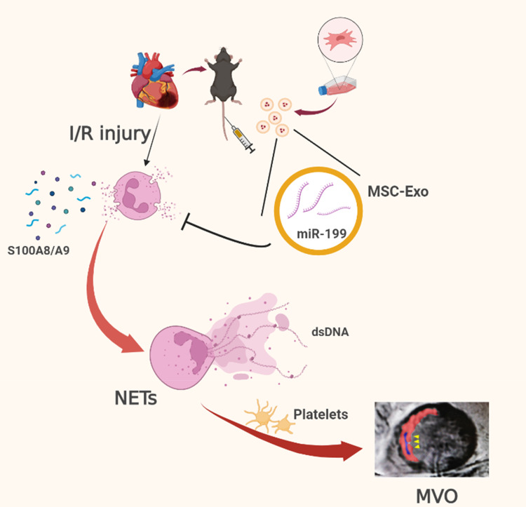
Introduction: Acute inflammatory storm is a major cause of myocardial ischemia/reperfusion (I/R) injury, with no effective treatment currently available. The excessive aggregation of neutrophils is correlated with an unfavorable prognosis in acute myocardial infarction (AMI) patients. Exosomes derived from mesenchymal stromal cells (MSC-Exo) have certain immunomodulatory potential and might be a therapeutic application. Therefore, we investigated the protective role of MSC-Exo in modulating neutrophil infiltration and formation of neutrophil extracellular traps (NETs) following myocardial I/R injury.

Methods: Exosomes were isolated from the supernatant of MSCs using a gradient centrifugation method. We used flow cytometry, histochemistry, and immunofluorescence to detect the changes of neutrophils post-intravenous MSC-Exo injection. Additionally, cardiac magnetic resonance (CMR) and thioflavin S experiments were applied to detect microvascular obstruction (MVO). The NLR family pyrin domain containing 3 (NLRP3) inflammasome was examined for mechanism exploration. Primary neutrophils were extracted for in vitro experiment. Antibody of Ly6G was given to depleting the neutrophils in mice for verification the effect of MSC-Exo. Finally, we analyzed the MiRNA sequence of MSC-Exo and verified it in vitro.

Results: MSC-Exo administration reduced neutrophil infiltration and NETs formation after myocardial I/R. MSC-Exo treatment also could attenuate the activation of NLRP3 inflammasome both in vivo and in vitro. At the same time, the infarction size and MVO following I/R injury were reduced by MSC-Exo. Moreover, systemic depletion of neutrophils partly negated the therapeutic effects of MSC-Exo. Up-regulation of miR-199 in neutrophils has been shown to decrease the expression of NETs formation after stimulation.

Discussion: Our results demonstrated that MSC-Exo mitigated myocardial I/R injury in mice by modulating neutrophil infiltration and NETs formation. This study provides novel insights into the potential therapeutic application of MSC-Exo for myocardial ischemia/reperfusion injury.

Keywords: exosomes; mesenchymal stromal cells; mir-199; myocardial ischemia/reperfusion injury; neutrophil extracellular traps.


Conflict of interest statement

The authors report no conflicts of interest in this work.

Figures

None
Graphical abstract
Figure 1
Figure 1
Increased levels of neutrophils are associated with MVO and LV function in patients with STEMI. (A and B) Correlation between EF% measured by echocardiography and the proportion (A), counts (B) of neutrophils in blood routine of AMI patients 6 hours post-PCI (n=71). (C) Correlation between plasma S100A8/a9 concentration and EF% in AMI patients (n=43). (D) Correlation between plasma dsDNA concentration and EF% in AMI patients (n=67). (E) Correlation between the percentage of enhanced volume measured by cardiac magnetic resonance imaging and counts of neutrophils in blood routine of AMI patients (n=18). (F) Plasma creatine CK-MB, cTNT, and BNP concentrations in AMI patients with MVO (n=32) and without MVO (n=31). (GI) EF% (G), the proportion (H), and counts (I) of neutrophils in AMI patients with MVO (n=32) and without MVO (n=33). (J) Concentration of plasma S100A8/A9 in control group (n=18), AMI patients with MVO (n=22) and without MVO (n=20). (K) Concentration of plasma dsDNA in control group (n=35), AMI patients with MVO (n=33) and without MVO (n=34). In panels A-E, p value as shown (Pearson’s correlation). In panels F-I, *p < 0.05, ***P<0.001 (unpaired two-tailed Student’s t-test). In panels (J and K), *P<0.05, ***P<0.001, ****P<0.0001 (one-way ANOVA with Bonferroni correction).
Figure 2
Figure 2
MSC-Exo reduced neutrophil infiltration and NETs release in vivo. (A) Schematic of the I/R model establishment, treatment, and tissue harvest for detection. (BD) Representative flow cytometry scatter plot and statistical analysis of CD45+CD11b+Ly6G+neutrophils (C) and Ly6G+CD11b+CD41+ neutrophil-platelet complex (D) in the heart from mice treated with either MSC-Exo or PBS at 3, 6, 12, 24.72 hours after I/R or sham operation (n=5). (E) Representative images of HE staining in murine heart sections 24 hours after I/R injury in each group. Scale bar = 100 μm (n=4). (F) Representative immunohistochemical staining for Ly6G+ neutrophils and H3Cit+ NETs in heart sections 24 hours after I/R injury. Scale bar = 100 μm (n=5). (G) Representative immunofluorescence images for NETs formation staining with MPO (green), H3Cit (red) and DAPI (blue). Scale bar = 100 μm. Scale bar in Enlarge= 25 μm (n=5). (H and I) Relative gene expression of CXCL1 and CXCL2 in the hearts of mice sacrificed 24 hours after myocardial I/R injury (n=4). (J and K) Representative immune blots and the corresponding analysis of NETs pathways in the hearts of mice treated with PBS or MSC-Exo 24 hours after I/R (n=5). (L) Concentration of plasma dsDNA in Sham group (n=6), PBS-treated I/R group (n=12), MSC-Exo-treated I/R group (n=10). In panels (C and D), *P<0.05; **P<0.01 (two-way ANOVA followed by Bonferroni’s multiple comparisons test). In panels (H, I, K and L), *P<0.05; **P<0.01; ***P<0.001; ****P < 0.0001 (one-way ANOVA with Bonferroni correction).
Figure 3
Figure 3
MSC-Exo reduced NETs formation in vitro. (A) Representative images of the uptake of DiD-labelled MSC-Exo (red) by primary neutrophils staining with DAPI (blue). DiD only group, PBS negative control stained DiD. Exo only group, background neutrophils fluorescence with non-stained MSC-Exo. Scale bar = 30 μm. (B) GO enrichment of the biological process category in primary neutrophils treated with PBS or MSC-Exo 12 hours after PMA stimulation. The top 7 down-regulated pathways after MSC-Exo treatment were presented and ranked. Sets were listed only if p <0.05. The extracellular matrix organization was found to be among the top down-regulated pathways in the MSC-Exo group (n=3 each group). (C) Representative immunofluorescence images for NETs formation staining with MPO (green), H3Cit (red) and DAPI (blue) in primary neutrophils treated with PBS or MSC-Exo 12 hours after PMA stimulation. Scale bar in Merge = 100μm.Scale bar in Enlarge= 30 μm. (D and E) Representative time-lapse differential interference contrast (DIC) spinning-disk confocal microscopy images (D) at indicated time intervals and arithmetic means (n=3) of the percentage of time course (E) of plasma membrane rupture (NETosis) in neutrophils treated with PBS or MSC-Exo after PMA stimulation. Red, DNA (siR-DNA); Red arrows indicate the area of plasma membrane rupture. Scale bar = 100 μm. (FH) Representative immune blots and the corresponding analysis of NETs pathways in the hearts of mice treated with PBS or MSC-Exo 24 hours after I/R (n=3). In panel (E), p value as shown (two-way ANOVA followed by Bonferroni’s multiple comparisons test). In panel (G and H), **P<0.01; ****P < 0.0001 (one-way ANOVA with Bonferroni correction).
Figure 4
Figure 4
MSC-Exo suppressed NLRP3 signal pathway and S100A8/A9 release in vivo and in vitro. (A and B) Relative gene expression of NLRP3 and IL-1βin the hearts of mice sacrificed 24 hours after myocardial I/R injury (n=3). (C) Concentration of plasma IL-1β in Sham group (n=7), PBS-treated I/R group (n=7) and MSC-Exo-treated group (n=9). (D) Representative immune blots of the NLRP3 pathway in the hearts of mice treated with PBS or MSC-Exo 24 hours after I/R (n=3). (E and F) Relative gene expression of S100A9and S100A8in the hearts of mice sacrificed 24 hours after myocardial I/R injury (n=3). (G) Concentration of plasma S100A8/A9 in Sham group (n=5), PBS-treated I/R group (n=8) and MSC-Exo-treated group (n=8). (H and I) Relative gene expression of NLPR3and IL-1βin primary neutrophils treated with PBS or MSC-Exo 12 hours after PMA stimulation (n=5). (J) Representative immune blots of the NLRP3 pathway in primary neutrophils treated with PBS or MSC-Exo 24 hours after PMA stimulation (n=3). (K) Concentration of IL-1β in the supernatant of mouse neutrophils treated with PBS (n=8) or MSC-Exo (n=8) 24 hours after PMA stimulation or control (n=6). (L and M) Relative gene expression of S100A9and S100A8 in primary neutrophils treated with PBS or MSC-Exo 12 hours after PMA stimulation (n=5). (N) Concentration of S100A8/A9 in the supernatant of primary neutrophils treated with PBS or MSC-Exo 24 hours after PMA stimulation or control (n=5). In panels (AC, EI, KN), *P<0.05; **P<0.01; ***P<0.001; ****P < 0.0001 (one-way ANOVA with Bonferroni correction).
Figure 5
Figure 5
MSC-Exo reduced MVO formation and preserved cardiac function after myocardial I/R injury. (A) Representative end-diastolic static frames from LGE in midventricular short-axis in mice 7 days after I/R injury. Yellow arrowheads indicate MVO in the LV wall. (B) Representative thioflavin S staining under ultraviolet light in the hearts of mice sacrificed 3 days after myocardial I/R injury and the statistical comparisons of the ratio of the no-reflow area (NRA) to the total heart section area (TA) (n=5). (C) Representative FITC-dextran (green) angiographic micrographs in murine heart 3 days after I/R injury. (D) Quantification of fluorescence intensity in (C) (n=5). (E) Echocardiography measurements of cardiac functions of mice at 24 hours, 3 days, 7 days and 1 month after I/R or sham operation, LVEF values were measured (n=5). (F) Representative immunofluorescence images for TUNEL staining with TUNEL (red) and DAPI (blue). Scale bar = 100 μm. (G) Quantitative analysis of TUNEL-positive nuclei. (H) Images illustrate representative sections of Evan’s blue and TTC double-stained hearts from mice treated with PBS or MSC-Exo 3 days after I/R. (I) Quantitative analysis of the percentage AAR and percentage infarct of hearts (n=5 each group). (J) Representative images and quantitative analysis of Masson staining in heart sections 1 month post-I/R (n=5 each group). In panels B and I, **P<0.01; ns, not significant (unpaired two-tailed Student’s t-test). In panels (D and G), *P<0.05; **P<0.01; ***P < 0.001; ****P < 0.0001 (one-way ANOVA with Bonferroni correction). In panel (E), *P<0.05; **P<0.01; ***P < 0.001; ****P < 0.0001 (two-way ANOVA followed by Bonferroni’s multiple comparisons test).
Figure 6
Figure 6
Systemic depletion of neutrophils reduced the efficacy of MSC-Exo treatment after I/R injury. (A and B) Flow cytometry statistical analysis of CD45+CD11b+Ly6G+ neutrophils (A) and Ly6G+CD11b+CD41+ neutrophil-platelet complex (B) in the heart from mice sacrificed 3 days after injection of either MSC-Exo or PBS with (n=4) or without (n=5) neutrophil depletion prior to myocardial I/R injury or in Sham group (n=4). (C) Echocardiography measurements of cardiac functions of mice at 3 days after I/R (n=4). (D) Images illustrate representative sections of Evan’s blue and TTC double-stained hearts from mice 3 days after I/R. (E and F) Quantitative analysis of the ratio of AAR (E)and infarct area of hearts (F) (n=4). (G) Statistical comparisons of the ratio of the no-reflow area (NR) to the total heart section area (TA) by thioflavin S staining (n=4). (H) Representative FITC-dextran (green) angiographic micrographs in murine heart 3 days after I/R injury. (I) Quantification of fluorescence intensity in (H) (n=4). In panels (AC, EG and I), *P<0.05; **P<0.01; ***P<0.001; ****P < 0.0001; ns, not significant (one-way ANOVA with Bonferroni correction).
Figure 7
Figure 7
Mir-199a contributed to the therapeutic effect mediated by MSC-Exo. (A) Relative gene expression of miR-199-5p and mir-199-3p levels in neutrophils treated with mimic or inhibitor (n=7). The expression levels of the miRNAs were normalized to U6. (B) Representative immune blots of PAD4, MPO and H3Cit in neutrophils after 24 hours of PMA stimulation following mimic or inhibitor treatment (n=3). (C) Relative gene expression of miR-199-5p and mir-199-3p levels in MSC-Exo isolated from MSCs treated with inhibitor (n=3). The expression levels of the miRNAs were normalized to U6. (D) Representative immunofluorescence images for NETs formation staining with MPO (green), H3Cit (red) and DAPI (blue) in primary neutrophils with MSC-Exo, NC-inhibitor MSC-Exo, mir-199a-5p inhibitor MSC-Exo or mir-199a-3p MSC-Exo treatment 12 hours after PMA stimulation. Scale bar in Merge = 50μm (n=3). (E and F) Representative immune blots of PAD4, MPO and H3Cit in neutrophils after 24 hours of PMA stimulation following MSC-Exo, NC-inhibitor MSC-Exo, mir-199a-5p inhibitor MSC-Exo or mir-199a-3p MSC-Exo inhibitor treatment (n=3). In panels (A and C), *P<0.05; **P<0.01; ***P<0.001; ****P < 0.0001 (one-way ANOVA with Bonferroni correction).

Similar articles

Cited by

References

    1. Heusch G. Myocardial ischaemia-reperfusion injury and cardioprotection in perspective. Nat Rev Cardiol. 2020;17(12):773–789. doi:10.1038/s41569-020-0403-y - DOI - PubMed
    1. Algoet M, Janssens S, Himmelreich U, et al. Myocardial ischemia-reperfusion injury and the influence of inflammation. Trends Cardiovasc Med. 2022;33:357–366. doi:10.1016/j.tcm.2022.02.005 - DOI - PubMed
    1. Zhang RYK, Cochran BJ, Thomas SR, Rye KA. Impact of reperfusion on temporal immune cell dynamics after myocardial infarction. J Am Heart Assoc. 2023;12:4. - PMC - PubMed
    1. Piccolo EB, Thorp EB, Sumagin R. Functional implications of neutrophil metabolism during ischemic tissue repair. Curr Opin Pharmacol. 2022;63:102191. doi:10.1016/j.coph.2022.102191 - DOI - PMC - PubMed
    1. Sreejit G, Johnson J, Jaggers RM, et al. Neutrophils in cardiovascular disease, warmongers, peacemakers, or both? Cardiovasc Res. 2022;118(12):2596–2609. doi:10.1093/cvr/cvab302 - DOI - PMC - PubMed