

Terahertz in vivo imaging of human skin: Toward detection of abnormal skin pathologies
- PMID: 38476403
- PMCID: PMC10932572
- DOI: 10.1063/5.0190573
Terahertz in vivo imaging of human skin: Toward detection of abnormal skin pathologies
Abstract
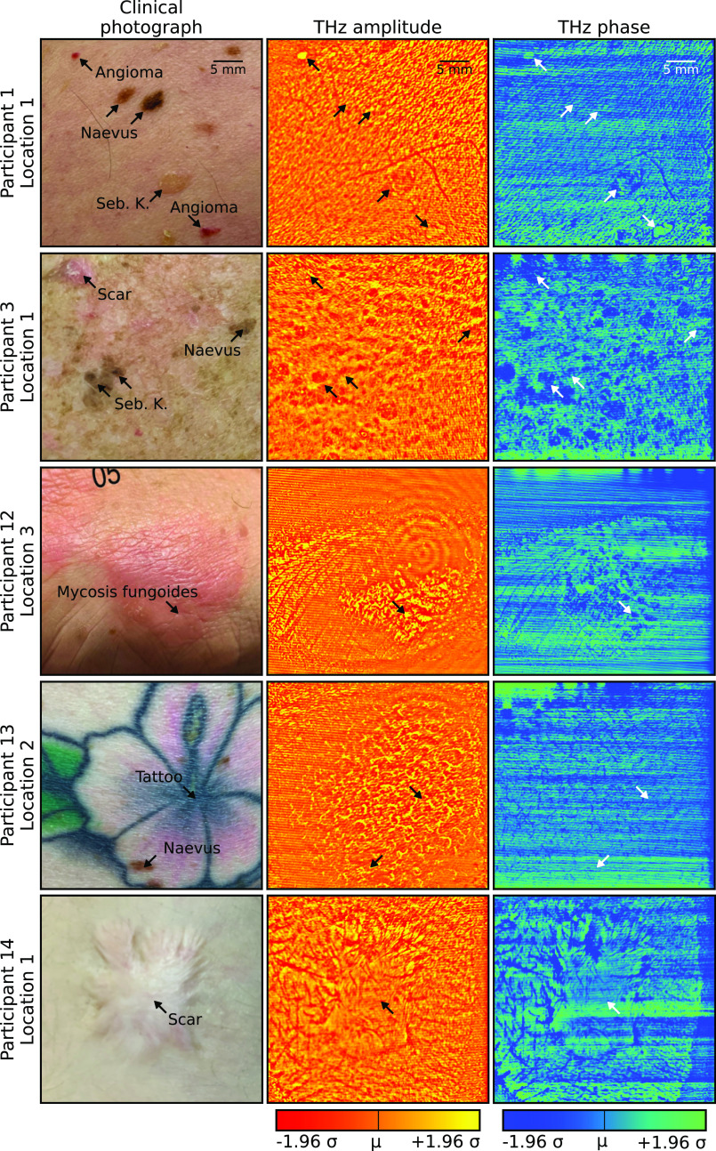
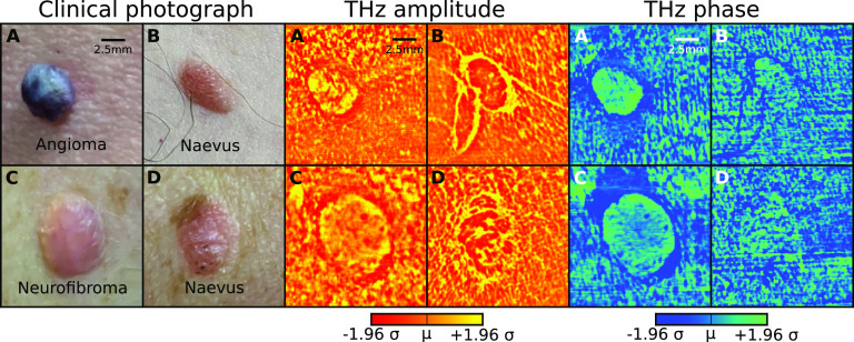
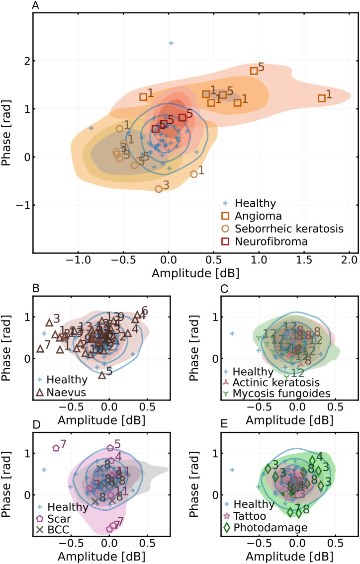
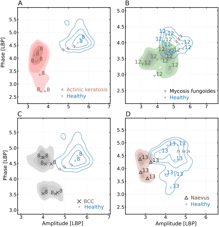
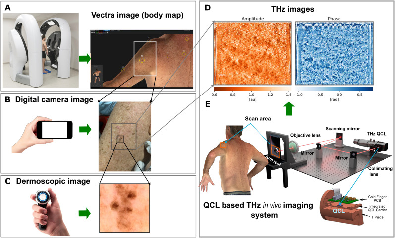
Terahertz (THz) imaging has long held promise for skin cancer detection but has been hampered by the lack of practical technological implementation. In this article, we introduce a technique for discriminating several skin pathologies using a coherent THz confocal system based on a THz quantum cascade laser. High resolution in vivo THz images (with diffraction limited to the order of 100 μm) of several different lesion types were acquired and compared against one another using the amplitude and phase values. Our system successfully separated pathologies using a combination of phase and amplitude information and their respective surface textures. The large scan field (50 × 40 mm) of the system allows macroscopic visualization of several skin lesions in a single frame. Utilizing THz imaging for dermatological assessment of skin lesions offers substantial additional diagnostic value for clinicians. THz images contain information complementary to the information contained in the conventional digital images.
© 2024 Author(s).
Conflict of interest statement
X.Q., K.B., J.T., T.G., P.D., D.I., E.H.L., M.S.S., H.P.S., and A.D.R. are authors of relevant patents. T.M. is an executive of OscillaDx Pty Ltd. H.P.S. is a shareholder of MoleMap NZ Limited and e-derm consult GmbH and undertakes regular teledermatological reporting for both companies. H.P.S. is a Medical Consultant for Canfield Scientific Inc, Blaze Bioscience Inc, and a Medical Advisor for First Derm. The other authors declare that there are no conflicts of interest related to this article. The research was conducted to refine and develop the imaging technology. The authors declare that this study received funding from OscillaDx Pty Ltd, Brisbane, with involvement in the study design.
Figures
Similar articles
-
Terahertz imaging of human skin pathologies using laser feedback interferometry with quantum cascade lasers.Biomed Opt Express. 2023 Mar 2;14(4):1393-1410. doi: 10.1364/BOE.480615. eCollection 2023 Apr 1. Biomed Opt Express. 2023. PMID: 37078035 Free PMC article.
-
Subwavelength 3D terahertz imaging with a single-pixel laser transceiver.Opt Express. 2025 May 19;33(10):21938-21950. doi: 10.1364/OE.561713. Opt Express. 2025. PMID: 40515078
-
Early detection of gastric cancer via high-resolution terahertz imaging system.Front Bioeng Biotechnol. 2022 Dec 14;10:1052069. doi: 10.3389/fbioe.2022.1052069. eCollection 2022. Front Bioeng Biotechnol. 2022. PMID: 36588946 Free PMC article.
-
Terahertz radiation and the skin: a review.J Biomed Opt. 2021 Feb;26(4):043005. doi: 10.1117/1.JBO.26.4.043005. J Biomed Opt. 2021. PMID: 33583155 Free PMC article. Review.
-
Three-dimensional terahertz wave imaging.Philos Trans A Math Phys Eng Sci. 2004 Feb 15;362(1815):283-98; discussion 298-9. doi: 10.1098/rsta.2003.1317. Philos Trans A Math Phys Eng Sci. 2004. PMID: 15306520 Review.
Cited by
-
In vivo terahertz sensing of psoriasis and eczema patients.Sci Rep. 2024 Jul 30;14(1):17546. doi: 10.1038/s41598-024-68106-2. Sci Rep. 2024. PMID: 39079961 Free PMC article.
-
Towards autonomous robotic THz-based in vivo skin sensing: the PicoBot.Sci Rep. 2025 Feb 7;15(1):4568. doi: 10.1038/s41598-025-88718-6. Sci Rep. 2025. PMID: 39915605 Free PMC article.
-
Review of Non-Invasive Imaging Technologies for Cutaneous Melanoma.Biosensors (Basel). 2025 May 7;15(5):297. doi: 10.3390/bios15050297. Biosensors (Basel). 2025. PMID: 40422036 Free PMC article. Review.
References
-
- Gordon L. G., Leung W., Johns R., McNoe B., Lindsay D., Merollini K. M., Elliott T. M., Neale R. E., Olsen C. M., Pandeya N. et al., “ Estimated healthcare costs of melanoma and keratinocyte skin cancers in Australia and Aotearoa New Zealand in 2021,” Int. J. Environ. Res. Public Health 19, 3178 (2022).10.3390/ijerph19063178 - DOI - PMC - PubMed
LinkOut - more resources
Full Text Sources