Synchronous presentation of prolactinoma and supratentorial tanycytic ependymoma
- PMID: 38476418
- PMCID: PMC10927033
- DOI: 10.25259/JNRP_217_2023
Synchronous presentation of prolactinoma and supratentorial tanycytic ependymoma
Abstract
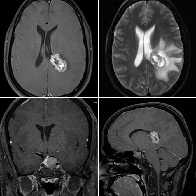
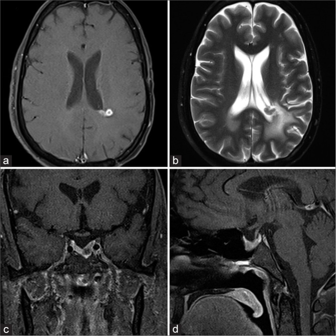
Tanycytic ependymomas mostly occur in the spinal cord and it is the rarest histological subtype of ependymoma. A 29-year-old male was referred from the infertility clinic after serum prolactin levels were found to be elevated. Magnetic resonance imaging (MRI) brain showed an irregular necrotic lesion in the periventricular region of the left parietal lobe which had an intraventricular component and associated perilesional edema. In addition, a sellar mass with suprasellar extension was also found on the MRI. He was started on cabergoline therapy for macroprolactinoma and underwent a left parietal craniotomy, and microsurgical excision of the tumor using intraoperative neurosonographic guidance. Histologically, the tumor showed spindle cytologic features and poorly developed inconspicuous pseudorosettes, with areas of rounded nuclear profiles and perinuclear cytoplasmic clearing. Tumor cells were positive for vimentin, glial fibrillary acidic protein and S100, and negative for epithelial membrane antigen. Ki67 was <7%. He was diagnosed with tanycytic ependymoma and a coexistent prolactinoma. He received 10 cycles of image-guided radiotherapy. Post-operative imaging showed minimal residual tumor the size of which remained stable at 1-year follow-up scan. The pituitary macroadenoma regressed with cabergoline therapy and he clinically improved. This presentation of synchronous macroprolactinoma and tanycytic ependymoma has not been reported in the literature previously. An exhaustive literature review showed only 18 previously reported cases of supratentorial tanycytic ependymoma.
Keywords: Cabergoline; Craniotomy; Prolactinoma; Synchronous intracranial tumors; Tanycytic ependymoma.
© 2024 Published by Scientific Scholar on behalf of Journal of Neurosciences in Rural Practice.
Conflict of interest statement
There are no conflicts of interest.
Figures
Similar articles
-
Features of intraventricular tanycytic ependymoma: report of a case and review of literature.Int J Clin Exp Pathol. 2014 May 15;7(6):3399-407. eCollection 2014. Int J Clin Exp Pathol. 2014. PMID: 25031767 Free PMC article. Review.
-
The clinical features and surgical outcomes of intracranial tanycytic ependymomas: a single-institutional experience.J Neurooncol. 2017 Sep;134(2):339-347. doi: 10.1007/s11060-017-2531-8. Epub 2017 Jun 26. J Neurooncol. 2017. PMID: 28653235
-
Supratentorial tanycytic ependymoma.J Clin Neurosci. 2004 Nov;11(8):928-30. doi: 10.1016/j.jocn.2004.03.022. J Clin Neurosci. 2004. PMID: 15519882
-
Spinal tanycytic ependymomas.Acta Neuropathol. 2001 Jan;101(1):43-8. doi: 10.1007/s004010000265. Acta Neuropathol. 2001. PMID: 11194940
-
Intraventricular tanycytic ependymoma: case report and review of the literature.J Neurooncol. 2005 Jan;71(2):189-93. doi: 10.1007/s11060-004-1371-5. J Neurooncol. 2005. PMID: 15690137 Review.
References
Publication types
LinkOut - more resources
Full Text Sources
Molecular Biology Databases