. 2024 Mar 12;6(1):zcae011.
doi: 10.1093/narcan/zcae011. eCollection 2024 Mar.

The AsiDNA™ decoy mimicking DSBs protects the normal tissue from radiation toxicity through a DNA-PK/p53/p21-dependent G1/S arrest

Affiliations

The AsiDNA™ decoy mimicking DSBs protects the normal tissue from radiation toxicity through a DNA-PK/p53/p21-dependent G1/S arrest

Anouk Sesink et al. NAR Cancer. .

Abstract

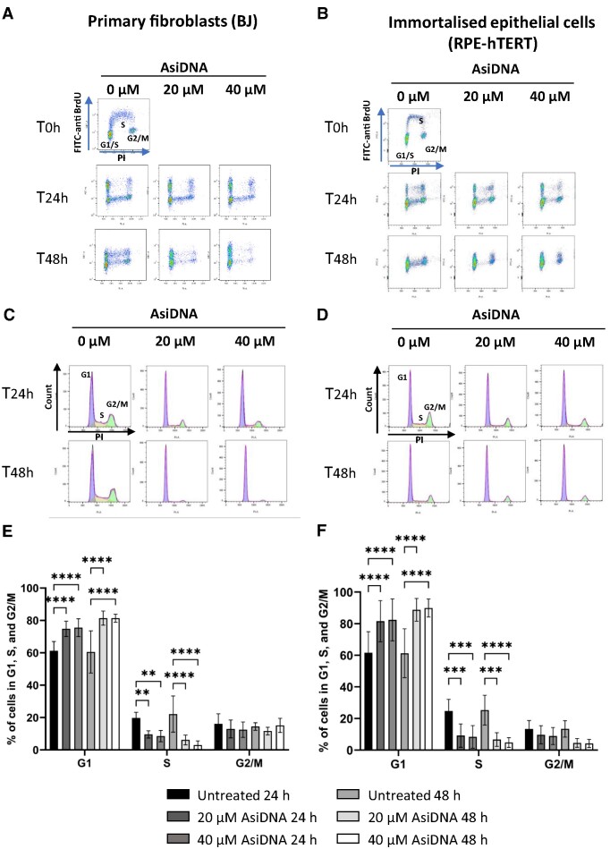
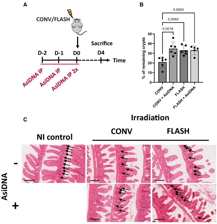
AsiDNA™, a cholesterol-coupled oligonucleotide mimicking double-stranded DNA breaks, was developed to sensitize tumour cells to radio- and chemotherapy. This drug acts as a decoy hijacking the DNA damage response. Previous studies have demonstrated that standalone AsiDNA™ administration is well tolerated with no additional adverse effects when combined with chemo- and/or radiotherapy. The lack of normal tissue complication encouraged further examination into the role of AsiDNA™ in normal cells. This research demonstrates the radioprotective properties of AsiDNA™. In vitro, AsiDNA™ induces a DNA-PK/p53/p21-dependent G1/S arrest in normal epithelial cells and fibroblasts that is absent in p53 deficient and proficient tumour cells. This cell cycle arrest improved survival after irradiation only in p53 proficient normal cells. Combined administration of AsiDNA™ with conventional radiotherapy in mouse models of late and early radiation toxicity resulted in decreased onset of lung fibrosis and increased intestinal crypt survival. Similar results were observed following FLASH radiotherapy in standalone or combined with AsiDNA™. Mechanisms comparable to those identified in vitro were detected both in vivo, in the intestine and ex vivo, in precision cut lung slices. Collectively, the results suggest that AsiDNA™ can partially protect healthy tissues from radiation toxicity by triggering a G1/S arrest in normal cells.


Conflict of interest statement

Wael Jdey is employed by Valerio Therapeutics (former ONXEO). Valerio Therapeutics owns the patent for AsiDNA.

Figures

Graphical Abstract
Graphical Abstract
Figure 1.
Figure 1.
AsiDNA™ induces a G1/S arrest in healthy cells in vitro. Cells were pulse-labelled with BrdU following incubation with 20 and 40 μM of AsiDNA™ for 24 and 48 h. Representative images of the bivariate analysis by flow cytometry of BrdU incorporation versus DNA content (PI) in (A) BJ and (B) RPE-hTERT cells. The deconvolution of the cellular DNA content frequency histograms allows the identification of G1 phase (purple), S-phase (orange), and G2/M-phase (green) in (C) BJ, and (D) RPE-hTERT cells. The percentage of cells in G1, S, and G2/M is shown in (E) for BJ, and in (F) for RPE-hTERT cells. Data are expressed as mean ± standard deviation (n = 8–9) with significance given by two-way ANOVA, Tukey's multiple comparison tests, and represented above the bar plots. Statistical significance was set at * P value < 0.05, ** P value < 0.01, *** P value < 0.001 and **** P value < 0.0001.
Figure 2.
Figure 2.
AsiDNA™-induced G1/S arrest in vitro is dependent on DNA-PK, p53, and p21. (A) RPE-hTERT cells were pre-treated with 1 μM olaparib (Ola) or 10 μM NU7026 (NU) for 1 h before addition of 20 μM AsiDNA™. The percentage of cells in G1, S and G2/M was analysed by flow cytometry 48 h post-AsiDNA™ treatment based on PI staining. (B) RPE-hTERT cells were transiently transfected with small inhibitory RNA (siRNA) silencing DNA-PKcs, p53, or p21 before being exposed to AsiDNA™ for 48 h. The percentage of cells in G1, S and G2/M was analysed by flow cytometry based on PI staining. (C) MRC-5V1, (D) RPE-hTERT shp53 and (E) RPE-hTERT p21−/− cells were treated with 20 and 40 μM of AsiDNA™ for 24 and 48 h. The percentage of cells in G1, S and G2/M was analysed by flow cytometry at the end of AsiDNA™ treatment based on PI staining. All the data are expressed as mean ± standard deviation (n = 3–9) with significance given by two-way ANOVA, Tukey's multiple comparison tests, and represented above the bar plots. Statistical significance was set at * P value < 0.05, ** P value < 0.01, *** P value < 0.001 and **** P value < 0.0001.
Figure 3.
Figure 3.
No effect of AsiDNA™ treatment on cell cycle progression in p53 proficient tumour cells in vitro. (A) A549, (B) HCT116 and (C) U2OS tumour cells were exposed to 20 and 40 μM of AsiDNA™ for 24 and 48 h. The percentage of cells in G1, S and G2/M was analysed by flow cytometry at the completion of AsiDNA™ treatment, based on PI staining. Data are expressed as mean ± standard deviation (n = 5–7) with significance given by two-way ANOVA, Tukey's multiple comparison tests, and represented above the bar plots. Statistical significance was set at *** P value < 0.001.
Figure 4.
Figure 4.
AsiDNA™ treatment protects p53-proficient normal cells, but not p53-proficient tumour cells, from radiation-induced toxicity in vitro. (A) p53 proficient normal cells (BJ and RPE-hTERT), (B) p53 deficient normal cells (RPE-hTERT shp53) and (C) p53 proficient tumour (HCT116 and A549) cells were pre-treated with AsiDNA™ 24 h before being co-exposed to increased doses of ionizing radiation (0–6 Gy). The survival fraction was determined 8–12 days post-treatment using a clonogenic survival assay. Data are expressed as mean ± standard deviation (n = 3 for BJ, RPE-hTERT shp53, HCT116 and A549; n = 4 for RPE-hTERT), fitted to the linear-quadratic model as a function of dose with significance given by nonlinear fit using GraphPad Prism.
Figure 5.
Figure 5.
AsiDNA™ delayed the onset of radiation-induced pulmonary fibrosis in vivo. (A) Scheme of the experimental protocol. C57BL6/J mice were treated for 3 consecutive days with AsiDNA™ followed by 13 Gy CONV or FLASH irradiation of the thorax on the final day. Retro-orbital blood sampling was performed from 1 week up to 5 months post-treatment. CT scans were recorded from 4 to 6.5 months (sacrifice) post-treatment, and lobes of the lung were collected at the day of sacrifice (day 200) for histologic analysis. (B) Representative CT scans of lung from untreated mice (NI) or 5 months post-irradiation at 13 Gy CONV or FLASH radiotherapy alone or combined with AsiDNA™. Images are obtained using micro-CT imaging, high resolution, and 100 μm reconstruction by Molecubes software (Molecubes, Belgium). Representative images are shown with the CT axial slice (top), CT coronal slice (middle) and 3D lung reconstruction of connected Hounsfield Units −800 to −100 (bottom). Images were obtained using VivoQuant software (Konica Minolta Company, Japan). (C) Kaplan–Meier representation of animal surviving fraction displayed in days post-treatment. Data are expressed with significance given by survival, curve comparison, and Logrank test. Significance: not significant, ns; * P< 0.05; ** P< 0.01, *** P< 0.001, **** P< 0.0001. The statistical analysis gave NT versus CONV is < 0.0001, NT versus CONV AsiDNA is 0.003, NT versus FLASH 0.002, NT versus FLASH AsiDNA is 0.0015, CONV versus CONV AsiDNA 0.0178, CONV versus FLASH 0.0053 and FLASH versus FLASH AsiDNA is not significant.
Figure 6.
Figure 6.
Single-cell RNA sequencing after irradiation and AsiDNA™ treatment. Identification of cell populations represented by (A) Dot plot of marker expression utilized for cell population identification. (B) UMAP visualizing the identified cell clusters separating representation of the Control (red), CONV (green), AsiDNA™ + CONV (blue), and FLASH (purple) treated samples. (C) UMAP visualizing the identified cell types in all samples. The individual dots signify single cells. Additionally, the created clusters are established on transcriptome resemblances. (D) Cell population proportions after Control, CONV, AsiDNA™ + CONV and FLASH treatment. Fibroblast populations represented by (E) UMAP visualizing the identified fibroblast cluster separating representation of the Control (red), CONV (green), AsiDNA™ CONV (blue) and FLASH (purple) treated samples. Pro-fibrotic markers were examined using Violin plots with myofibroblast signature genes Hp (F) and Pla1a (G), and healthy collagen homeostasis Nr1d1 (H). CONV irradiation upregulates the expression of Hp and Pla1a and decreases the expression of Nr1d1 compared to FLASH, CONV + AsiDNA™, and NI control, significance given by Wilcox test. (NS, P-value > 0.05; *, P-value < 0.05; **, P-value < 0.01; ***, P-value < 0.001; ****, P-value < 0.0001).
Figure 7.
Figure 7.
AsiDNA™ induces a DNA-PK and p53-dependent cell cycle arrest in the ex vivo model of precision cut lung slices. Precision cut lung slices (PCLS) were derived from C57BL6/J WT or p53 knock-out mice and treated with EdU upon 24 h of AsiDNA™ or Nol8 treatment. (A) Representative images of PCLS derived from WT mice (N = 2) with DAPI and EdU detection after AsiDNA™ or Nol8 treatment. (B) EdU positive cells detected after AsiDNA™ or Nol8 treatment (N = 2). EdU was detected in 6 slices per condition with 5 readouts per slice. (C) Representative Images of PCLS derived from WT (N = 2) and p53 knock-out mice (N = 2) with DAPI and EdU detection after AsiDNA™ treatment. (D) EdU positive cells detected after AsiDNA™ or Nol8 treatment of two WT mice. EdU was detected in 8–10 slices per condition with 5 readouts per slice. Data are expressed as mean ± standard deviation with significance given by two-way ANOVA, Tukey's multiple comparison test and represented above the bar plots. Scale bar = 100 μm.
Figure 8.
Figure 8.
AsiDNA™ protects intestinal crypts from radiation toxicity. (A) Scheme of the experimental treatment timeline. C57BL6/J mice were treated for 3 consecutive days with AsiDNA™ followed by 10 Gy CONV or FLASH irradiation of the abdomen on the final day. (B) Small intestinal crypt survival of C57BL6/J mice 4 days after abdominal radiation, normalised to non-irradiated control mice. Data are expressed as mean ± standard deviation (n = 5–6) with significance given by one-way ANOVA, Tukey's multiple comparison test and represented above the bar plots. (C) Representative images of intestinal rolls stained with H&E from each treatment group. Arrows point to the intestinal crypts. Scale bar = 200 μm.
Figure 9.
Figure 9.
AsiDNA™-induced cell cycle arrest is reversible upon release of AsiDNA™ in intestinal normal tissue. (A) Scheme of the experimental treatment timeline. C57BL6/J mice were treated for 3 consecutive days with AsiDNA™ followed by 0–72 h of recovery. Thereafter, mice received EdU for 4 h prior to sacrifice. (B) Representative images of intestinal rolls stained with DAPI and Click-iTTM EdU AlexaFluorTM 488 for each treatment group. (C) EdU positive cells per 1000 detected cells in small intestinal crypts after AsiDNA™ treatment. A total number of 14 000–16 000 cells per mouse were scored. Data are expressed as mean ± standard deviation (n = 3) with significance given by one-way ANOVA, Brown-Forsythe and Welch tests. Scale bar = 50 μm. (D) p21 positive cells per 100 detected cells in small intestinal crypts after AsiDNA™ treatment. A total number of 6000–11 000 cells per mouse were scored. Data are expressed as mean ± standard deviation (n = 3) with significance given by one-way ANOVA, Tukey's multiple comparison test and represented above the bar plots. (E) Representative images of intestinal rolls stained by immunohistochemistry to detect p21 expression in each treatment group. Scale bar = 200 μm.

Similar articles

Cited by

References

    1. Barnett G.C., West C.M., Dunning A.M., Elliott R.M., Coles C.E., Pharoah P.D., Burnet N.G. Normal tissue reactions to radiotherapy: towards tailoring treatment dose by genotype. Nat. Rev. Cancer. 2009; 9:134–142. - PMC - PubMed
    1. De Ruysscher D., Niedermann G., Burnet N.G., Siva S., Lee A.W.M., Hegi-Johnson F. Radiotherapy toxicity. Nat. Rev. Dis. Primers. 2019; 5:13. - PubMed
    1. Al-Mahayri Z.N., Patrinos G.P., Ali B.R. Toxicity and pharmacogenomic biomarkers in breast cancer chemotherapy. Front. Pharmacol. 2020; 11:445. - PMC - PubMed
    1. Hellman S. Improving the therapeutic index in breast cancer treatment: the Richard and Hinda Rosenthal Foundation Award lecture. Cancer Res. 1980; 40:4335–4342. - PubMed
    1. Quanz M., Berthault N., Roulin C., Roy M., Herbette A., Agrario C., Alberti C., Josserand V., Coll J.L., Sastre-Garau X. et al. . Small-molecule drugs mimicking DNA damage: a new strategy for sensitizing tumors to radiotherapy. Clin. Cancer Res. 2009; 15:1308–1316. - PubMed

Publication types

MeSH terms

Substances