

Anti-ulcerative colitis effects of chemically characterized extracts from C alliandra haematocephala in acetic acid-induced ulcerative colitis
- PMID: 38476652
- PMCID: PMC10927971
- DOI: 10.3389/fchem.2024.1291230
Anti-ulcerative colitis effects of chemically characterized extracts from C alliandra haematocephala in acetic acid-induced ulcerative colitis
Abstract
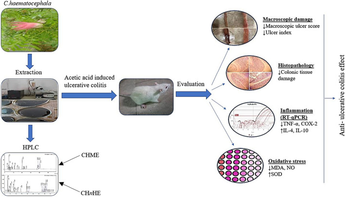
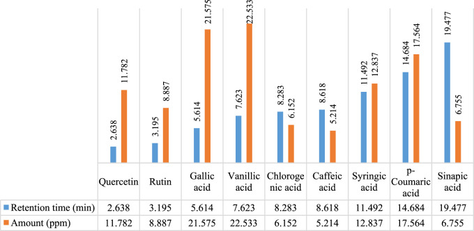
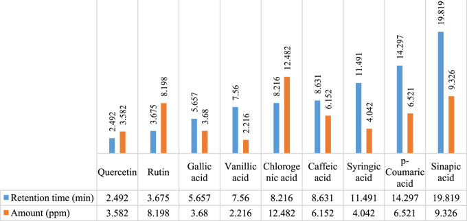
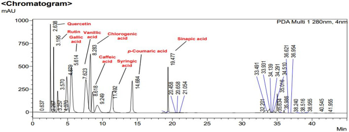
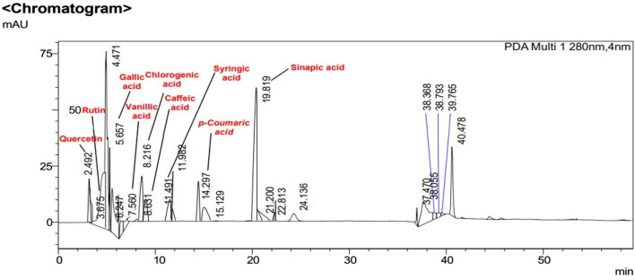
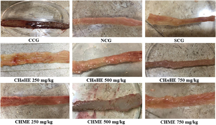
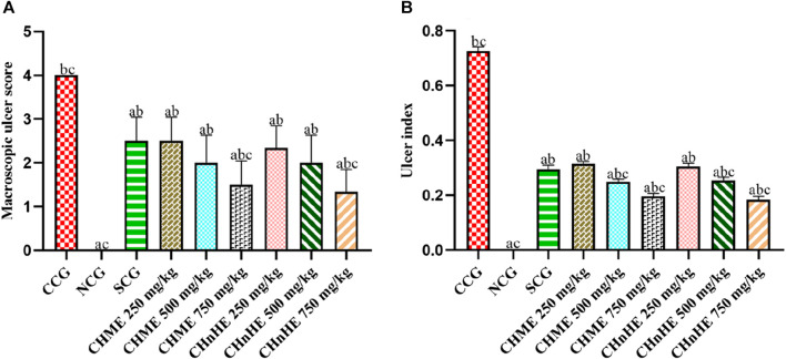
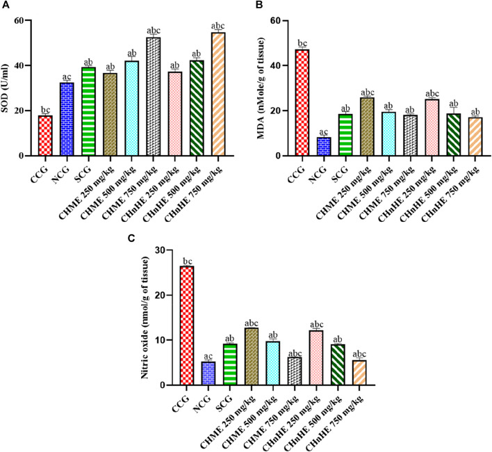
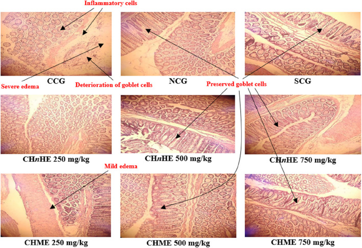
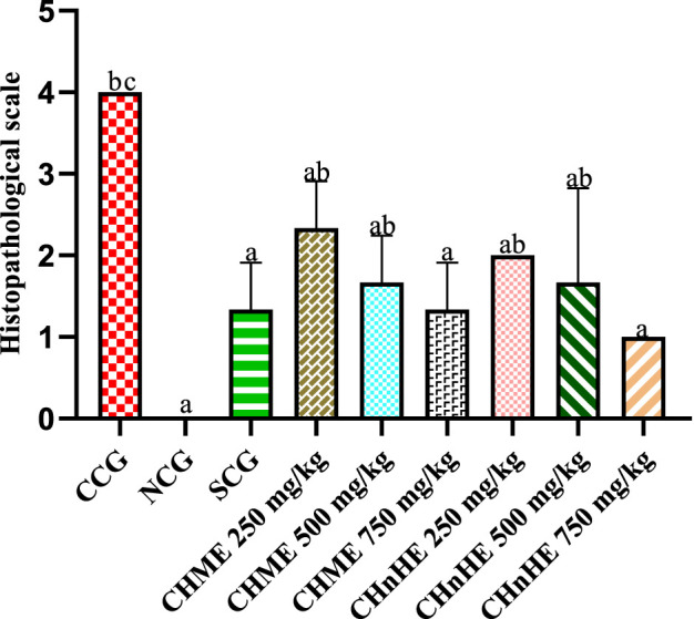
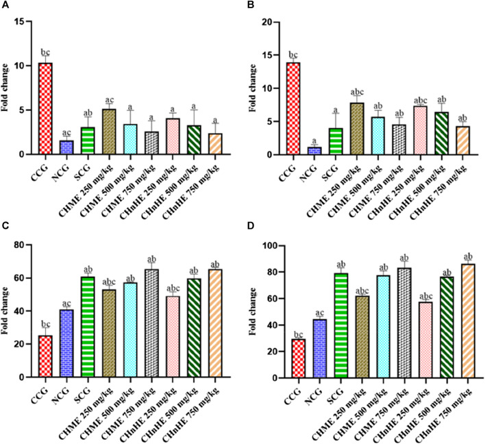
Background: Ulcerative colitis is a chronic immune-mediated inflammatory bowel disease that involves inflammation and ulcers of the colon and rectum. To date, no definite cure for this disease is available. Objective: The objective of the current study was to assess the effect of Calliandra haematocephala on inflammatory mediators and oxidative stress markers for the exploration of its anti-ulcerative colitis activity in rat models of acetic acid-induced ulcerative colitis. Methods: Methanolic and n-hexane extracts of areal parts of the plant were prepared by cold extraction method. Phytochemical analysis of both extracts was performed by qualitative analysis, quantitative methods, and high-performance liquid chromatography (HPLC). Prednisone at 2 mg/kg dose and plant extracts at 250, 500, and 750 mg/kg doses were given to Wistar rats for 11 days, which were given acetic acid on 8th day through the trans-rectal route for the induction of ulcerative colitis. A comparison of treatment groups was done with a normal control group and a colitis control group. To evaluate the anti-ulcerative colitis activity of Calliandra haematocephala, different parameters such as colon macroscopic damage, ulcer index, oxidative stress markers, histopathological examination, and mRNA expression of pro and anti-inflammatory mediators were evaluated. mRNA expression analysis was carried out by reverse transcription quantitative real-time polymerase chain reaction (RT-qPCR). Results: The phytochemical evaluation revealed polyphenols, flavonoids, tannins, alkaloids, and sterols in both extracts of the plant. Results of the present study exhibited that both extracts attenuated the large bowel inflammation and prevented colon ulceration at all tested doses. Macroscopic damage and ulcer scoreswere significantly decreased by both extracts. Malondialdehyde (MDA) levels and nitrite/nitrate concentrations in colon tissues were returned to normal levels while superoxide dismutase (SOD) activity was significantly improved by all doses. Histopathological examination exhibited that both extracts prevented the inflammatory changes, cellular infiltration, and colon thickening. Gene expression analysis by RT-qPCR revealed the downregulation of pro-inflammatory markers such as tumor necrosis factor-alpha (TNF-α) and cyclooxygenase-2 (COX-2) whereas the anti-inflammatory cytokines including Interleukin-4 (IL-4) and Interleukin-10 (IL-10) were found to be upregulated in treated rats. Conclusion: It was concluded based on study outcomes that methanolic and n-hexane extracts of Calliandra haematocephala exhibited anti-ulcerative colitis activity through modulation of antioxidant defense mechanisms and the immune system. In this context, C. haematocephala can be considered as a potential therapeutic approach for cure of ulcerative colitis after bioassay-directed isolation of bioactive phytochemicals and clinical evaluation.
Keywords: Calliandra haematocephala; HPLC; anti-inflammatory cytokines; oxidative stress; pro-inflammatory markers; ulcerative colitis.
Copyright © 2024 Rehman, Saleem, Raza, Bashir, Muhammad, Asghar, Qamar, Shah, Bin Jardan, Mekonnen and Bourhia.
Conflict of interest statement
The authors declare that the research was conducted in the absence of any commercial or financial relationships that could be construed as a potential conflict of interest.
Figures
Similar articles
-
Piper umbellatum L. (Piperaceae): Phytochemical profiles of the hydroethanolic leaf extract and intestinal anti-inflammatory mechanisms on 2,4,6 trinitrobenzene sulfonic acid induced ulcerative colitis in rats.J Ethnopharmacol. 2020 May 23;254:112707. doi: 10.1016/j.jep.2020.112707. Epub 2020 Feb 27. J Ethnopharmacol. 2020. PMID: 32112897
-
Effect of piperine on inhibition of FFA induced TLR4 mediated inflammation and amelioration of acetic acid induced ulcerative colitis in mice.J Ethnopharmacol. 2015 Apr 22;164:239-46. doi: 10.1016/j.jep.2015.01.039. Epub 2015 Feb 13. J Ethnopharmacol. 2015. PMID: 25683300
-
Evaluation of Anti-Inflammatory and Antioxidant Effects of Ferulic Acid and Quinic Acid on Acetic Acid-Induced Ulcerative Colitis in Rats.J Biochem Mol Toxicol. 2025 Feb;39(2):e70169. doi: 10.1002/jbt.70169. J Biochem Mol Toxicol. 2025. PMID: 39957712
-
Juglone Mediates Inflammatory Bowel Disease Through Inhibition of TLR-4/NF KappaB Pathway in Acetic Acid-induced Colitis in Rats.Antiinflamm Antiallergy Agents Med Chem. 2023;22(2):92-103. doi: 10.2174/1871523022666230825105223. Antiinflamm Antiallergy Agents Med Chem. 2023. PMID: 37936449
-
Protective effect of Averrhoa bilimbi L. fruit extract on ulcerative colitis in wistar rats via regulation of inflammatory mediators and cytokines.Biomed Pharmacother. 2017 Jul;91:1113-1121. doi: 10.1016/j.biopha.2017.05.057. Epub 2017 May 16. Biomed Pharmacother. 2017. PMID: 28531922
Cited by
-
Candida utilis Ameliorates Dextran Sulfate Sodium-Induced Colitis in Mice via NF-κB/MAPK Suppression and Gut Microbiota Modulation.Int J Mol Sci. 2025 Feb 25;26(5):1993. doi: 10.3390/ijms26051993. Int J Mol Sci. 2025. PMID: 40076616 Free PMC article.
-
Garlic Peel-Derived Phytochemicals Using GC-MS: Antioxidant, Anti-Inflammatory, and Anti-Apoptotic Effects in Ulcerative Colitis Rat Model.Pharmaceuticals (Basel). 2025 Jun 27;18(7):969. doi: 10.3390/ph18070969. Pharmaceuticals (Basel). 2025. PMID: 40732258 Free PMC article.
-
Indoximod Attenuates Inflammatory Responses in Acetic Acid-Induced Acute Colitis by Modulating Toll-like Receptor 4 (TLR4) Signaling and Proinflammatory Cytokines in Rats.Medicina (Kaunas). 2025 Jun 3;61(6):1033. doi: 10.3390/medicina61061033. Medicina (Kaunas). 2025. PMID: 40572721 Free PMC article.
-
Natural approaches for the management of ulcerative colitis: evidence of preclinical and clinical investigations.Nat Prod Bioprospect. 2024 Jul 30;14(1):42. doi: 10.1007/s13659-024-00463-x. Nat Prod Bioprospect. 2024. PMID: 39078427 Free PMC article. Review.
References
-
- Abdallah H. M., Ammar N. M., Abdelhameed M. F., Gendy A., Ragab T. I., Abd-Elgawad A. M., et al. (2020). Protective mechanism of Acacia saligna butanol extract and its nano-formulations against ulcerative colitis in rats as revealed via biochemical and metabolomic assays. Biol. (Basel) 9, 195. 10.3390/biology9080195 - DOI - PMC - PubMed
-
- Abdul M. M., Sarker A. A., Saiful I. M., Muniruddin A., Research P. (2010). Cytotoxic and antimicrobial activity of the crude extract of Abutilon indicum. Res. Article 2, 1–4.
-
- Adjouzem C. F., Gilbert A., Mbiantcha M., Yousseu Nana W., Matah Marthe Mba V., Djuichou Nguemnang S. F., et al. (2020). Effects of aqueous and methanolic extracts of stem bark of Alstonia boonei de wild. (Apocynaceae) on dextran sodium sulfate-induced ulcerative colitis in wistar rats. Evidence-Based Complementary Altern. Med. 2020, 1–15. 10.1155/2020/4918453 - DOI - PMC - PubMed
LinkOut - more resources
Full Text Sources
Research Materials