

Comparison of Computed Tomography and Clinical Features Between Patients Infected with the SARS-CoV-2 Omicron Variant and the Original Strain
- PMID: 38476766
- PMCID: PMC10929164
- DOI: 10.2147/IDR.S448713
Comparison of Computed Tomography and Clinical Features Between Patients Infected with the SARS-CoV-2 Omicron Variant and the Original Strain
Abstract
Purpose: To investigate potential differences in clinical and computed tomography (CT) features between patients with the SARS-CoV-2 Omicron variant and the original strain.
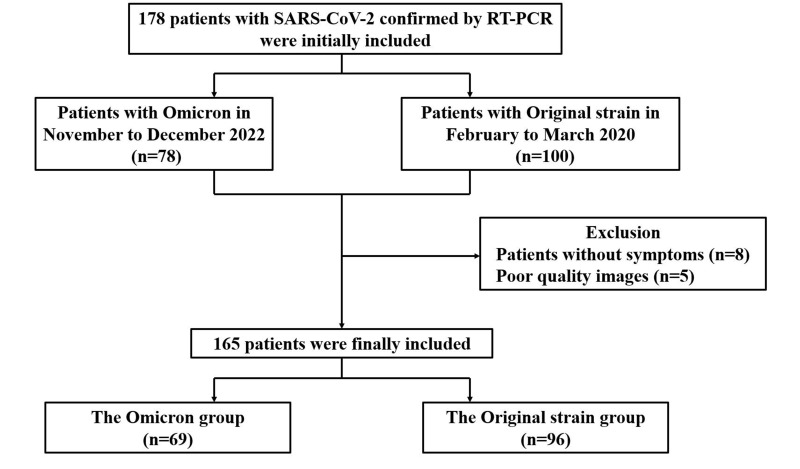
Patients and methods: This retrospective study included 69 hospitalized patients infected with Omicron variant from November to December 2022, and 96 hospitalized patients infected with the original strain from February to March 2020 in Chongqing, China. The clinical features, CT manifestations, degrees of lung involvement in different stages on CT, and imaging changes after the reverse-transcription polymerase chain reaction (RT-PCR) results turned negative were compared between the two groups.
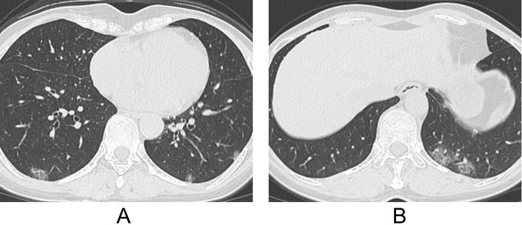
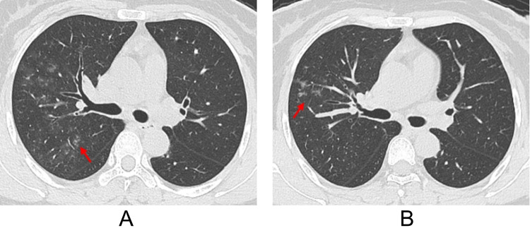
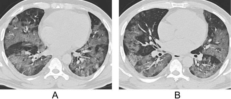
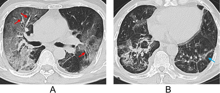
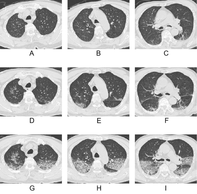
Results: For clinical features, patients with Omicron were predominantly old people and females, without manifestation of any clinical symptoms, who had low serum levels of C-reactive protein and procalcitonin. Shorter interval from symptoms onset to initial CT scan was observed in Omicron patients compared to patients with the original strain (all P < 0.05). For CT features, patients with Omicron were more likely to present with round-like opacities and tree-in-bud pattern (all P < 0.05), but less likely to exhibit a diffuse distribution, patchy and linear opacities, as well as vascular enlargement pattern (all P < 0.05). The Omicron group was more susceptible to exhibiting lower CT involvement scores in each stage (all P < 0.05) and imaging progression after the RT-PCR results turned negative (P < 0.001).
Conclusion: Patients infected with the Omicron variant exhibited less severe changes on chest CT compared to those infected with the original strain. Furthermore, imaging progression under low viral load conditions was more common in patients with Omicron than in those with the original strain.
Keywords: computed tomography; pneumonia; severe acute respiratory syndrome coronavirus-2.
© 2024 Zhang et al.
Conflict of interest statement
The authors report no conflicts of interest in this work.
Figures
Similar articles
-
Comparison between original SARS-CoV-2 strain and omicron variant on thin-section chest CT imaging of COVID-19 pneumonia.Radiologie (Heidelb). 2023 Nov;63(Suppl 2):55-63. doi: 10.1007/s00117-023-01147-2. Epub 2023 Jun 6. Radiologie (Heidelb). 2023. PMID: 37280418 Free PMC article.
-
Imaging Progression Under Low Respiratory Viral Load of SARS-CoV-2 Omicron Variant Infection: A Retrospective Study in China.Infect Drug Resist. 2023 Oct 25;16:6795-6806. doi: 10.2147/IDR.S417062. eCollection 2023. Infect Drug Resist. 2023. PMID: 37904829 Free PMC article.
-
Clinical and Pulmonary CT Characteristics of Patients Infected With the SARS-CoV-2 Omicron Variant Compared With Those of Patients Infected With the Alpha Viral Strain.Front Public Health. 2022 Jul 12;10:931480. doi: 10.3389/fpubh.2022.931480. eCollection 2022. Front Public Health. 2022. PMID: 35903393 Free PMC article.
-
SARS-CoV-2 Omicron (B.1.1.529) Variant: A Challenge with COVID-19.Diagnostics (Basel). 2023 Feb 2;13(3):559. doi: 10.3390/diagnostics13030559. Diagnostics (Basel). 2023. PMID: 36766664 Free PMC article. Review.
-
Similarities and Differences of Early Pulmonary CT Features of Pneumonia Caused by SARS-CoV-2, SARS-CoV and MERS-CoV: Comparison Based on a Systemic Review.Chin Med Sci J. 2020 Sep 30;35(3):254-261. doi: 10.24920/003727. Chin Med Sci J. 2020. PMID: 32972503 Free PMC article.
Cited by
-
Changes in Clinical Features and Severity of COVID-19 with the Emergence of Omicron Variants: A Shift Towards a Common Disease.Infect Drug Resist. 2024 Dec 18;17:5595-5603. doi: 10.2147/IDR.S492816. eCollection 2024. Infect Drug Resist. 2024. PMID: 39711829 Free PMC article.
-
Differences in chest imaging between Omicron and non-Omicron coronavirus disease 2019 (COVID-19) patients: a systematic review and meta-analysis.BMC Infect Dis. 2025 Apr 29;25(1):631. doi: 10.1186/s12879-025-11032-z. BMC Infect Dis. 2025. PMID: 40301746 Free PMC article.
References
LinkOut - more resources
Full Text Sources
Research Materials
Miscellaneous
