Review
. 2024 Jan 31;128(9):3632-3640.
doi: 10.1021/acs.jpcc.3c07367. eCollection 2024 Mar 7.

Quantum Dot Fluorescent Imaging: Using Atomic Structure Correlation Studies to Improve Photophysical Properties

Affiliations
Review

Quantum Dot Fluorescent Imaging: Using Atomic Structure Correlation Studies to Improve Photophysical Properties

Ruben Torres et al. J Phys Chem C Nanomater Interfaces. .

Abstract

Efforts to study intricate, higher-order cellular functions have called for fluorescence imaging under physiologically relevant conditions such as tissue systems in simulated native buffers. This endeavor has presented novel challenges for fluorescent probes initially designed for use in simple buffers and monolayer cell culture. Among current fluorescent probes, semiconductor nanocrystals, or quantum dots (QDs), offer superior photophysical properties that are the products of their nanoscale architectures and chemical formulations. While their high brightness and photostability are ideal for these biological environments, even state of the art QDs can struggle under certain physiological conditions. A recent method correlating electron microscopy ultrastructure with single-QD fluorescence has begun to highlight subtle structural defects in QDs once believed to have no significant impact on photoluminescence (PL). Specific defects, such as exposed core facets, have been shown to quench QD PL in physiologically accurate conditions. For QD-based imaging in complex cellular systems to be fully realized, mechanistic insight and structural optimization of size and PL should be established. Insight from single QD resolution atomic structure and photophysical correlative studies provides a direct course to synthetically tune QDs to match these challenging environments.


Conflict of interest statement

The authors declare no competing financial interest.

Figures

Figure 1
Figure 1
(a) Chemical structures and schematic depictions of coated QD surfaces using a multidentate zwitterionic polymer ligand (left) or an amphiphilic copolymer (right). (b) Surface plot of an amphiphilic copolymer coated QD point spread function imaged 50 μm into a live mouse brain slice. (c) Stitched image of amphiphilic copolymer coated QD conjugates dispersed in a brain tissue (scale bar = 50 μm). (d and e) 10× magnification of various regions captured in the stitched image in (c) (scale bar = 5 μm). Adapted with permission from ref (60). Copyright 2020 Royal Society of Chemistry.
Figure 2
Figure 2
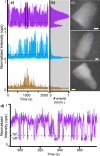
Correlated fluorescence and structural data for individual QDs. (a) Full fluorescence intensity transients with (b) corresponding photoluminescence intermittency histograms for the 3 QDs whose structures are shown in (c). (d) The boxed truncated portion of the topmost fluorescence intensity transient in (a) shows the “on” and “off” states of a fluorescent QD. Scale bars in (c) are 2 nm. Reprinted with permission from ref (36). Copyright 2014 American Chemical Society.
Figure 3
Figure 3
Characteristic defects for QDs with a low on-fraction. (a) QD exhibiting an exposed core Cd-rich facet at (101) and (b) (001). (c) Representative QD structure indicating Cd-rich facets onto which lack of shell growth results in a lower on-fraction. (d) Distribution of all observed QDs as a function of on-fraction with QDs containing surface defects (a) and (b) overlaid. Scale bars are 2 nm. Reprinted with permission from ref (36). Copyright 2014 American Chemical Society.
Figure 4
Figure 4
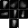
Structures of permanently nonradiative, or “dark”, CdSe/CdS core/shell QDs. Structural defects such as stacking fault in core region (a), asymmetrical shell growth (b), little or no shell material (c). (d–f) “Dark” QDs that do not show obvious surface or internal defects. Scale bars are 2 nm. Reprinted with permission from ref (36). Copyright 2014 American Chemical Society.
Figure 5
Figure 5
Time series and blinking behavior of single QDs. (a) Representative fluorescence intensity transients for symm-shelled QDs and QD655s in both HEPES and oxygenated aCSF. (b) Comparison of ON fraction populations under each condition (N ≥ 40 QDs). (c) Comparison of photobleaching profiles for symm-shelled QDs vs QD655s under each condition (N > 40 QDs. Reprinted with permission from ref (60). Copyright 2020 Royal Society of Chemistry.
Figure 6
Figure 6
Hypothetical schematic of amphiphilic copolymer coated QDs of four different emission wavelengths with hydrodynamic diameters small enough to access a synaptic cleft. Each QD is functionalized to specifically label four different arbitrary membrane proteins and then tracked using optical microscopy. Reconstructed hypothetical trajectories of QDs corresponding to dynamic movement of membrane proteins are plotted on the right.

Similar articles

Cited by

References

    1. Guo Z.; Park S.; Yoon J.; Shin I. Recent Progress in the Development of Near-Infrared Fluorescent Probes for Bioimaging Applications. Chem. Soc. Rev. 2014, 43 (1), 16–29. 10.1039/C3CS60271K. - DOI - PubMed
    1. Jeong S.; Widengren J.; Lee J. C. Fluorescent Probes for STED Optical Nanoscopy. Nanomaterials 2022, Vol. 12, Page 21 2022, 12 (1), 21.10.3390/nano12010021. - DOI - PMC - PubMed
    1. Resch-Genger U.; Grabolle M.; Cavaliere-Jaricot S.; Nitschke R.; Nann T. Quantum Dots versus Organic Dyes as Fluorescent Labels. Nat. Methods 2008, 5 (9), 763–775. 10.1038/nmeth.1248. - DOI - PubMed
    1. Mann V. R.; Powers A. S.; Tilley D. C.; Sack J. T.; Cohen B. E. Azide-Alkyne Click Conjugation on Quantum Dots by Selective Copper Coordination. ACS Nano 2018, 12 (5), 4469–4477. 10.1021/acsnano.8b00575. - DOI - PMC - PubMed
    1. Pedroso C. C. S.; Mann V. R.; Zuberbühler K.; Bohn M. F.; Yu J.; Altoe V.; Craik C. S.; Cohen B. E. Immunotargeting of Nanocrystals by SpyCatcher Conjugation of Engineered Antibodies. ACS Nano 2021, 15 (11), 18374–18384. 10.1021/acsnano.1c07856. - DOI - PMC - PubMed