. 2024 Feb 13;27(4):137.
doi: 10.3892/etm.2024.12425. eCollection 2024 Apr.

Biochanin A inhibits endothelial dysfunction induced by IL‑6‑stimulated endothelial microparticles in Perthes disease via the NFκB pathway

Affiliations

Biochanin A inhibits endothelial dysfunction induced by IL‑6‑stimulated endothelial microparticles in Perthes disease via the NFκB pathway

Jianhong Liu et al. Exp Ther Med. .

Abstract

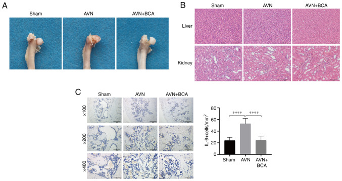
Endothelial dysfunction caused by the stimulation of endothelial microparticles (EMPs) by the inflammatory factor IL-6 is one of the pathogenic pathways associated with Perthes disease. The natural active product biochanin A (BCA) has an anti-inflammatory effect; however, whether it can alleviate endothelial dysfunction in Perthes disease is not known. The present in vitro experiments on human umbilical vein endothelial cells showed that 0-100 pg/ml IL-6-EMPs could induce endothelial dysfunction in a concentration-dependent manner, and the results of the Cell Counting Kit 8 assay revealed that, at concentrations of <20 µM, BCA had no cytotoxic effect. Reverse transcription-quantitative PCR demonstrated that BCA reduced the expression levels of the endothelial dysfunction indexes E-selectin and intercellular cell adhesion molecule-1 (ICAM-1) in a concentration-dependent manner. Immunofluorescence and western blotting illustrated that BCA increased the expression levels of zonula occludens-1 and decreased those of ICAM-1. Mechanistic studies showed that BCA inhibited activation of the NFκB pathway. In vivo experiments demonstrated that IL-6 was significantly increased in the rat model of ischemic necrosis of the femoral head, whereas BCA inhibited IL-6 production. Therefore, in Perthes disease, BCA may inhibit the NFκB pathway to suppress IL-6-EMP-induced endothelial dysfunction, and could thus be regarded as a potential treatment for Perthes disease.

Keywords: IL-6; NFκB signaling pathway; Perthes disease; biochanin A; endothelial dysfunction; endothelial microparticles.


Conflict of interest statement

The authors declare that they have no competing interests.

Figures

Figure 1
Figure 1
EMPs stimulated by IL-6 induce endothelial dysfunction. (A) EMPs labeled with red fluorescent Dil were endocytosed by HUVECs. Scale bar=200 µm. (B) EMPs stimulated by 100 pg/ml IL-6 significantly promoted the levels of ICAM-1 and VCAM-1 in HUVECs but did not affect the levels of E-Selectin. (C) Monocyte adhesion is increased after endothelial cell injury. The endothelial-monocyte adhesion assay showed that IL-6 promoted monocyte adhesion to HUVECs in a concentration-dependent manner in the concentration range of 0-100 pg/ml IL-6. Green fluorescence represents monocytes. Scale bar=50 µm. (D) EMPs stimulated by 100 pg/ml IL-6 decreased the expression of ZO-1 (n=3). Scale bar=50 µm. (E) EMPs stimulated by 100 pg/ml IL-6 promoted the expression of ICAM-1 and promoted endothelial dysfunction (n=3). Scale bar=50 µm. *P<0.05, **P<0.01, ***P<0.001, ****P<0.0001. EMPs, endothelial microparticles; HUVECs, human umbilical vein endothelial cells; ICAM-1, intercellular cell adhesion molecule-1; VCAM-1, vascular cell adhesion molecule-1; ZO-1, zonula occludens-1.
Figure 2
Figure 2
BCA inhibits endothelial dysfunction induced by IL-6-EMPs in the non-cytotoxic concentration range. (A) Chemical structure of BCA. (B) CCK8 assay results showed that BCA had no toxic effect on cells in the concentration range of 0-20 µM, IL-6-EMPs had no toxic effect on cells in the concentration range of 0-1,000 pg/ml, and 100 pg/ml IL-6-EMPs + 0-10 µM BCA had no toxic effect on cells (n=3). (C) After treatment with BCA, the indexes of endothelial dysfunction ICAM-1 and E-selectin were decreased with the increase in BCA concentration, indicating that BCA can effectively reduce the endothelial dysfunction induced by IL-6-EMPs (n=3). (D) Adhesion assay showed that BCA could inhibit the injury of endothelial cells induced by IL-6-EMPs and reduce the adhesion of monocytes to human umbilical vein endothelial cells. Green fluorescence represents monocytes (n=3). Scale bar=100 µm. (E) IL-6-EMPs (100 pg/ml) decreased the expression of ZO-1. After treatment with BCA, the endothelial dysfunction induced by IL-6-EMPs was inhibited and the expression of ZO-1 was increased (n=3). Scale bar=50 µm. *P<0.05, **P<0.01, ***P<0.001, ****P<0.0001. BCA, biochanin A; CCK8, Cell Counting Kit 8; EMPs, endothelial microparticles; ICAM-1, intercellular cell adhesion molecule-1; ZO-1, zonula occludens-1.
Figure 3
Figure 3
BCA inhibits endothelial dysfunction via the NFκB signaling pathway. (A) After HUVECs were treated with different concentrations of IL-6-EMPs (0, 100 and 1,000 pg/ml), the expression levels of ICAM-1 and ZO-1 were analyzed by western blotting. Representative images showed that the expression levels of ICAM-1 were increased with the increase in IL-6-EMPs concentration, whereas the expression levels of ZO-1 were decreased with the increase in IL-6-EMPs concentration (n=3). (B) Relative expression levels of ICAM-1 and ZO-1 were calculated. (C) HUVECs were treated with 100 pg/ml IL-6-EMPs + BCA (0, 5 and 10 µM). After treatment, the expression levels of NFκB, IκB, ICAM-1, ZO-1 and VE-cadherin were analyzed by western blotting. Representative images showed that the expression levels of NFκB and ICAM-1 were decreased with the increase in BCA concentration, and the expression levels of IκB, ZO-1 and VE-cadherin were increased with the increase in BCA concentration (n=3). (D) Relative expression levels of NFκB, IκB, ICAM-1, ZO-1 and VE-cadherin were calculated. *P<0.05, **P<0.01, ***P<0.001, ****P<0.0001. BCA, biochanin A; EMPs, endothelial microparticles; HUVECs, human umbilical vein endothelial cells; ICAM-1, intercellular cell adhesion molecule-1; ns, not significant; ZO-1, zonula occludens-1.
Figure 4
Figure 4
BCA inhibits endothelial dysfunction and alleviates deformation of the femoral head in in vivo experiments. (A) Macroscopic images showed that the femoral head in the sham operation group was in good shape, whereas an obvious collapse and deformity of the femoral head was observed in the femoral head necrosis group, which were reduced in the BCA group (n=6). (B) Hematoxylin and eosin staining sections showed no marked histological differences in the liver and kidney of the three groups of rats (n=6). Scale bar=100 µm. (C) Immunohistochemistry and statistical analysis of pathological sections of the femoral head showed that the expression levels of IL-6 were decreased after treatment with BCA (n=6). Scale bar=200, 100, 50 µm for ‘x100’, ‘x200’, ‘x400’, respectively. ****P<0.0001. BCA, biochanin A. AVN, avascular necrosis.
Figure 5
Figure 5
Mechanism underlying the effects of BCA on the treatment of Perthes disease. BCA inhibits endothelial dysfunction induced by IL-6-EMPs via the NFκB pathway, and may therefore be considered a potential drug for the treatment of Perthes disease. Created with BioRender.com. BCA, biochanin A; EMPs, endothelial microparticles; ICAM, intercellular cell adhesion molecule; p, phosphorylated.

Similar articles

Cited by

References

    1. Leroux J, Abu Amara S, Lechevallier J. Legg-Calvé-Perthes disease. Orthop Traumatol Surg Res. 2018;104:S107–S112. doi: 10.1016/j.otsr.2017.04.012. - DOI - PubMed
    1. Zhao Y, Liao S, Lu R, Dang H, Zhao J, Ding X. Endothelial nitric oxide synthase gene polymorphism is associated with Legg-Calvé-Perthes disease. Exp Ther Med. 2016;11:1913–1917. doi: 10.3892/etm.2016.3111. - DOI - PMC - PubMed
    1. Kim S, Oh H, Lim J, Cho S, Jung S. Results of early proximal femoral osteotomy at skeletal maturity in Legg-Calvé-Perthes disease: Implication for the bypass of fragmentation stage. J Pediatr Orthop. 2021;41:e768–e773. doi: 10.1097/BPO.0000000000001809. - DOI - PubMed
    1. Little D, McDonald M, Sharpe I, Peat R, Williams P, McEvoy T. Zoledronic acid improves femoral head sphericity in a rat model of perthes disease. J Orthop Res. 2005;23:862–868. doi: 10.1016/j.orthres.2004.11.015. - DOI - PubMed
    1. Kim HKW, Randall TS, Bian H, Jenkins J, Garces A, Bauss F. Ibandronate for prevention of femoral head deformity after ischemic necrosis of the capital femoral epiphysis in immature pigs. J Bone Joint Surg Am. 2005;87:550–557. doi: 10.2106/JBJS.D.02192. - DOI - PubMed