. 2024 Feb 15;27(4):142.
doi: 10.3892/etm.2024.12430. eCollection 2024 Apr.

Fluorofenidone attenuates renal fibrosis by inhibiting lysosomal cathepsin‑mediated NLRP3 inflammasome activation

Affiliations

Fluorofenidone attenuates renal fibrosis by inhibiting lysosomal cathepsin‑mediated NLRP3 inflammasome activation

Linfeng Zheng et al. Exp Ther Med. .

Abstract

Currently, no antifibrotic drug in clinical use can effectively treat renal fibrosis. Fluorofenidone (AKFPD), a novel pyridone agent, significantly reduces renal fibrosis by inhibiting the activation of the NOD-like receptor thermal protein domain associated protein 3 (NLRP3) inflammasome; however, the underlying mechanism of this inhibition is not fully understood. The present study aimed to reveal the molecular mechanism underlying the suppression of NLRP3 inflammasome activation by AKFPD. It investigated the effect of AKFPD on NLRP3 activation and lysosomal cathepsins in a unilateral ureteral obstruction (UUO) rat model, and hypoxia/reoxygenation (H/R)-treated HK-2 cells and murine peritoneal-derived macrophages (PDMs) stimulated with lipopolysaccharide (LPS) and ATP. The results confirmed that AKFPD suppressed renal interstitial fibrosis and inflammation by inhibiting NLRP3 inflammasome activation in UUO rat kidney tissues. In addition, AKFPD reduced the production of activated caspase-1 and maturation of IL-1β by suppressing NLRP3 inflammasome activation in H/R-treated HK-2 cells and murine PDMs stimulated with LPS and ATP. AKFPD also decreased the activities of cathepsins B, L and S both in vivo and in vitro. Notably, AKFPD downregulated cathepsin B expression and NLRP3 colocalization in the cytoplasm after lysosomal disruptions. Overall, the results suggested that AKFPD attenuates renal fibrosis by inhibiting lysosomal cathepsin-mediated activation of the NLRP3 inflammasome.

Keywords: antifibrotic therapy; fluorofenidone; inflammation; lysosomal cathepsin; renal fibrosis.


Conflict of interest statement

The authors declare they have no competing interests.

Figures
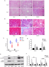
Figure 1
Figure 1
AKFPD ameliorates renal fibrosis in the kidney tissue of UUO rats on days 3 and 7 after surgery. (A) H&E staining of representative rat kidney tissue sections (scale bar, 100 µm). (B) Masson's trichrome staining of representative rat kidney tissue sections. (scale bar, 100 µm). (C) H&E score for kidney damage. (D) The tubulointerstitial fibrosis index is indicated. (E) Representative western blots of FN and α-SMA in the rat kidney tissue. (F) Quantitative analysis of FN and α-SMA protein expression in the rat kidney tissue. H&E scores are expressed using median and interquartile range, with the rest of the data are expressed as mean ± standard deviation (n=5 per group). *P<0.05 vs. sham group, **P<0.01 vs. sham group, ##P<0.01 vs. UUO group. AKFPD, fluorofenidone; UUO, unilateral ureteral obstruction; H&E, hematoxylin-eosin; FN, fibronectin; α-SMA, α-smooth muscle actin.
Figure 2
Figure 2
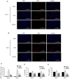
AKFPD suppresses renal inflammation in UUO rats. (A and B) Immunofluorescence of MPO-positive cells in rat kidney tissues (magnification, x200; scale bars, 100 µm). (C) Quantification of MPO-positive cells in five fields. (D) ELISA of IL-1β expression in the rat kidney tissues. (E) ELISA of TNF-α expression in the rat kidney tissue. Data are presented as mean ± standard deviation (n=5 per group). *P<0.05 vs. sham group, #P<0.05 vs. UUO group. AKFPD, fluorofenidone; UUO, unilateral ureteral obstruction; MPO, myeloperoxidase.
Figure 3
Figure 3
AKFPD inhibits NLRP3 inflammasome activation in UUO rats. (A) Representative western blot of NLRP3, ASC, pro-caspase-1, pro-IL-1β, caspase-1 and IL-1β expression in the rat kidney tissues on days 3 and 7 post-surgery. (B-E) Quantitative analysis of (B) NLRP3, pro-caspase-1, (C) ASC, (D) pro-caspase-1 and caspase-1, and (E) pro-IL-1β and IL-1β protein expression in rat kidney tissues. Data are presented as mean ± standard deviation (n=5 per group). *P<0.05 vs. sham group, #P<0.05 vs. UUO group. AKFPD, fluorofenidone; NLRP3, NOD-like receptor thermal protein domain associated protein 3; UUO, unilateral ureteral obstruction; ASC, apoptosis-associated speck-like protein containing a caspase recruitment domain.
Figure 4
Figure 4
AKFPD inhibits NLRP3 inflammasome activation in vitro. (A) Representative western blot and (B) quantitative analysis of NLRP3, pro-IL-1β, caspase-1 and IL-1β expression in LPS/ATP-stimulated PDMs. (C) Representative western blot and (D) quantitative analysis of NLRP3, pro-IL-1β, caspase-1 and IL-1β expression in H/R-treated HK-2 cells. (E) Colocalization of NLRP3 and ASC in PDMs visualized using immunofluorescent staining. The white arrows indicate overlap (yellow). (magnification, x400; scale bar, 30 µm). (F) Colocalization of NLRP3 and ASC in HK-2 cells visualized using immunofluorescent staining. The white arrows indicate overlap (yellow) (magnification, x400; scale bar, 50 µm). (G) Quantification of the immunofluorescent co-localization of NLRP3 and ASC in PDMs. The quantitative changes were measured using Image-Pro Plus 6.0. #P<0.05. (H) Quantification of the immunofluorescent co-localization of NLRP3 and ASC in HK-2 cells. Data are presented as the mean ± standard deviation (n=3 per group). *P<0.05, **P<0.01 vs. N group; #P<0.05 vs. H/R group or vs. LPS+ATP group, ##P<0.01 vs. H/R group. AKFPD, fluorofenidone; NLRP3, NOD-like receptor thermal protein domain associated protein 3; PDMs, peritoneal-derived macrophages; N, normal; H/R, hypoxia/reoxygenation; ASC, apoptosis-associated speck-like protein containing a caspase recruitment domain.
Figure 5
Figure 5
AKFPD downregulates cathepsin activity in vitro. (A) Effects of AKFPD on cathepsin (CTSB, CTSS and CTSL) activities in PDMs. (B) Effects of AKFPD on the accumulation of lysosomes visualized using LysoTracker Red dye in PDMs (magnification, x400; scale bar, 50 µm). (C) Effects of AKFPD on cathepsin (CTSB, CTSS and CTSL) activities in HK-2 cells. (D) Effects of AKFPD on the accumulation of lysosomes visualized using LysoTracker Red dye in HK-2 cells (magnification, x400; scale bar, 50 µm). (E) Quantitative analysis of the effect of AKFPD on lysosome accumulation visualized in PDMs using LysoTracker red dye. (F) Quantitative analysis of the effect of AKFPD on lysosome accumulation visualized in HK-2 cells using LysoTracker red dye. Data are presented as mean ± standard deviation (n=3 per group). *P<0.05, **P<0.01 vs. N group; #P<0.05 ##P<0.01 vs. H/R group or vs. LPS + ATP group. AKFPD, fluorofenidone; CTSB, cathepsin B; CTSS, cathepsin S; CTSL cathepsin L; N, normal; PDMs, peritoneal-derived macrophages; H/R, hypoxia/reoxygenation.
Figure 6
Figure 6
AKFPD inhibits NLRP3 inflammasome activation by CTSB expression in vitro. (A) Representative western blot and quantitative data of CTSB in LPS + ATP-stimulated PDMs. (B) Effects of AKFPD on the colocalization of NLRP3 and CTSB in activated PDMs visualized using immunofluorescent staining. White arrows indicate overlap (yellow) (magnification, x400; scale bar, 30 µm). (C) Effects of AKFPD on the colocalization of CTSB and NLRP3 in H/R-treated HK-2 cells visualized using immunofluorescent staining. White arrows represent overlap (yellow) (magnification, x400; scale bar, 50 µm). (D) Representative western blot and quantitative data of CTSB in H/R-treated HK-2 cells. (E) Quantitative analysis of the effect of AKFPD on NLRP3 and CTSB co-localization in activated PDM visualized using immunofluorescent staining. The quantitative changes were measured using Image-Pro Plus 6.0. (F) Quantitative analysis of the effect of AKFPD on the co-localization of CTSB and NLRP3 in H/R-treated HK-2 cells observed using immunofluorescent staining. The quantitative changes were measured using Image-Pro Plus 6.0. Data are presented as mean ± standard deviation (n=3 per group). *P<0.05, **P<0.01 vs. N group; #P<0.05 vs. H/R group or vs. LPS + ATP group. AKFPD, fluorofenidone; NLRP3, NOD-like receptor thermal protein domain associated protein 3; CTSB, cathepsin B; PDMs, peritoneal-derived macrophages; N, normal; H/R, hypoxia/reoxygenation.
Figure 7
Figure 7
AKFPD inhibits CTSB-mediated NLRP3 inflammasome activation in UUO rats. (A) Effects of AKFPD on cathepsin (CTSB, CTSS and CTSL) activities in the rat kidney tissues. (B) Effects of AKFPD on the colocalization of NLRP3 and CTSB in the rat kidney tissues visualized using immunostaining in UUO rats on day 3 (magnification, x400; scale bar, 30 µm). (C) Representative western blot of CTSB in the rat kidney tissues. (D) Effects of AKFPD on the colocalization of NLRP3 and CTSB in the rat kidney tissues visualized using immunostaining in UUO rats on day 7 (magnification, x400; scale bar, 30 µm). (E) Quantitative data of CTSB in the rat kidney tissues. Data are presented as mean ± standard deviation (n=5 per group). *P<0.05 vs. sham group, #P<0.05 vs. UUO group. AKFPD, fluorofenidone; CTSB, cathepsin B; NLRP3, NOD-like receptor thermal protein domain associated protein 3; UUO, unilateral ureteral obstruction; CTSS, cathepsin S; CTSL cathepsin L.

References

    1. Hackl MJ, Burford JL, Villanueva K, Lam L, Suszták K, Schermer B, Benzing T, Peti-Peterdi J. Tracking the fate of glomerular epithelial cells in vivo using serial multiphoton imaging in new mouse models with fluorescent lineage tags. Nat Med. 2013;19:1661–1666. doi: 10.1038/nm.3405. - DOI - PMC - PubMed
    1. Lovisa S, LeBleu VS, Tampe B, Sugimoto H, Vadnagara K, Carstens JL, Wu CC, Hagos Y, Burckhardt BC, Pentcheva-Hoang T, et al. Epithelial-to-mesenchymal transition induces cell cycle arrest and parenchymal damage in renal fibrosis. Nat Med. 2015;21:998–1009. doi: 10.1038/nm.3902. - DOI - PMC - PubMed
    1. Li X, Pan J, Li H, Li G, Liu X, Liu B, He Z, Peng Z, Zhang H, Li Y, et al. DsbA-L mediated renal tubulointerstitial fibrosis in UUO mice. Nat Commun. 2020;11(4467) doi: 10.1038/s41467-020-18304-z. - DOI - PMC - PubMed
    1. Meng XM, Nikolic-Paterson DJ, Lan HY. Inflammatory processes in renal fibrosis. Nat Rev Nephrol. 2014;10:493–503. doi: 10.1038/nrneph.2014.114. - DOI - PubMed
    1. Kimura T, Isaka Y, Yoshimori T. Autophagy and kidney inflammation. Autophagy. 2017;13:997–1003. doi: 10.1080/15548627.2017.1309485. - DOI - PMC - PubMed