

Secondary bladder stone caused by delayed penetration of the bladder by a pubic fracture: A case report and literature review
- PMID: 38476919
- PMCID: PMC10928999
- DOI: 10.3892/etm.2024.12455
Secondary bladder stone caused by delayed penetration of the bladder by a pubic fracture: A case report and literature review
Abstract
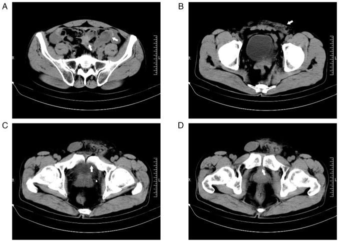
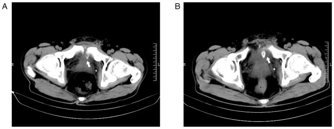
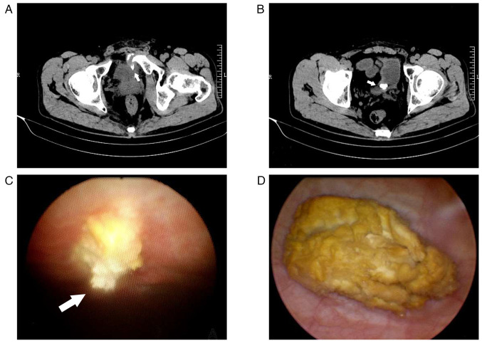
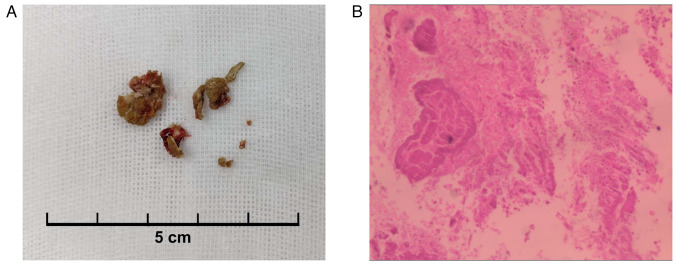
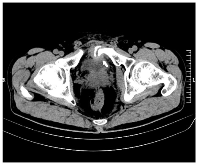
Pelvic fractures sometimes lead to injuries of the urinary bladder, which commonly present as gross hematuria, dysuria and lower abdominal pain. As a type of urinary stone, bladder stones are usually secondary to lower urinary tract obstruction, such as benign prostatic hyperplasia, urethral stricture, and neurogenic bladder. The present case report examines an unusual case of a delayed pubic fracture penetrating the bladder, which caused a secondary bladder stone. A 53-year-old man was first hospitalized at The Second Hospital of Jiaxing (Jiaxing, China) in January 2020 because of trauma-induced bleeding in the scalp and abdominal pain. The patient underwent abdominal exploration and partial bowel resection, and his condition stabilized after surgery. After discharge, the patient had regular outpatient check-ups every 2-3 weeks. However, after 3 months, in April 2020, the patient was readmitted to the hospital because of frequent urination, an urgent need for urination and dysuria. Abdominal computed tomography imaging and cystoscopy revealed a pubic fracture that had penetrated the bladder wall, accompanied by a bladder stone. Subsequently, cystolithotomy was performed, which provided significant relief of symptoms once the catheter was removed after 2 weeks. Since then, the patient has been followed up until January 2023 and had remained asymptomatic. Bladder stones caused by necrotic bone fragmentation are rare. Bladder injuries resulting from pelvic fractures can have delayed onset; therefore, clinicians should be aware of the possibility of urogenital injury in such patients. It is crucial for clinicians to comprehend the potential mechanisms involved, analyze the clinical data of patients, closely monitor their condition and implement appropriate treatment measures when necessary.
Keywords: bladder injury; bladder stone; cystolithotomy; ischemic necrosis; pubic fracture.
Copyright: © 2024 Wang et al.
Conflict of interest statement
The authors declare that they have no competing interests.
Figures
Similar articles
-
Delayed Bladder Perforation Resulting From Large Bladder Stone and Gluteal Abscess Caused by Pelvic Plate Loosening: A Case Report.Cureus. 2024 Jun 2;16(6):e61543. doi: 10.7759/cureus.61543. eCollection 2024 Jun. Cureus. 2024. PMID: 38957240 Free PMC article.
-
Giant urinary bladder stone: A rare case report.Int J Surg Case Rep. 2024 Sep;122:110174. doi: 10.1016/j.ijscr.2024.110174. Epub 2024 Aug 14. Int J Surg Case Rep. 2024. PMID: 39146668 Free PMC article.
-
A case report of a giant bladder stone (12 × 8 cm, 610 g).Radiol Case Rep. 2023 Dec 20;19(3):970-973. doi: 10.1016/j.radcr.2023.11.081. eCollection 2024 Mar. Radiol Case Rep. 2023. PMID: 38204936 Free PMC article.
-
Bladder Stone in Pregnancy: A Case Report and Review of the Literature.Am J Case Rep. 2018 Dec 30;19:1546-1549. doi: 10.12659/AJCR.912614. Am J Case Rep. 2018. PMID: 30594944 Free PMC article. Review.
-
Osteochondroma of the pubic symphysis causing hematuria: a case report and literature review.BMC Urol. 2021 Jan 6;21(1):1. doi: 10.1186/s12894-020-00770-8. BMC Urol. 2021. PMID: 33407372 Free PMC article. Review.
References
-
- Koraitim MM. Pelvic fracture urethral injuries: The unresolved controversy. J Urol. 1999;161:1433–1441. - PubMed
Publication types
LinkOut - more resources
Full Text Sources
Research Materials
Miscellaneous