Longitudinal Free-Water Changes in Dementia with Lewy Bodies
- PMID: 38477399
- PMCID: PMC11102324
- DOI: 10.1002/mds.29763
Longitudinal Free-Water Changes in Dementia with Lewy Bodies
Abstract
Background: Diffusion-weighted magnetic resonance imaging (dMRI) examines tissue microstructure integrity in vivo. Prior dementia with Lewy bodies (DLB) diffusion tensor imaging studies yielded mixed results.
Objective: We employed free-water (FW) imaging to assess DLB progression and correlate with clinical decline in DLB.
Methods: Baseline and follow-up MRIs were obtained at 12 and/or 24 months for 27 individuals with DLB or mild cognitive impairment with Lewy bodies (MCI-LB). FW was analyzed using the Mayo Clinic Adult Lifespan Template. Primary outcomes were FW differences between baseline and 12 or 24 months. To compare FW change longitudinally, we included 20 cognitively unimpaired individuals from the Alzheimer's Disease Neuroimaging Initiative.
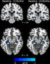
Results: We followed 23 participants to 12 months and 16 participants to 24 months. Both groups had worsening in Montreal Cognitive Assessment (MoCA) and Movement Disorder Society-Unified Parkinson's Disease Rating Scale (MDS-UPDRS) scores. We found significant FW increases at both time points compared to baseline in the insula, amygdala, posterior cingulum, parahippocampal, entorhinal, supramarginal, fusiform, retrosplenial, and Rolandic operculum regions. At 24 months, we found more widespread microstructural changes in regions implicated in visuospatial processing, motor, and cholinergic functions. Between-group analyses (DLB vs. controls) confirmed significant FW changes over 24 months in most of these regions. FW changes were associated with longitudinal worsening of MDS-UPDRS and MoCA scores.
Conclusions: FW increased in gray and white matter regions in DLB, likely due to neurodegenerative pathology associated with disease progression. FW change was associated with clinical decline. The findings support dMRI as a promising tool to track disease progression in DLB. © 2024 International Parkinson and Movement Disorder Society.
Keywords: Lewy body disease; dementia with Lewy bodies; diffusion‐weighted imaging; magnetic resonance imaging.
© 2024 International Parkinson and Movement Disorder Society.
Conflict of interest statement
S.Y. Chiu receives research support from NIH (K23 AG073525-01A1). R. Chen has no relevant disclosures. W. Wang has no relevant disclosures. M.J. Armstrong receives research support from the NIH (R01AG068128, P30AG047266, R01NS121099, R44AG062072), the Florida Department of Health (grant 20A08), and as the local PI of a Lewy Body Dementia Association Research Center of Excellence. She serves on the DSMBs for the Alzheimer’s Therapeutic Research Institute/Alzheimer’s Clinical Trial Consortium and the Alzheimer’s Disease Cooperative Study. She has provided educational content for Medscape, Vindico CME, and Prime Inc. B.F. Boeve receives honorarium for SAB activities for the Tau Consortium, institutional research grant support from the Lewy Body Dementia Association, American Brain Foundation, Alector, Biogen, Transposon, and GE Healthcare. R. Savica receives support from the National Institute on Aging, the National Institute of Neurological Disorders and Stroke, the Parkinson’s Disease Foundation, and Acadia Pharmaceuticals. V. Ramanan receives research support from NIH. J.A. Fields receives research support from NIH. N. Graff-Radford receives research support as the local PI of a Lewy Body Dementia Association Research Center of Excellence and from NIH. T.J. Ferman receives research support from NIH, the Mayo Clinic Dorothy and Harry T. Mangurian Jr. Lewy Body Dementia Program, and Ted Turner and Family LBD Functional Genomics Program. K. Kantarci receives research support from NIH, Avid Radiopharmaceuticals, Eli Lilly and consults for Biogen. D.E. Vaillancourt receives research support from NIH and is Co-Founder of Automated Imaging Diagnostics.
Figures


References
-
- Savica R, Boeve BF, Logroscino G. Epidemiology of alpha-synucleinopathies: from Parkinson disease to dementia with Lewy bodies. Handb Clin Neurol. 2016;138:153–8. - PubMed
MeSH terms
Substances
Grants and funding
- RF1 NS121099/NS/NINDS NIH HHS/United States
- U01 AG024904/AG/NIA NIH HHS/United States
- R01 NS121099/NS/NINDS NIH HHS/United States
- P30 AG62677/NH/NIH HHS/United States
- U01 AG024904/NH/NIH HHS/United States
- U19 AG024904/AG/NIA NIH HHS/United States
- R01 NS121099/NH/NIH HHS/United States
- U01 NS100620/NH/NIH HHS/United States
- U19 AG071754/AG/NIA NIH HHS/United States
- P30 AG062677/AG/NIA NIH HHS/United States
- P30 AG066506/AG/NIA NIH HHS/United States
- K23 AG073525-01A1/NH/NIH HHS/United States
- R44 AG062072/NH/NIH HHS/United States
- K23 AG073525/AG/NIA NIH HHS/United States
- R01 AG068128/AG/NIA NIH HHS/United States
- T32 NS082168/NS/NINDS NIH HHS/United States
- R01 NS058487/NS/NINDS NIH HHS/United States
- P30AG047266/NH/NIH HHS/United States
- P50 AG047266/AG/NIA NIH HHS/United States
- R01AG068128/NH/NIH HHS/United States
- R44 AG062072/AG/NIA NIH HHS/United States
- U01 NS100620/NS/NINDS NIH HHS/United States
- U19 AG71754/NH/NIH HHS/United States
- W81XWH-12-2-0012/U.S. Department of Defense
- R01 NS058487/NH/NIH HHS/United States
LinkOut - more resources
Full Text Sources
Medical
Research Materials
Miscellaneous

