Clinical Trial
. 2024 Apr 11;390(14):1290-1298.
doi: 10.1056/NEJMoa2314390. Epub 2024 Mar 13.

Intraventricular CARv3-TEAM-E T Cells in Recurrent Glioblastoma

Affiliations
Clinical Trial

Intraventricular CARv3-TEAM-E T Cells in Recurrent Glioblastoma

Bryan D Choi et al. N Engl J Med. .

Abstract

In this first-in-human, investigator-initiated, open-label study, three participants with recurrent glioblastoma were treated with CARv3-TEAM-E T cells, which are chimeric antigen receptor (CAR) T cells engineered to target the epidermal growth factor receptor (EGFR) variant III tumor-specific antigen, as well as the wild-type EGFR protein, through secretion of a T-cell-engaging antibody molecule (TEAM). Treatment with CARv3-TEAM-E T cells did not result in adverse events greater than grade 3 or dose-limiting toxic effects. Radiographic tumor regression was dramatic and rapid, occurring within days after receipt of a single intraventricular infusion, but the responses were transient in two of the three participants. (Funded by Gateway for Cancer Research and others; INCIPIENT ClinicalTrials.gov number, NCT05660369.).


Figures

Figure 1
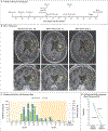
Figure 1. Treatment Response on MRI and Liquid Biopsy in Participant 1.
Panel A summarizes the timeline of events for Participant 1. CAR+ cells denote CARv3-TEAM-E T cells, which are chimeric antigen receptor (CAR) T cells engineered to target the epidermal growth factor receptor (EGFR) variant III (EGFRvIII) tumor-specific antigen, as well as the wild-type EGFR protein, through secretion of a T-cell–engaging antibody molecule (TEAM). Panel B shows contrast-enhanced T1-weighted magnetic resonance imaging (MRI) in Participant 1 immediately after resection, before infusion, and after infusion. The day numbers specified above the images are relative to the day of infusion (day 0). The yellow circles indicate the tumor. The top row shows axial views, and the bottom row shows coronal views. Panel C shows the copy numbers of EGFRvIII and EGFR over time, as detected in extracellular-vesicle RNA (evRNA) derived from cerebrospinal fluid (CSF). Asterisks indicate that the EGFRvIII and EGFR copy numbers were below the threshold of detection. Panel D shows the copy numbers of EGFRvIII and EGFR before and after infusion, as detected in evRNA derived from peripheral-blood samples.
Figure 2.
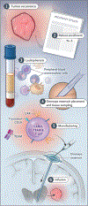
Figure 2.. Amended Workflow Used in Participants 2 and 3.
Shown is the amended workflow used in Participants 2 and 3, which enabled concomitant craniotomy, tissue sampling, and Ommaya reservoir placement during a single surgery.
Figure 3.
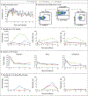
Figure 3.. Treatment Response on MRI and Liquid Biopsy in Participants 2 and 3.
Panel A shows an axial, contrast-enhanced T1-weighted MRI scan in Participant 2 before and after infusion; tumor regression was observed at day 2 after infusion. The tumor continued to regress, and this regression was sustained at later time points. The number of the day above the images is relative to the day of infusion (day 0). The yellow circles indicate the tumor. Panel B shows EGFRvIII and EGFR copy number over time, as detected in evRNA derived from CSF. Panel C shows an axial, contrast-enhanced T1-weighted MRI scan in Participant 3 before and after a single infusion of CARv3-TEAM-E T cells. Asterisks indicate that the EGFRvIII and EGFR copy numbers were below the threshold of detection.
Figure 4.
Figure 4.. Correlative Data on All Three Participants.
Panel A shows the body temperature curves for all three participants over time. Panel B shows a representative flow cytometric analysis of the engineered T-cell product (CARv3-TEAM-E) in Participant 1, with gating for CAR-positive and TEAM-positive T cells among all CD3+ T cells. The numbers in the plots indicate the corresponding percentages. APC, PE, and PE-Cy7 are fluorophore labels on the indicated antibodies. Panel C shows quantification of CAR-positive and TEAM-positive T cells by means of vector copy number (VCN) analysis and flow cytometry in all three participants. Panel D shows changes in inflammatory cytokines in the CSF samples obtained from the participants in the first month after infusion. Panel E shows detection of CAR-positive and TEAM-positive T cells in the peripheral-blood samples. TNF-α denotes tumor necrosis factor α.

Comment in

References

    1. O’Rourke DM, Nasrallah MP, Desai A, et al. A single dose of peripherally infused EGFRvIII-directed CAR T cells mediates antigen loss and induces adaptive resistance in patients with recurrent glioblastoma. Sci Transl Med 2017;9:eaaa0984. - PMC - PubMed
    1. Choi BD, Yu X, Castano AP, et al. CAR-T cells secreting BiTEs circumvent antigen escape without detectable toxicity. Nat Biotechnol 2019;37:1049–58. - PubMed
    1. Choi BD, Gedeon PC, Herndon JE II, et al. Human regulatory T cells kill tumor cells through granzyme-dependent cytotoxicity upon retargeting with a bispecific antibody. Cancer Immunol Res 2013;1:163. - PMC - PubMed
    1. Batool SM, Muralidharan K, Hsia T, et al. Highly sensitive EGFRvIII detection in circulating extracellular vesicle RNA of glioma patients. Clin Cancer Res 2022;28:4070–82. - PMC - PubMed
    1. Ahmed N, Brawley V, Hegde M, et al. HER2-specific chimeric antigen receptor-modified virus-specific T cells for progressive glioblastoma: a phase 1 dose-escalation trial. JAMA Oncol 2017;3:1094–101. - PMC - PubMed

Publication types

MeSH terms

Associated data