. 2024 Mar 13;22(3):e3002555.
doi: 10.1371/journal.pbio.3002555. eCollection 2024 Mar.

Specification of distinct cell types in a sensory-adhesive organ important for metamorphosis in tunicate larvae

Affiliations

Specification of distinct cell types in a sensory-adhesive organ important for metamorphosis in tunicate larvae

Christopher J Johnson et al. PLoS Biol. .

Abstract

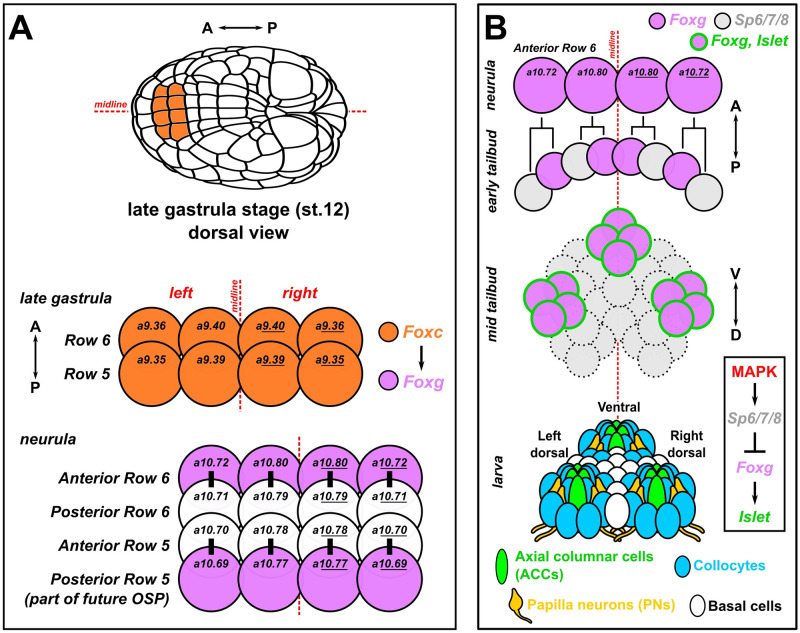
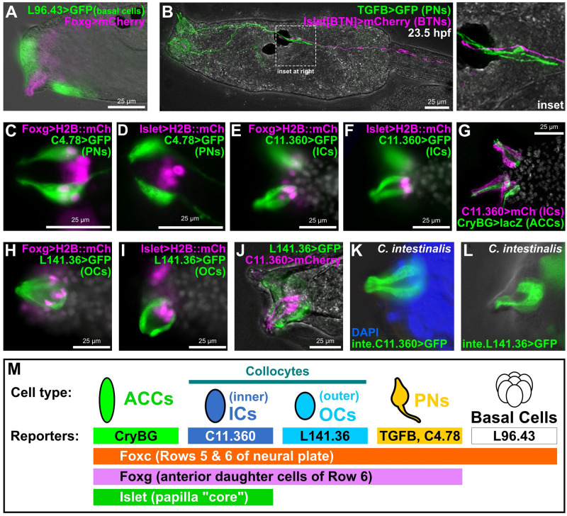
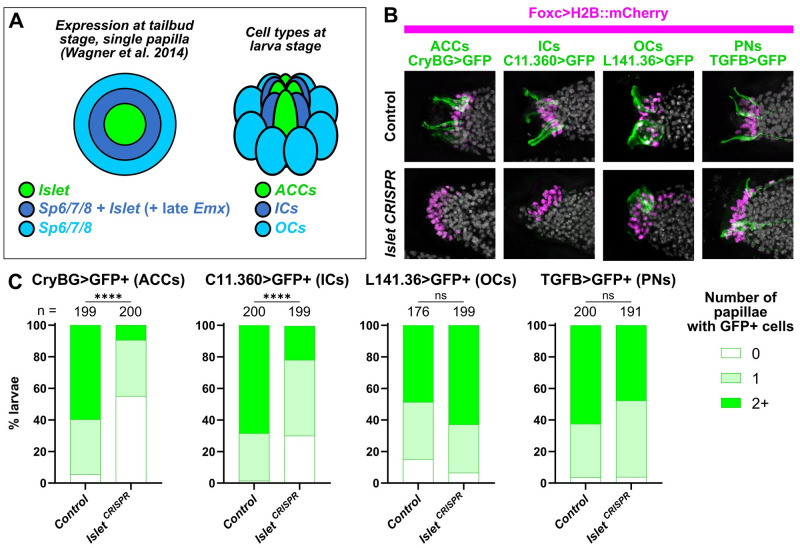
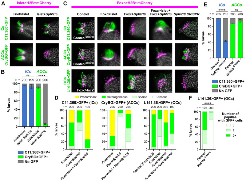
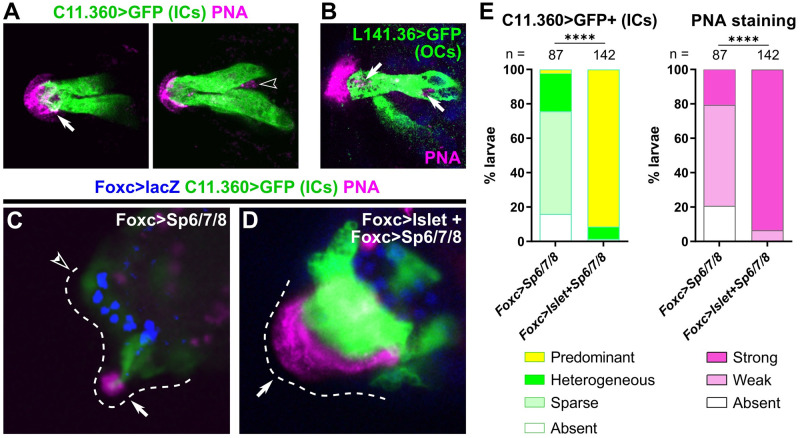
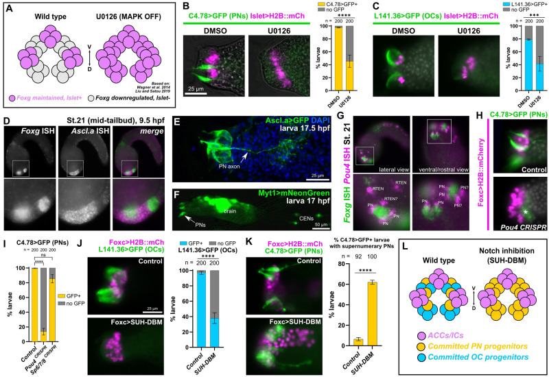
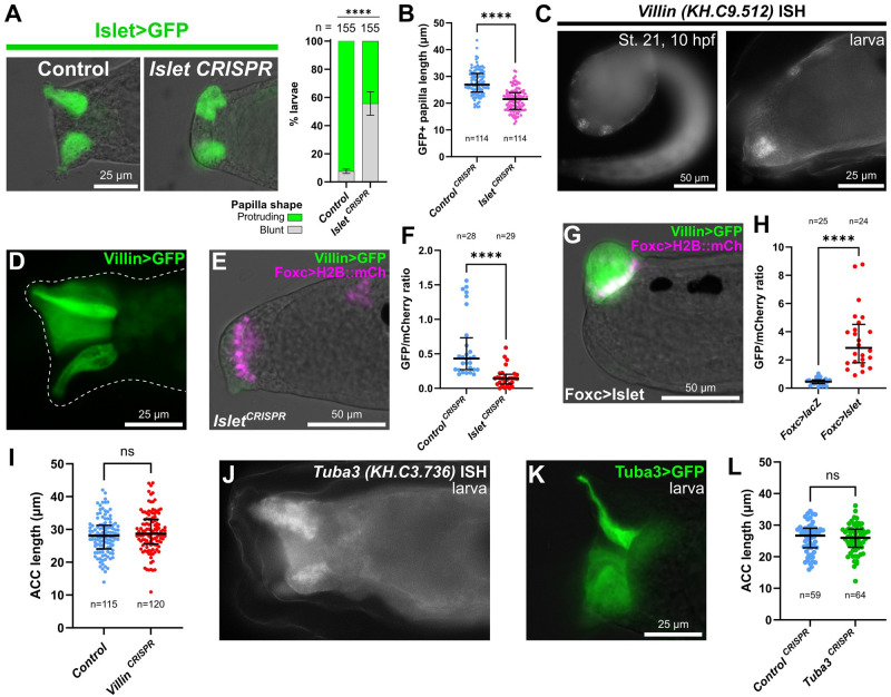
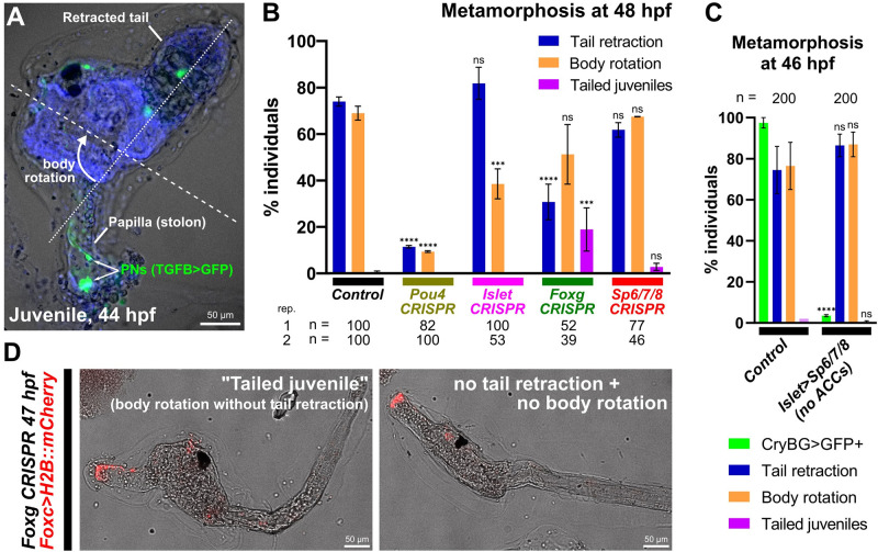
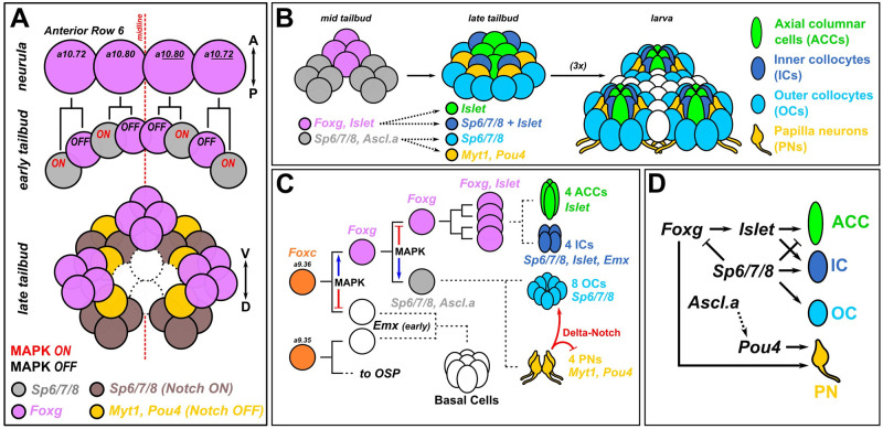
The papillae of tunicate larvae contribute sensory, adhesive, and metamorphosis-regulating functions that are crucial for the biphasic lifestyle of these marine, non-vertebrate chordates. We have identified additional molecular markers for at least 5 distinct cell types in the papillae of the model tunicate Ciona, allowing us to further study the development of these organs. Using tissue-specific CRISPR/Cas9-mediated mutagenesis and other molecular perturbations, we reveal the roles of key transcription factors and signaling pathways that are important for patterning the papilla territory into a highly organized array of different cell types and shapes. We further test the contributions of different transcription factors and cell types to the production of the adhesive glue that allows for larval attachment during settlement, and to the processes of tail retraction and body rotation during metamorphosis. With this study, we continue working towards connecting gene regulation to cellular functions that control the developmental transition between the motile larva and sessile adult of Ciona.


Conflict of interest statement

The authors have declared that no competing interests exist.

Figures

Fig 1
Fig 1. Development of the papillae of Ciona.
(A) Diagram showing the early cell lineages that give rise to the papillae. The papillae invariantly derive from Foxc+ cells in the anterior neural plate, more specifically the anterior daughter cells of “Row 6” of the neural plate, which activate Foxg downstream of Foxc. Foxg is also activated in the posterior daughter cells of “Row 5,” which go on to give rise to part of the OSP. Numbers in each cell indicate their invariant identity according to the Conklin cell lineage nomenclature. Black bars indicate sibling cells born from the same mother cell. (B) Diagram of what is currently known about the later lineage and fates of the Foxg+ “Anterior Row 6” cells shown in panel A. As the cells divide mediolaterally, some cells up-regulate Sp6/7/8 and down-regulate Foxg (gray cells). Those cells that maintain Foxg expression turn on Islet and coalesce as 3 clusters of cells (pink with green outline): 1 medial, more ventral cluster, and 2 left/right, more dorsal clusters. Later, these 3 clusters organize the territory into the 3 protruding papillae of the larva, which contains several cell types described in detail by TEM [9]. Dashed cell outlines indicate uncertain number/provenance of cells. A-P: anterior-posterior. D-V: dorsal-ventral. Lineages and gene networks are based mostly on: [21,22,86,87]. OSP, oral siphon primordium; TEM, transmission electron microscopy.
Fig 2
Fig 2. Novel genetic markers label distinct cell types of the papillae.
(A) GFP reporter plasmid (green) constructed using the cis-regulatory sequences from the KH.L96.43 gene labels basal cells in between and surrounding the protruding papillae labeled by Foxg reporter plasmid (pink). (B) TGFB>GFP reporter (green) labels PNs, the axons of which make contacts with BTN axons labeled by a BTN-specific Islet reporter (pink), at 23.5 h hpf, approximately corresponding to Hotta stage 30. (C) A KH.C4.78 reporter (C4.78>GFP) also labels PNs, which are also labeled by Foxg>H2B::mCherry (mCh) reporter (pink nuclei). (D) Lack of overlap between expression of C4.78>GFP (green) and a papilla-specific Islet reporter plasmid (pink nuclei) showing that PNs do not arise from Islet+ cells. (E, F) Co-electroporation of C11.360>GFP (green) with H2B::mCherry reporter plasmids (pink nuclei) indicates these cells come from Foxg-expressing cells that also express Islet. (G) C11.360>mCherry reporter (pink) labels centrally located ICs adjacent to ACCs labeled by CryBG>LacZ reporter (green). (H, I) L141.36>GFP reporter (green) labels OCs that arise from Foxg+ cells (pink nuclei) but do not express Islet (pink nuclei). (J) ICs and OCs are distinct cells as there is no overlap between C11.360 (green) and L141.36 (pink) reporter plasmid expression. (K) Ciona intestinalis (Type B) larva ICs labeled with a reporter plasmid made from the corresponding cis-regulatory sequence of the C. intestinalis Chr11.1038 gene, orthologous to C. robusta KH.C11.360. (L) C. intestinalis larva OCs labeled by a Chr7.130 reporter, corresponding to the C. robusta ortholog KH.L141.36. (M) Summary of the main marker genes and corresponding reporter plasmids used in this study to label different subsets of papilla progenitors and their derivative cell types. All GFP and mCherry reporters fused to the Unc-76 tag, unless specified (see Methods and supplement for details). Weaker Foxg -2863/-3 promoter used in panel A, all other Foxg reporters used the improved Foxg -2863/+54 sequence instead. All Islet reporters shown correspond to the Islet intron 1 + bpFOG>H2B::mCherry plasmid. White channel shows either DAPI (nuclei) and/or larva outline in brightfield, depending on the panel. All C. robusta raised at 20 °C to 18 hpf (roughly st. 28) except: panel B (23.5 hpf, ~st. 30); panels C–F (17 hpf, ~st. 27); panels H–J (20 hpf, ~st. 29). C. intestinalis raised at 18 °C to 20–22 hpf (Hotta stage 28). ACC, axial columnar cell; BTN, bipolar tail neuron; hpf, hours post-fertilization; IC, inner collocyte; OC, outer collocyte; PN, papilla neuron.
Fig 3
Fig 3. The transcription factor Islet is required for specification of ACCs and ICs.
(A) Diagram depicting a partially overlapping expression patterns of Islet and Sp6/7/8, as originally shown by in situ mRNA hybridizations (Wagner and colleagues), and the correlation of these patterns with the later arrangement of ACCs, ICs, and OCs in the papillae. “Late” Emx expression in a ring of cells expressing both Islet and Sp6/7/8 appears to be distinct from earlier Emx expression in Foxg-negative cells (see text and S2 Fig for details). (B) Papilla lineage-specific CRISPR/Cas9-mediated mutagenesis of Islet using Foxc>Cas9 and a the U6>Islet.2 sgRNA plasmid shows reduction of larvae showing expression of reporters labeling ACCs and ICs, but not OCs or PNs. Results compared to a negative “control” condition using a negative control sgRNA (U6>Control, see text for details). Nuclei counterstained with DAPI (white). (C) Scoring data for larvae represented in panel B, averaged between biological duplicates. Foxc>H2B::mCherry+ larvae were scored for quantity of papillae showing visible expression of the corresponding GFP reporter plasmid. Due to mosaic uptake or retention of the plasmids after electroporation, number of papillae with GFP fluorescence is variable and rarely seen in all 3 papillae even in control larvae. Normally larvae have 3 papilla (GFP+ or not), but some mutants have more/fewer than 3. ACC/IC/OC subpanels in panel B at 20 hpf/20 °C (~st. 29), PN subpanels at 21 hpf/20 °C (~st. 29). Same applies to scoring data in Panel C. All experiments were performed in duplicate, with number of embryos ranging from 76 to 100 per condition per replicate. **** p < 0.0001 in both duplicates, as determined by chi-square test. ns = not statistically significant in at least 1 duplicate, also by chi-square test. See S4 Data for the data underlying the graphs and for statistical test details. ACC, axial columnar cell; IC, inner collocyte; OC, outer collocyte; PN, papilla neuron; sgRNA, single-chain guide RNA.
Fig 4
Fig 4. Specification of ACCs, ICs, and OCs by a combinatorial logic of Islet and Sp6/7/8.
(A) Overexpression of Sp6/7/8 (using the Islet>Sp6/7/8 plasmid) in all Islet+ papilla cells results in loss of ACCs (assayed by expression of CryBG>Unc-76::GFP, green), but not of ICs (assayed by expression of C11.360>Unc-76::GFP, green). Islet overexpression (with Islet>Flag::Islet-rescue) does not significantly impact the specification of ACCs or ICs. Larvae at 20 hpf/20 °C (~st. 29). (B) Scoring data showing presence or absence of ICs or ACCs in Foxc>H2B::mCherry+ larvae, as represented in panel A. Experiments were performed in duplicate with 99 or 100 larvae in each duplicate. (C) Cell type specification assayed by reporter plasmid expression (green) in larvae subjected to various Islet and/or Sp6/7/8 perturbation conditions (see main text for details). For ICs and ACCs, the “control” condition is negative control CRISPR (U6>Control), while for OCs it is Foxc>lacZ. Overexpression ACC/IC subpanels are at 18.5 hpf/20 °C (~st. 28), all CRISPR and OC panels at 20 hpf/20 °C (~st. 29). (D) Scoring data for most larvae represented in panel C. Foxc>H2B::mCherry+ larvae were scored for cell type-specific GFP reporter expression that was “heterogeneous” (mixed on/off GFP expression, with all “wild type” patterns of expression falling under this category), “predominant” (ectopic/supernumerary GFP+ cells), “sparse” (reduced frequency/intensity of GFP expression), or “absent” (no GFP visible). (E) IC or ACC reporter (C11.360>Unc-76::GFP, CryBG>Unc-76::GFP) expression scored in Foxc>H2B::mCherry+ larvae represented in top 2 panels of right-most column in C. Experiment was performed and scored in duplicate, with number of larvae in each duplicate ranging from 100 to 105. (F) OC-specific reporter (L141.36>Unc-76::GFP) expression scored in Foxc>H2B::mCherry+ larvae represented by the bottom/right-most subpanel in panel B. Scoring strategy same as in Fig 3. Asterisk denotes when a duplicate of the negative control condition was also used for plots in Fig 3, as multiple CRISPR experiments were performed in parallel. All experiments were performed in duplicate, with number of embryos ranging from 76 to 100 per duplicate. Foxc>Cas9 used for all CRISPR/Cas9 experiments. The Islet cis-regulatory sequence used (panels A and B) was always Islet intron 1 + -473/-9. For overexpression conditions, Foxc>lacZ or Islet>LacZ were used to normalize total amount of DNA (see S1 File for detailed electroporation recipes). All error bars indicate upper and lower limits. **** p < 0.0001 in both duplicates as determined by Fisher’s exact test (panels B and E) or chi-square test (panel F). ns = not statistically significant in at least 1 duplicate. See S4 Data for the data underlying the graphs and statistical test details. ACC, axial columnar cell; IC, inner collocyte; OC, outer collocyte.
Fig 5
Fig 5. Both types of collocytes contribute to production of adhesive material.
(A) PNA-stained granules (pink) are seen in the hyaline cap and the apical tip of ICs (left panel, white arrow) in a C. intestinalis larva labeled by the C. intestinalis C11.360>Unc-76::GFP reporter (green). PNA-stained granules are also seen in cells not labeled by the IC reporter (right subpanel, hollow arrowhead), suggesting they are localized in a different cell type. Left and right subpanels are from different focal planes of the same papilla. (B) OCs labeled with C. robusta L141.36>Unc-76::GFP (green) in a C. robusta larva, with PNA-stained granules (pink) in both apical and basal positions within the cell (white arrows). DAPI in blue. (C) PNA staining (pink) in C. robusta upon overexpression of Sp6/7/8 alone, showing reduction of IC specification as assayed by C11.360>Unc-76::GFP expression (green). Weak PNA staining and GFP expression are still visible in some papillae (solid arrow), but not others (open arrowhead). (D) PNA staining (pink) and C11.360>Unc-76::GFP expression (green) in C. robusta upon overexpression of both Islet and Sp6/7/8, showing expansion of IC fate in a single large papilla (arrow). PNA staining is similarly expanded over the entire IC cluster, confirming that ICs produce the adhesive glue. Foxc>lacZ expression (β-galactosidase immunostaining) shown in blue in both C and D. (E) Scoring of larvae represented in panels C and D, averaged across duplicates. Weak PNA staining is observed upon partial suppression of IC fate, but strong PNA staining is seen upon expansion of supernumerary ICs, confirming that this cell type is one of the major contributors of PNA-positive adhesive glue. Total larvae (duplicate 1) or β-galactosidase+ larvae (duplicate 2) were scored. **** p < 0.0001 in both duplicates, as determined by chi-square test. See S4 Data for sample size, statistical test details, and for the data underlying the graphs. C. intestinalis raised to 20–22 hpf at 18 °C (~st. 28), C. robusta raised to 20 hpf at 20 °C (~st. 29). See Supplemental Movies for full confocal stacks and S6 Fig for single-channel images. IC, inner collocyte; OC, outer collocyte; PNA, peanut agglutinin.
Fig 6
Fig 6. Specification of PNs and OCs from Islet-negative cells by MAPK and Notch pathways.
(A) Diagram showing effect of MAPK inhibition with the pharmacological MEK inhibitor U0126, based on findings from Wagner and colleagues and Liu and Satou. Inhibition of FGF/MAPK results in expansion of Foxg and Islet from 3 discrete foci to a large “U-shaped” swath, transforming 3 papillae into a single, enlarged papilla (similar results reported with BMP inhibition by Roure and colleagues). (B) The 10 μm U0126 treatment at 7.5 hpf/20 °C (st. 16) results in loss of PNs (assayed by C4.78>Unc-76::GFP at 17 hpf/20 °C, ~st. 27, green) upon expansion of Islet+ cells (pink nuclei), relative to DMSO alone. (C) The same treatment results in loss of OCs (L141.36>Unc-76::GFP at either 17 or 20 hpf/20 °C, ~st. 27–29 green) upon expansion of Islet+ cells (pink nuclei). Both U0126 experiments were performed in duplicate, with 100 larvae per condition per duplicate. (D) Two-color, whole-mount mRNA in situ hybridization for Foxg (green in merged image) and Ascl.a (KH. L9.13, pink). (E) Larva electroporated with Ascl.a>Unc-76::GFP labeling several papilla cells including PNs. (F) Myt1>mNeonGreen labeling PNs and other neurons including CENs. (G) Two-color in situ hybridization of Foxg (green) and Pou4 (pink), the latter labeling adjacent PNs and possibly RTENs. (H) Lineage-specific CRISPR/Cas9-mediated mutagenesis of Pou4 results in loss of PN reporter expression (C4.78>Unc-76::GFP, green). Asterisk denoted background/leaky expression in mesenchyme. Larvae at 17 hpf/20 °C (~st. 27). (I) Scoring of Foxc>H2B::mCherry+ larvae represented in panel H and in Sp6/7/8 CRISPR mutagenesis condition showing Sp6/7/8 does not appear to play a major role in PN reporter expression like Pou4. Experiments repeated in duplicate with 100 larvae in each. (J) Inhibition of Delta/Notch signaling using Foxc>SUH-DBM results in reduced expression of OC reporter (L141.36>Unc-76::GFP, green) at 21 hpf/20 °C (~st. 29). Experiment was repeated in duplicate with 100 larvae in each. (K) Notch inhibition also results in concomitant expansion of supernumerary PNs at 17 hpf/20 °C (~st. 27, labeled by C4.78>Unc-76::GFP, green) relative to Foxc>lacZ control. Experiment was repeated in duplicate, with 42 to 50 larvae in each. (L) Summary diagram and model of effects of Delta/Notch inhibition on PN/OC fate choice in Islet-negative (but formerly Foxg+) papilla progenitor cells. All Islet reporters are the Islet intron 1 + bpFOG>H2B::mCherry. All error bars indicate upper and lower limits. **** p < 0.0001, *** p = 0.0003 in both duplicates as determined by Fisher’s exact test, ns = not statistically significant in at least 1 duplicate. See S4 Data for the data underlying the graphs and for statistical test details. CEN, caudal epidermal neuron; OC, outer collocyte; PN, papilla neuron; RTEN, rostral trunk epidermal neuron.
Fig 7
Fig 7. Islet is also required for papilla morphogenesis.
(A) Papilla shape is shortened and blunt at the apical end upon tissue-specific CRISPR/Cas9-mediated mutagenesis of Islet. Embryos were electroporated with Islet intron 1 + -473/-9>Unc-76::GFP and Foxc>Cas9. Islet CRISPR was performed using U6>Islet.2 sgRNA plasmid and the negative control used U6>Control. Larvae were imaged at 20 hpf/20 °C (~st. 28). Right: Scoring of percentage of GFP+ larvae classified as having normal “protruding” or blunt papillae, as represented to the left. Experiment was performed and scored in duplicate, using 2 different GFP fusions: Unc-76::GFP and DcxΔC::GFP [88]. Replicate 1: n = 100 for either condition; replicate 2: n = 55 for either condition **** p < 0.0001 in both duplicates as determined by Fisher’s exact test. (B) Quantification of papilla cell (Islet intron 1 +-473/-9>Unc-76::GFP+) lengths along apical-basal axis in negative control and Islet CRISPR larvae at 18 hpf/20 °C (~st. 28). Both Islet.1 and Islet.2 sgRNAs used in combination. Statistical significance tested by unpaired t test (two-tailed). See S8 Fig for duplicate experiment. (C) In situ mRNA hybridization of Villin, showing expression in Foxg+/Islet+ central papilla cells at 10 hpf/20 °C (st. 21, left) and at larval stage (~st. 27, right). (D) Villin -1978/-1>Unc-76::GFP showing expression in the papilla territory of electroporated larvae (~st. 28), strongest in the central cells. (E) Villin -1978/-1>Unc-76::GFP in st. 28 larvae is down-regulated by tissue-specific CRISPR/Cas9 mutagenesis of Islet (Foxc>Cas9 + U6>Islet.1 + U6>Islet.2, see text for details). (F) Quantification of effect of Islet CRISPR (as in panel E) on Villin -1978/-1>Unc 76::GFP/Foxc>H2B::mCherry mean fluorescence intensity ratios in ROIs defined by the mCherry+ nuclei (see Methods for details). Significance determined by Mann–Whitney test (two-tailed). (G) Villin reporter is up-regulated in st. 28 larvae by overexpressing Islet (Foxc>Islet, see text for details). (H) GFP/mCherry ratio quantification done in identical manner as in F, but comparing Islet overexpression (as in panel G) and control lacZ larvae. (I) Quantification of ACC lengths measured in negative control and papilla-specific Villin CRISPR larvae at 17 hpf/20 °C (~st. 27). Significance tested by unpaired t test (two-tailed). Although no statistically significant difference between control and CRISPR larvae was observed in this replicate, average ACC length was significantly shorter in the CRISPR condition in an additional replicate (S8 Fig). (J) mRNA in situ hybridization for Tuba3, showing enrichment in the central cells of the papillae in st. 27 larvae. (K) Tuba3>Unc-76::GFP reporter plasmid is broadly expressed in the papillae of st. 27 larvae but stronger in central cells. (L) Papilla-specific CRISPR knockout of Tuba3 does not result in decrease of average ACC apical-basal cell length compared to negative control CRISPR using U6>Control sgRNA instead. Significance tested by unpaired t test (two-tailed). ns = not significant. All large bars indicate medians and smaller bars indicate interquartile ranges. See S3 and S4 Data for the data underlying the graphs and for statistical test details. ACC, axial columnar cell; ROI, region of interest; sgRNA, single-chain guide RNA.
Fig 8
Fig 8. Genetic perturbations of metamorphosis.
(A) Ciona robusta juvenile undergoing metamorphosis, showing the retracted tail and rotated anterior-posterior body axis (dashed lines). PNs in the former papilla (now substrate attachment stolon, or holdfast) labeled by TGFB>Unc-76::GFP (green). Animal counterstained with DAPI (blue). (B) Scoring of Foxc>H2B::mCherry+ individuals showing tail retraction and/or body rotation at 48 hpf/20 °C in various papilla territory-specific (using Foxc>Cas9) CRISPR-based gene knockouts. Experiments were performed and scored in duplicate and percentages averaged, except for Foxg CRISPR for which a third replicate was performed (see S9 Fig). Number scored individuals in each replicate indicated underneath. “Tailed juveniles” have undergone body rotation but not tail retraction, whereas normally body rotation follows tail retraction. The sgRNA plasmids used for each condition were as follows- Control: U6>Control; Pou4: U6>Pou4.3.21 + U6>Pou4.4.106; Islet: U6>Islet.2; Foxg: U6>Foxg.1.116 + U6>Foxg.5.419; Sp6/7/8: U6>Sp6/7/8.4.29 + U6>Sp6/7/8.8.117. (C) Plot showing lack of any discernable metamorphosis defect after eliminating ACCs using Islet intron 1 + bpFOG>Sp6/7/8 (images not shown). Only Islet intron 1 + bpFOG>H2B::mCherry+ individuals were scored. Experiment was performed and scored in duplicate and averaged (n = 100 each duplicate). ACC specification was scored using the CryBG>Unc-76::GFP reporter. (D) Example of “tailed juveniles” at 47 hpf/20 °C compared to a larva in which no tail retraction or body rotation has occurred, elicited by tissue-specific Foxg CRISPR (Foxc>Cas9 + U6>Foxg.1.116 + U6>Foxg5.419). See S9 Fig for scoring. All error bars denote upper and lower limits. **** p < 0.0001, *** p < 0.0015 in both duplicates, ns = not significant in at least 1 duplicate, as determined by chi-square test comparing to the control conditions. See S4 Data for the data underlying the graphs and for statistical test details. ACC, axial columnar cell; PN, papilla neuron; sgRNA, single-chain guide RNA.
Fig 9
Fig 9. Summary diagram.
(A) Updated diagram of the development of the anterior descendants of Row 6 in the neural plate to show the proposed patterns of MAPK and Delta/Notch signaling that set up the 3 Foxg+ clusters and interleaved Foxg-negative neurogenic cells. (B) Diagram proposing the contributions of Foxg+ and Foxg-negative cells to later patterns of transcription factors that specify the different cell types found in each papilla, which is in turn is repeated 3 times, thanks to the process shown in panel A. (C) Papilla development shown as cell lineages, with dashed lines indicating uncertain cell divisions and lineage history. MAPK regulates binary fate choices, promoting Foxg expression in the papillae proper at first but later suppressing it (and Islet). Lastly, Delta-Notch signaling promotes OC fate and limits PN specification through lateral inhibition. Cell type numbers based on [9]. (D) Provisional gene regulatory network diagram of the transcription factors involved in specification and differentiation of the different papilla cell types. Arrowheads indicate activating gene expression or promoting cell fate, while blunt ends indicate repression of gene expression of cell fate. Solid lines indicate regulatory links (direct or indirect) that are supported by the current data and literature. Dashed lines indicate regulatory links that have not been tested, or need to be investigated in more detail. A-P, anterior-posterior; D-V, dorsal-ventral; OC, outer collocyte; OSP, oral siphon primordium; PN, papilla neuron.

Similar articles

Cited by

References

    1. Fodor ACA, Liu J, Turner L, Swalla BJ. Transitional chordates and vertebrate origins: Tunicates. Curr Top Dev Biol. 2021;139:325–374. doi: 10.1016/bs.ctdb.2020.10.001 - DOI - PubMed
    1. Lemaire P. Evolutionary crossroads in developmental biology: the tunicates. Development. 2011;138(11):2143–2152. doi: 10.1242/dev.048975 - DOI - PubMed
    1. Satoh N. Developmental genomics of ascidians. John Wiley & Sons; 2013.
    1. DeBiasse MB, Colgan WN, Harris L, Davidson B, Ryan JF. Inferring tunicate relationships and the evolution of the tunicate Hox cluster with the genome of Corella inflata. Genome Biol Evol. 2020;12(6):948–964. doi: 10.1093/gbe/evaa060 - DOI - PMC - PubMed
    1. Kocot KM, Tassia MG, Halanych KM, Swalla BJ. Phylogenomics offers resolution of major tunicate relationships. Mol Phylogenet Evol. 2018;121:166–73. doi: 10.1016/j.ympev.2018.01.005 - DOI - PubMed