. 2024 Mar 15;10(11):eadk6906.
doi: 10.1126/sciadv.adk6906. Epub 2024 Mar 13.

Mechanosensing regulates tissue repair program in macrophages

Affiliations

Mechanosensing regulates tissue repair program in macrophages

Matthew L Meizlish et al. Sci Adv. .

Abstract

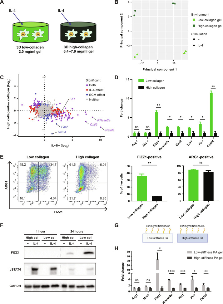
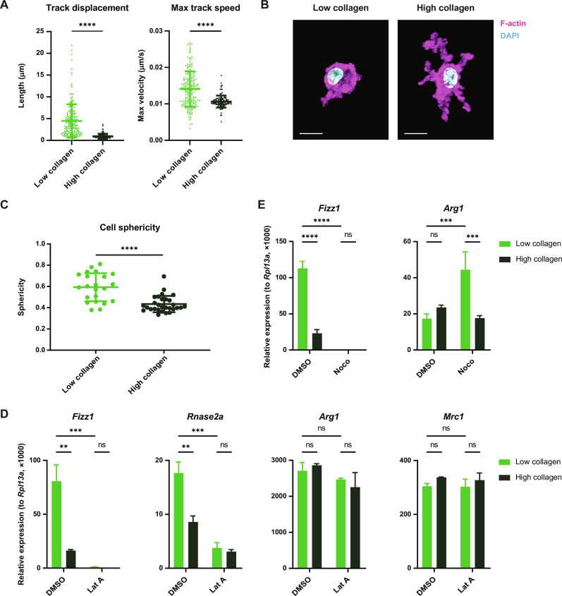
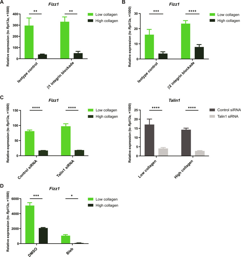
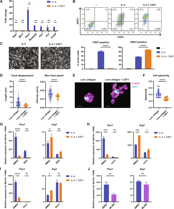
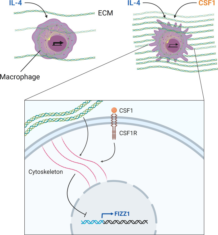
Tissue-resident macrophages play important roles in tissue homeostasis and repair. However, how macrophages monitor and maintain tissue integrity is not well understood. The extracellular matrix (ECM) is a key structural and organizational component of all tissues. Here, we find that macrophages sense the mechanical properties of the ECM to regulate a specific tissue repair program. We show that macrophage mechanosensing is mediated by cytoskeletal remodeling and can be performed in three-dimensional environments through a noncanonical, integrin-independent mechanism analogous to amoeboid migration. We find that these cytoskeletal dynamics also integrate biochemical signaling by colony-stimulating factor 1 and ultimately regulate chromatin accessibility to control the mechanosensitive gene expression program. This study identifies an "amoeboid" mode of ECM mechanosensing through which macrophages may regulate tissue repair and fibrosis.


Figures

Fig. 1.
Fig. 1.. Sensing of ECM stiffness controls a tissue repair–associated gene expression program in macrophages.
(A) Schematic overview of the experimental setup for macrophage culture within 3D collagen hydrogels. (B and C) Principal components analysis (B) and plot of all individual genes (C) in RNA-seq data from BMDMs cultured in low- or high-collagen gels with or without IL-4. Plots in (C) are colored according to the statistical significance of gene expression changes caused by culture in high-collagen gels versus low-collagen gels, as well as treatment with IL-4 versus control (q < 0.2). Retnla = Fizz1, Chil3 = Ym1. (D) Fold change of relative expression of selected genes in BMDMs cultured in low-collagen gels compared to high-collagen gels. Mean gene expression in high-collagen gels is shown as 1. (E) FIZZ1 and ARG1 protein expression in BMDMs cultured in low- or high-collagen gels with IL-4, analyzed by flow cytometry following the gating strategy displayed in fig. S1E. Representative plots (left) and the percentage of FIZZ1-positive and ARG1-positive cells (right) are shown. (F) Western blot showing FIZZ1 and phospho-STAT6 (pSTAT6) protein 1 hour and 24 hours after control or IL-4 treatment in low- or high-collagen gels. (G) Schematic overview of the experimental setup for PA-fibronectin hydrogel culture. (H) Fold change of relative gene expression in BMDMs cultured on low-stiffness PA gels compared to high-stiffness PA gels. Mean gene expression on high-stiffness gels is shown as 1. Data are represented as means ± SD. Multiple t test (D and H) and Student’s t test (E) were used for statistical analysis. *P < 0.05, **P < 0.01, ***P < 0.001, and ****P < 0.0001. ns, not significant.
Fig. 2.
Fig. 2.. Macrophage mechanosensing is mediated by cytoskeletal remodeling.
(A) Quantification of migration distance and speed of BMDMs cultured in low- or high-collagen gels after stimulation with IL-4. (B and C) Representative confocal images (B) and quantification of the sphericity (C) of BMDMs cultured in low- or high-collagen gels and stimulated with IL-4. Cells were stained with phalloidin (magenta) to visualize F-actin and 4′,6-diamidino-2-phenylindole (DAPI) (blue) to visualize the nucleus. Scale bars, 10 μm. (D) Relative gene expression in IL-4–stimulated BMDMs cultured in low- or high-collagen gels with dimethyl sulfoxide (DMSO) or latrunculin A (Lat A). (E) Relative gene expression in IL-4–stimulated BMDMs cultured in low- or high-collagen gels with DMSO or nocodazole (Noco). Data are represented as means ± SD. Student’s t test [(A) and (C)] and two-way analysis of variance (ANOVA) [(D) and (E)] are used for statistical analysis. **P < 0.01, ***P < 0.001, and ****P < 0.0001.
Fig. 3.
Fig. 3.. Macrophage mechanosensing does not require integrins or myosin II.
(A and B) Relative gene expression in BMDMs treated with β1 integrin blocking antibody (A), β2 integrin blocking antibody (B), or isotype-matched controls [(A) and (B)] and cultured in low- or high-collagen gels in the presence of IL-4. (C) Relative gene expression in BMDMs treated with control siRNA or Talin1 siRNA and cultured in low- or high-collagen gels in the presence of IL-4. (D) Relative gene expression in BMDMs treated with DMSO or blebbistatin (Bleb) and cultured in low- or high-collagen gels in the presence of IL-4. Data are represented as means ± SD. Two-way ANOVA was used for statistical analysis. *P < 0.05, **P < 0.01, ***P < 0.001, and ****P < 0.0001.
Fig. 4.
Fig. 4.. CSF1 sensing by macrophages induces cytoskeletal remodeling to regulate the mechanosensitive gene expression program.
(A) Fold change of relative gene expression in BMDMs cultured on TC plates and stimulated with IL-4 only, compared to both IL-4 and CSF1. Mean gene expression in cells treated with both IL-4 and CSF1 is shown as 1. (B) FIZZ1 and ARG1 protein expression in BMDMs cultured on TC plates and left unstimulated, stimulated with IL-4 only, or stimulated with both IL-4 and CSF1. Protein expression was analyzed by flow cytometry following the gating strategy displayed in fig. S1E. Representative plots (top) and the percentage of FIZZ1-positive and ARG1-positive cells (bottom) are shown. (C) Representative microscopic images of BMDMs cultured on TC plates in the presence of IL-4 with or without CSF1. (D) Quantification of migration distance and speed of BMDMs cultured in low-collagen gels in the presence of IL-4 with or without CSF1. (E and F) Representative confocal images (E) and quantification of the sphericity (F) of BMDMs cultured in low-collagen gels in the presence of IL-4 with or without CSF1. Cells were stained with phalloidin (magenta) to visualize F-actin and DAPI (blue) to visualize the nucleus. Scale bars, 10 μm. (G to I) Relative gene expression in IL-4–stimulated BMDMs cultured on TC plates and treated with or without CSF1 in the presence of DMSO or blebbistatin (G), latrunculin A (H), or nocodazole (I). (J) Relative gene expression in IL-4–stimulated BMDMs cultured on TC plates and treated with DMSO or ML099. Data are represented as means ± SD. Student’s t test [(A), (D), and (F)], one-way ANOVA (B), and two-way ANOVA [(G) to (I)] are used for statistical analysis. *P < 0.05, **P < 0.01, ***P < 0.001, and ****P < 0.0001.
Fig. 5.
Fig. 5.. Cytoskeletal remodeling controls chromatin accessibility in a site-specific fashion.
(A and B) ATAC-seq signal in genomic regions upstream of and including the Retnla (Fizz1) (A) and Arg1 (B) genes, analyzed in BMDMs cultured on TC plates and treated with control, IL-4, IL-4 and latrunculin A, or IL-4 and CSF1. Retnla and Arg1 promoter regions are highlighted by blue rectangles. Known Arg1 enhancer region is highlighted by a green rectangle. (C and D) Transcription factor-binding motifs enriched at genomic sites closed by both latrunculin A and CSF1 in the presence (C) or absence (D) of IL-4. The top 15 motifs, ranked by statistical significance, are shown. The shared motifs between (C) and (D) are highlighted as red.
Fig. 6.
Fig. 6.. ECM deposition during tissue repair in vivo induces cytoskeletal remodeling and suppresses mechanosensitive gene expression in macrophages.
(A and B) Representative microscopic images of hematoxylin and eosin staining (A) and Masson’s trichrome staining (B) of lungs at 3, 6, 9, and 12 days after oropharyngeal phosphate-buffered saline (PBS) or bleomycin administration to mice (n = 3 per group per time point). Scale bars, 100 μm. (C) Relative gene expression in whole lungs from PBS- or bleomycin-treated mice (n = 6 to 7 per group per time point). Statistically significant differences between time points in the bleomycin-treated group are shown. (D) FIZZ1 and ARG1 protein expression in lung macrophages in PBS- or bleomycin-treated mice, analyzed by flow cytometry following the gating strategy in fig. S5. The percentages of FIZZ1-positive and ARG1-positive cells are shown (n = 4 to 5 per group per time point). Statistically significant differences between time points in the bleomycin-treated group are indicated. (E) Representative snapshots of 3D reconstructed confocal images of lungs from bleomycin-treated mice (n = 3 per time point). Surfaces of F4/80+ cells (magenta) and collagen I (green) are shown. Scale bars, 20 μm. (F) Quantification of the sphericity of F4/80+ cells in (E). Two-way ANOVA [(C) and (D)] and one-way ANOVA (F) are used for statistical analysis. *P < 0.05, **P < 0.01, ***P < 0.001, and ****P < 0.0001.
Fig. 7.
Fig. 7.. Model: ECM mechanics and CSF1 signaling are integrated through cytoskeletal remodeling to regulate tissue repair–associated gene expression in macrophages.
Macrophages sense ECM mechanics through integrin-independent cytoskeletal remodeling to regulate the expression of a subset of tissue repair–associated genes induced by IL-4, including Fizz1. Signaling by CSF1, the lineage-restricted growth factor for macrophages, also converges on these cytoskeletal dynamics to regulate this mechanosensitive gene expression program. Macrophage cytoskeletal dynamics ultimately control chromatin accessibility at regulatory elements of target genes, translating environmental sensing into transcriptional regulation. These mechanisms may allow for appropriate regulation of tissue repair: As ECM stiffness and CSF1 production increase over the course of repair, macrophages undergo cytoskeletal remodeling and suppress genes that drive repair, which may be critical for resolving tissue repair and avoiding fibrosis.

References

    1. Magie C. R., Martindale M. Q., Cell-cell adhesion in the cnidaria: Insights into the evolution of tissue morphogenesis. Biol. Bull. 214, 218–232 (2008). - PubMed
    1. Tyler S., Epithelium – The primary building block for metazoan complexity. Integr. Comp. Biol. 43, 55–63 (2003). - PubMed
    1. Fidler A. L., Darris C. E., Chetyrkin S. V., Pedchenko V. K., Boudko S. P., Brown K. L., Gray Jerome W., Hudson J. K., Rokas A., Hudson B. G., Collagen IV and basement membrane at the evolutionary dawn of metazoan tissues. eLife 6, e24176 (2017). - PMC - PubMed
    1. Frantz C., Stewart K. M., Weaver V. M., The extracellular matrix at a glance. J. Cell Sci. 123 (Pt 24), 4195–4200 (2010). - PMC - PubMed
    1. Duffield J. S., Lupher M., Thannickal V. J., Wynn T. A., Host responses in tissue repair and fibrosis. Annu. Rev. Pathol. 8, 241–276 (2013). - PMC - PubMed

Publication types