. 2024 May:71:103116.
doi: 10.1016/j.redox.2024.103116. Epub 2024 Mar 6.

GSTP alleviates acute lung injury by S-glutathionylation of KEAP1 and subsequent activation of NRF2 pathway

Affiliations

GSTP alleviates acute lung injury by S-glutathionylation of KEAP1 and subsequent activation of NRF2 pathway

Xiaolin Sun et al. Redox Biol. 2024 May.

Abstract

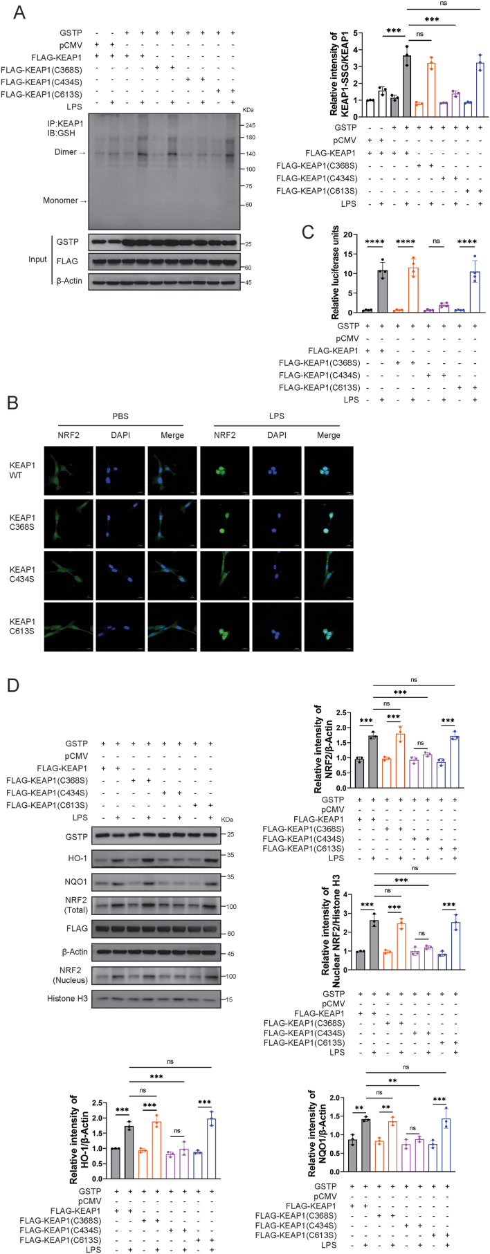
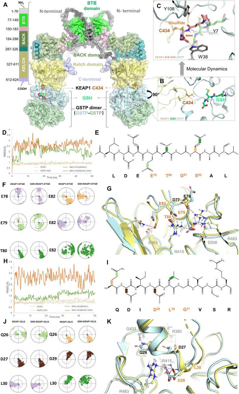
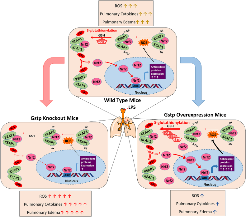
Oxidative stress plays an important role in the pathogenesis of acute lung injury (ALI). As a typical post-translational modification triggered by oxidative stress, protein S-glutathionylation (PSSG) is regulated by redox signaling pathways and plays diverse roles in oxidative stress conditions. In this study, we found that GSTP downregulation exacerbated LPS-induced injury in human lung epithelial cells and in mice ALI models, confirming the protective effect of GSTP against ALI both in vitro and in vivo. Additionally, a positive correlation was observed between total PSSG level and GSTP expression level in cells and mice lung tissues. Further results demonstrated that GSTP inhibited KEAP1-NRF2 interaction by promoting PSSG process of KEAP1. By the integration of protein mass spectrometry, molecular docking, and site-mutation validation assays, we identified C434 in KEAP1 as the key PSSG site catalyzed by GSTP, which promoted the dissociation of KEAP1-NRF2 complex and activated the subsequent anti-oxidant genes. In vivo experiments with AAV-GSTP mice confirmed that GSTP inhibited LPS-induced lung inflammation by promoting PSSG of KEAP1 and activating the NRF2 downstream antioxidant pathways. Collectively, this study revealed the novel regulatory mechanism of GSTP in the anti-inflammatory function of lungs by modulating PSSG of KEAP1 and the subsequent KEAP1/NRF2 pathway. Targeting at manipulation of GSTP level or activity might be a promising therapeutic strategy for oxidative stress-induced ALI progression.

Keywords: Acute lung injury; GSTP; KEAP1; NRF2; Oxidative stress; S-glutathionylation.


Conflict of interest statement

Declaration of competing interest The authors declare that they have no known competing financial interests or personal relationships that could have appeared to influence the work reported in this paper.

Figures

Image 1
Graphical abstract
Fig. 1
Fig. 1
Down regulation of GSTP-mediated PSSG aggravates LPS-induced human pulmonary cells injury. Human pulmonary cells, HPAEpiCs and BEAS-2B, were transfected with NC-siRNA or GSTP-siRNA and further treated with LPS (200 ng/mL). (A) GSH, GSSG, and the GSH/GSSG ratio in HPAEpiCs. (B) Intracellular Fe2+ and total iron levels in HPAEpiCs. (C) Total PSSG in HPAEpiCs with manipulated GSTP expression levels via non-reducing Western blot. (D) Time- and GSTP-dependency of total PSSG in BEAS-2B cells via non-reducing Western blot. Data are expressed as means ± SD. Experimental data for each quantitative analysis were replicated at least three times. *P < 0.05, **P < 0.01, ***P < 0.001, ****P < 0.0001, ns = no significance.
Fig. 2
Fig. 2
GSTP alleviated the oxidative damage, inflammation and lung tissue injury in mice with ALI. Gstp−/− mice were i. t. Administrated with 3 mg/kg of LPS. After LPS challenge for 24 h, all mice were euthanized and their lungs and BALF were collected. (A–B) MDA and MPO activity in lung tissues were measured. (C–D) Total cell counts and neutrophils percentage from the BALF were measured using a multispecies hematology analyzer. (E–H) Levels of total protein and cytokines (IL-1β, IL-6 and TNF-α) secretion in BALF were measured. (I) The lung coefficient was calculated. (J) H&E staining of lung tissues. (K) Semiquantitative analysis of lung injury scores. Data are expressed as means ± SD. n = 8 or 9 mice/group. *P < 0.05, **P < 0.01, ***P < 0.001, ****P < 0.0001.
Fig. 3
Fig. 3
Effects of GSTP on LPS-induced KEAP1/NRF2 pathway activation. (A) HPAEpiCs transfected with NC-siRNA and GSTP-siRNA (n = 3), both groups were treated with LPS (200 ng/mL) for 60 min, then co-immunoprecipitation of KEAP1 and immunoblotting analysis were performed to assess the binding of NRF2. (B) HEK293 transfected with pCMV and GSTP plasmids (n = 3), both groups were treated with LPS (200 ng/mL) for 60 min, and co-immunoprecipitation of KEAP1 and immunoblotting analysis were performed to assess the binding of NRF2. (C) Immunofluorescence staining and confocal microscopy imaging of BEAS-2B cells transfected with GSTP-siRNA after exposure to LPS (200 ng/mL) for 2 h. (D) Expression levels of KEAP1, HO-1, total and nuclear NRF2 in GSTP-knocked down and control HPAEpiCs were measured after LPS (200 ng/mL) treatment for 30 min, n = 3. (E) Time- and GSTP-dependency of KEAP1, HO-1, NQO1, total and nuclear NRF2 expression levels in GSTP-knocked down and control BEAS-2B were measured after LPS treatment (200 ng/mL), n = 3. (F) The lung tissues were collected and lysed for protein analyzed of HO-1, NQO1 and NRF2 levels. Gstp−/− mice were i. t. Administrated with 3 mg/kg of LPS. n = 8 or 9 mice/group. (G) HPAEpiCs cells were co-transfected with the antioxidant response element reporter plasmid and the indicated siRNA for 48 h and were harvested for luciferase assay, n = 4. Data are expressed as means ± SDs. *P < 0.05, **P < 0.01, ***P < 0.001, ****P < 0.0001, ns = no significance.
Fig. 4
Fig. 4
KEAP1 is S-glutathionylated and catalyzed by GSTP under oxidative stress conditions. (A) Co-IP showing PSSG of KEAP1 in HPAEpiCs was reduced by GSTP-siRNA transfection via non-reducing Western blot, after exposure to LPS (200 ng/mL) for 30 min, n = 3. Whole cell lysates confirmed the expression of GSTP, KEAP1, and β-Actin via reducing Western blot. (B) Total PSSG level via non-reducing Western blot in HEK293 transfected with pCMV and GSTP expression plasmids, after exposure to LPS (200 ng/mL) for 30 min, n = 3. Whole cell lysates confirmed the expression of GSTP and β-Actin via reducing Western blot. (C) Co-IP showing PSSG of KEAP1 in HEK293 was enhanced by GSTP overexpression via non-reducing Western blot, after exposure to LPS (200 ng/mL) for 30 min, n = 3. Whole cell lysates confirmed the expression of GSTP, KEAP1, and β-Actin via reducing Western blot. (D) Non-reducing Western blot of PSSG of KEAP1 in lung tissues from Gstp−/− and WT mice following i. t. Administration of PBS or LPS. n = 8 or 9 mice/group. (E) Immunofluorescence staining and confocal microscopy imaging of HPAEpiCs showing the colocalization of GSTP and KEAP1. (F) GSTP concentration dependency of human recombinant KEAP1 PSSG in dimer and monomer status via non-reducing Western blot. The level of KEAP1 was conformed via reducing Western blot. Data are expressed as means ± SDs. *P < 0.05, **P < 0.01, ***P < 0.001, ****P < 0.0001, ns = no significance.
Fig. 5
Fig. 5
C434 PSSG of KEAP1 enhances NRF2 nuclear translocation and activated downstream pathways. (A) Co-immunoprecipitation of KEAP1 and GSH was performed, and relative KEAP1 PSSG levels were measured, n = 3. (B) Immunofluorescence staining and confocal microscopy imaging of HPAEpiCs cells with overexpression of KEAP1 WT, C368S, C434S and C613S after exposure to LPS (200 ng/mL) for 1 h. (C) HPAEpiCs were co-transfected with the antioxidant response element reporter plasmid and the indicated cDNA for 48 h and were harvested for luciferase assay, n = 4. (D) Levels of HO-1, NQO1, total and nuclear NRF2 in BEAS-2B were measured and calculated LPS (200 ng/mL), n = 3. Data are expressed as means ± SD. Experimental data for each quantitative analysis were replicated at least three times. *P < 0.05, **P < 0.01, ***P < 0.001, ****P < 0.0001, ns = no significance.
Fig. 6
Fig. 6
Computational modeling of GSTP-catalyzed KEAP1 PSSG at C434 and molecular dynamic analysis of C434 PSSG on KEAP1-NRF2 binding. (A) The binding mode of the full-length structure of KEAP1 (dimer) bound to GSTP dimers. Major domains of KEAP1 were shown in different colors. The BTB domain (PDB: 7X4X) and Kelch domain (PDB: 7K29) are crystal structures, while the other domains are predicted by Alphafold. The GSTP dimers are shown in pale blue and pale orange, respectively. Key residue C434 and GSH are shown in orange and green-cyan spheres. The whole complex is shown as a cartoon as well as transparent surfaces. (B) The detailed binding information of KEAP1 (pale yellow cartoon) bound to the G-site of GSTP (pale blue cartoon) with GSH (green-cyan ball and sticks). (C) The binding modes of GSH (pink-cyan sticks) to KEAP1 by forming a disulfide bond with C434 after molecular dynamics simulations. The un-bounded conformation of GSH is shown in transparent green-cyan sticks. Other key residues are shown in grey sticks, and key interactions are depicted as dotted lines. (D) The root-mean-square deviation (RMSD) values of the backbone (αC) of KEAP1 (pale green), KEAP1-GSH (pale orange), and the corresponding bound NRF2 ETGE motif (green and orange, respectively) were determined. The RMSD is used to measure the average displacement change of a group of atoms within a specific frame, relative to a reference frame, which was calculated for each frame in the trajectory. (E) The chemical structure of the ETGE motif is presented. 2D schematic NRF2 ETGE with color-coded rotatable bonds is presented, and each rotatable bond torsion is accompanied by a dial plot and bar plot of the same color in panel F. (F) The NRF2 ETGE motif torsion plot summarizes the conformational changes of every rotatable bond (RB) in NRF2 ETGE throughout the simulation trajectory (0.00–50.00 ns). The dial plots describe the torsion over the course of the simulation, with the center representing the beginning and the radial direction representing the time evolution. The dial plots on the left and right refer to the KEAP1-NRF2-ETGE and GSH-KEAP1-NRF2-ETGE system, respectively. (G) Comparative binding modes of ETGE motif of NRF2 with the KEAP1 Kelch domain before (pale-yellow) and after (pale-blue) molecular dynamics. Key residues are highlighted in corresponding colors, and key interactions are indicated by magenta dotted lines. The residues that correspond to the residues of KEAP1 are depicted as balls and sticks, and labelled in grey to differentiate them from the orange-labelled residues in ETGE motif. (H) The RMSD values of the backbone (αC) of KEAP1 (pale green), KEAP1-GSH (pale orange), and the corresponding bound NRF2 DLG motif (green and orange, respectively) were determined. (I) The chemical structure of the DLG motif is presented. 2D schematic of DLG motif with color-coded rotatable bonds is presented, and each rotatable bond torsion is accompanied by a dial plot and bar plot of the same color in panel J. (J) The DLG motif torsion plot summarizes the conformational changes of every RB in DLG motif throughout the simulation trajectory (0.00–50.00 ns). The dial plots on the left and right refer to the KEAP1-DLG and GSH-KEAP1-DLG system, respectively. (K) Comparative binding modes of DLG motif of NRF2 with the KEAP1 Kelch domain before (pale-yellow) and after (pale-blue) molecular dynamics is shown. Key residues are highlighted in corresponding colors, and key interactions are indicated by magenta dotted lines. The residues that correspond to the residues of KEAP1 are depicted as balls and sticks, and labelled in grey to differentiate them from the orange-labelled residues in DLG motif. (For interpretation of the references to color in this figure legend, the reader is referred to the Web version of this article.)
Fig. 7
Fig. 7
GSTP alleviated the oxidative damage, inflammation and lung tissue injury in mice with ALI via promoting KEAP1 PSSG. Mice pretreated with AAV-GSTP were i. t. Administrated with 3 mg/kg of LPS. After LPS challenge for 24 h, all mice were euthanized and their lungs and BALF were collected. (A) PSSG of KEAP1 in lung tissues from mice pretreated with AAV-GSTP. (B) The lung tissues were collected and lysed for the immunoblotting of HO-1, NQO-1 and NRF2 levels. (C–D) MDA and MPO activity in lung tissues were measured. (E–F) The total cell counts and neutrophils percentage from the BALF were measured counted using a multispecies hematology analyzer. (G–J) Levels of total protein and cytokines (IL-1β, IL-6 and TNF-α) secretion in BALF were measured. (K) The lung coefficient was calculated. (L) H&E staining of sections of lungs. (M) Semiquantitative analysis of lung injury scores. Data are expressed as means ± SD. n = 8 or 9 mice/group. *P < 0.05, **P < 0.01, ***P < 0.001, ****P < 0.0001, ns = no significance.
Fig. 8
Fig. 8
Proposed mechanism for alleviating inflammation in acute lung injury via GSTP-mediated KEAP1/NRF2 pathway activation. GSTP increased the dissociation of the KEAP1-NRF2 by catalyzing KEAP1 PSSG process, subsequently activated NRF2 downstream genes including HO-1 and NQO1 and alleviated the progression of inflammation in acute lung injury.

References

    1. Gallelli L., Zhang L., Wang T., Fu F. Severe acute lung injury related to COVID-19 infection: a review and the possible role for escin. J. Clin. Pharmacol. 2020;60:815–825. doi: 10.1002/jcph.1644. - DOI - PMC - PubMed
    1. Long M.E., Mallampalli R.K., Horowitz J.C. Pathogenesis of pneumonia and acute lung injury. Clin Sci (Lond). 2022;136:747–769. doi: 10.1042/CS20210879. - DOI - PMC - PubMed
    1. Rubenfeld G.D., Caldwell E., Peabody E., Weaver J., Martin D.P., Neff M., Stern E.J., Hudson L.D. Incidence and outcomes of acute lung injury. N. Engl. J. Med. 2005;353:1685–1693. doi: 10.1056/NEJMoa050333. - DOI - PubMed
    1. Schouten L.R., Veltkamp F., Bos A.P., Van Woensel J.B., Neto A.S., Schultz M.J., Wösten R.M., Van Asperen Incidence and mortality of acute respiratory distress syndrome in children: a systematic review and meta-analysis. Crit. Care Med. 2016;44:819–829. doi: 10.1097/CCM.0000000000001388. - DOI - PubMed
    1. Bezerra F.S., Lanzetti M., Nesi R.T., Nagato A.C., Silva C.P.E., Kennedy-Feitosa E., Melo A.C., Cattani-Cavalieri I., Porto L.C., Valenca S.S. Oxidative stress and inflammation in acute and chronic lung injuries. Antioxidants. 2023;12:548. doi: 10.3390/antiox12030548. - DOI - PMC - PubMed

MeSH terms