. 2024 May;300(5):107149.
doi: 10.1016/j.jbc.2024.107149. Epub 2024 Mar 11.

Acidity of persulfides and its modulation by the protein environments in sulfide quinone oxidoreductase and thiosulfate sulfurtransferase

Affiliations

Acidity of persulfides and its modulation by the protein environments in sulfide quinone oxidoreductase and thiosulfate sulfurtransferase

Dayana Benchoam et al. J Biol Chem. 2024 May.

Abstract

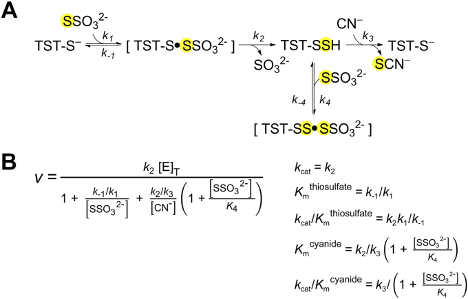
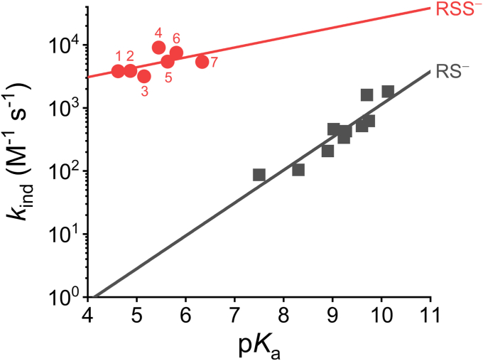
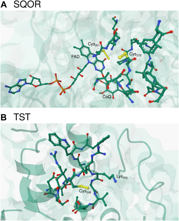
Persulfides (RSSH/RSS-) participate in sulfur metabolism and are proposed to transduce hydrogen sulfide (H2S) signaling. Their biochemical properties are poorly understood. Herein, we studied the acidity and nucleophilicity of several low molecular weight persulfides using the alkylating agent, monobromobimane. The different persulfides presented similar pKa values (4.6-6.3) and pH-independent rate constants (3.2-9.0 × 10³ M-1 s-1), indicating that the substituents in persulfides affect properties to a lesser extent than in thiols because of the larger distance to the outer sulfur. The persulfides had higher reactivity with monobromobimane than analogous thiols and putative thiols with the same pKa, providing evidence for the alpha effect (enhanced nucleophilicity by the presence of a contiguous atom with high electron density). Additionally, we investigated two enzymes from the human mitochondrial H2S oxidation pathway that form catalytic persulfide intermediates, sulfide quinone oxidoreductase and thiosulfate sulfurtransferase (TST, rhodanese). The pH dependence of the activities of both enzymes was measured using sulfite and/or cyanide as sulfur acceptors. The TST half-reactions were also studied by stopped-flow fluorescence spectroscopy. Both persulfidated enzymes relied on protonated groups for reaction with the acceptors. Persulfidated sulfide quinone oxidoreductase appeared to have a pKa of 7.8 ± 0.2. Persulfidated TST presented a pKa of 9.38 ± 0.04, probably due to a critical active site residue rather than the persulfide itself. The TST thiol reacted in the anionic state with thiosulfate, with an apparent pKa of 6.5 ± 0.1. Overall, our study contributes to a fundamental understanding of persulfide properties and their modulation by protein environments.

Keywords: alpha effect; hydrogen sulfide; pK(a); persulfide; rhodanese; sulfide quinone oxidoreductase; thiol; thiosulfate sulfurtransferase.


Conflict of interest statement

Conflict of interest The authors declare that they have no conflicts of interest with the contents of this article.

Figures

Figure 1
Figure 1
Proposed catalytic mechanisms for human SQOR and TST.A, the active site cysteine trisulfide in SQOR is attacked by H2S to form two persulfides, at Cys379 and at Cys201; the latter engages in a transient charge transfer (CT) complex with the FAD cofactor (reaction a). Then, a thiophilic acceptor (Acc) attacks Cys379SSH and the sulfane sulfur is transferred, generating AccSH and Cys379SH while the CT complex is proposed to evolve to a transient C4a adduct (reaction b). Then, Cys379SH attacks the adduct, regenerating the trisulfide and producing FADH2 (reaction c). Finally, FADH2 reduces CoQ to CoQH2, restoring SQOR to its resting state (reaction d) (21). B, the active site Cys248SH in TST attacks a sulfane sulfur donor (DS), resulting in the formation of Cys248SSH and release of the first product (D) (reaction e). Next, a thiophilic acceptor (Acc) attacks Cys248SSH, producing AccSH and restoring the resting enzyme (reaction f). SQOR, sulfide quinone oxidoreductase; TST, thiosulfate sulfurtransferase.
Figure 2
Figure 2
pKaof LMW persulfides and their reactivity with mBrB.A, representative stopped-flow fluorescence kinetic traces (λex = 396 nm, emission cut-off 435 nm) of the reaction of CysSSH-containing mixtures (0.5–3 μM) with mBrB (57.5 μM) in acetic/MES/Tris buffer (pH 3.65–8.15, 25 °C). Exponential plus straight line functions were fitted to the time courses over 10 half-lives. In some cases, where double exponential plus straight line functions were fitted, the exponential phase with the lower observed rate constant (kobs) and larger amplitude was attributed to the reaction of the persulfide with mBrB. B, linear dependence of kobs of CysSS with mBrB concentration. Circles are quintuplates of kobs obtained for every pH and mBrB concentration. The slope at each pH represents the apparent second-order rate constants, kpH. At the more alkaline pH values, a small negative y-intercept was observed, as seen previously with GSSH (13). C, for CysSS, a single-pKa function was fitted to the plot of kpHversus pH with data obtained in three independent experiments (black circles, blue squares, and green triangles). A pKa of 5.2 ± 0.1 and a pH-independent second-order rate constant, kind, of (3.2 ± 0.1) × 103 M−1 s−1 (parameters ± standard errors of the fit) were determined. As seen previously with GSSH (13), a small decrease of unknown origin in the kpH at the more alkaline pH values was observed. DG, the pKa and kind of the reactions with mBrB for other LMW persulfides were determined analogously. Plots of kpHversus pH for HcySS (D), CystSS (E), β-MESS (F), and CysOMeSS (G). In the case of CysOMeSS, a two-pKa function was fitted resulting in two sets of pKa and kind values. The obtained values are summarized in Table 1. CysOMeSS, cysteine methyl ester persulfide anion; CysSSH, cysteine persulfide; CystSS, cysteamine persulfide anion; HcySS, homocysteine persulfide anion; LMW, low molecular weight; mBrB, monobromobimane; β-MESS, β-mercaptoethanol persulfide anion.
Figure 3
Figure 3
pH-dependence of SQOR activity. The steady-state rate of reduction of CoQ1 was followed by the decrease in absorbance at 278 nm. The assays included 69 μM CoQ1, 0.03% DHPC, 0.06 mg/ml BSA, 150 μM H2S, and variable concentrations of sulfite or cyanide in MES/Tris/ethanolamine buffer (pH 5.65–9.93, 25 °C). A, the reactions with sulfite (0.01–8 mM) were started by the addition of 1 nM SQOR. Representative absorbance kinetic traces of CoQ1 reduction at pH 7.57. The steady-state rates were calculated from the linear fits to the data obtained 15 to 30 s after SQOR was added (subtracting the slopes before addition of SQOR). B, SQOR activity versus sulfite concentration at different pH values. Representative experiments at pH 8.68 (black circles), 7.57 (blue squares), 6.65 (green triangles), and 5.65 (red diamonds). Michaelis-Menten hyperbolas were fitted and yielded the kinetic parameters Kmsulfite (Fig. S1A), kcatsulfite (Fig. S1B), and kcat/Kmsulfite for each pH. C, pH-dependence of kcat/Kmsulfite. Equation 3 was fitted to the data obtaining two pKas, 6.8 ± 0.5 for the deprotonated species and 7.7 ± 0.4 for the protonated one. A pH-independent kcat/Kmsulfite of 2.9 ± 0.2 × 106 M−1 s−1 was obtained for the reaction of sulfite with the persulfidated SQOR. D, the reactions with cyanide (90 μM) contained 50 nM SQOR and were initiated by the addition of cyanide. Representative time courses at different pHs. To calculate the steady-state rates, the slopes in the absence of cyanide were subtracted from those obtained 4 to 20 s after the addition of cyanide. The kcat/Kmcyanide at each pH was calculated using Equation 2. E, pH-dependence of kcat/Kmcyanide. Equation 3 plus an offset was fitted to the data, yielding a pKa of 8.9 ± 0.2 for the deprotonated species, a pKa of 7.9 ± 0.1 for the protonated species, an offset of (−10 ± 5) × 103 M−1 s−1, and a pH-independent kcat/Kmcyanide of 1.5 ± 0.8 × 106 M−1 s−1 for the reaction of cyanide with the persulfidated SQOR. Values are parameters ± standard errors of the fits. DHPC, 1,2-diheptanoyl-sn-glycero-3-phosphocholine; SQOR, sulfide quinone oxidoreductase.
Figure 4
Figure 4
pH-dependence of TST activity. The steady-state activity of TST (5–100 nM) was measured by the formation of thiocyanate in the presence of thiosulfate (300 mM) and cyanide (300 μM, lower than Kmcyanide) in ACES/Tris/ethanolamine buffer in the pH range of 7.03 to 10.12 and 25 °C. A sigmoidal function was fitted to the data of two independent experiments (black circles and blue squares) and yielded an apparent pKa of 8.47 ± 0.06 and a maximum apparent rate constant of (4.0 ± 0.1) × 105 M−1 s−1 (parameters ± standard errors of the fit). TST, thiosulfate sulfurtransferase.
Figure 5
Figure 5
Stopped-flow kinetics of TST persulfide with sulfur acceptors.A and B, representative fluorescence time courses (λex = 295 nm, US 360 nm bandpass filter) of TST persulfide (0.8–1.0 μM) exposed to 75 μM sulfite (A) or 25 μM cyanide (B), in ACES/Tris/ethanolamine buffer (pH 5.60–10.38, 25 °C). Single exponential functions were fitted and the kobs was divided by the concentration of sulfite or cyanide to give the corresponding kpH. C and D, pH-dependence of kpH. Bell-shaped functions (Equation 3 or Equation 3 plus an offset) were fitted to the data, yielding pKa values of 6.89 ± 0.09 (deprotonated species) and 9.38 ± 0.07 (protonated species), an offset of (3.5 ± 0.9) × 104 M−1 s−1, and a kind of (2.5 ± 0.1) × 105 M−1 s−1 for the reaction with sulfite (C) and pKa values of 8.87 ± 0.06 (deprotonated species) and 9.37 ± 0.05 (protonated species) and a kind of (1.0 ± 0.1) × 107 M−1 s−1 for the reaction with cyanide (parameters ± standard errors of the fits). TST, thiosulfate sulfurtransferase.
Figure 6
Figure 6
TST mechanism and inhibition by thiosulfate.A, TST catalytic mechanism using thiosulfate and cyanide as substrates and depicting inhibition by high concentrations of thiosulfate (30, 31). B, steady-state rate equation assuming fast equilibrium for thiosulfate binding and steady-state for the persulfidated enzyme. TST, thiosulfate sulfurtransferase.
Figure 7
Figure 7
Stopped-flow kinetics of TST thiol with thiosulfate.A, representative fluorescence time courses (λex = 295 nm, US 360 nm bandpass filter) of TST thiol (0.9 μM) exposed to 200 μM thiosulfate in acetic/MES/Tris buffer (pH 3.68–8.75, 25 °C). Single or double exponential functions were fitted to the data. B, pH-dependence of kobs for a representative experiment. For the double exponential fits, the smaller kobs values, which corresponded to the larger amplitude, were used. Using data from three independent experiments, a two-pKa sigmoidal function was fitted yielding pKa values of 4.6 ± 0.1 and 6.5 ± 0.1 and maximal kobs of 5.9 ± 0.6 and 11.5 ± 0.3 s−1, respectively (parameters ± standard errors of the fit). C, pH-dependence of the kobs at alkaline pH values. The reactions were performed as described but in ACES/Tris/ethanolamine buffer (pH 6.68–10.34, 25 °C), and a single exponential plus straight line function was fitted to the data to obtain the kobs. Representative of two independent experiments, five replicates each. TST, thiosulfate sulfurtransferase.
Figure 8
Figure 8
Comparison of the reactivity of LMW persulfide anions and thiolates with mBrB. Brønsted plot exhibiting pH-independent rate constants (in logarithmic scale) versus pKa for the reactions of persulfide anions or thiolates with mBrB (I = 0.15 and 25 °C). Red circles: CysOMeSS(NH3+) (1), CystSS (2), CysSS (3), GSS (4), HcySS (5), β-MESS (6), and CysOMeSS(NH2) (7); βnuc = 0.2 ± 0.1 (R2 0.31). The values are depicted in Table 1. Black squares: reported data for LMW thiolates; βnuc = 0.52 ± 0.08 (R2 = 0.85) (16). LMW, low molecular weight; mBrB, monobromobimane.
Figure 9
Figure 9
Persulfides in SQOR and TST.A, close-up of the bis-persulfide in the SQOR structure (PDB 6OIB). Residues within 5 Å from Cys379SSH, the FAD cofactor, and the CoQ substrate are depicted in sticks. B, close-up of the persulfide in the bovine TST structure (PDB 1RHD). Residues within 5 Å from Cys248 are depicted in sticks. Figures were constructed with Mol∗ (65). SQOR, sulfide quinone oxidoreductase; TST, thiosulfate sulfurtransferase.

Similar articles

Cited by

References

    1. Wood J.L. Sulfane sulfur. Methods Enzymol. 1987;143:25–29. - PubMed
    1. Bouillaud F., Blachier F. Mitochondria and sulfide: a very old story of poisoning, feeding, and signaling? Antioxid. Redox Signal. 2011;15:379–391. - PubMed
    1. Filipovic M.R., Zivanovic J., Alvarez B., Banerjee R. Chemical biology of H2S signaling through persulfidation. Chem. Rev. 2018;118:1253–1337. - PMC - PubMed
    1. Mueller E.G. Trafficking in persulfides: delivering sulfur in biosynthetic pathways. Nat. Chem. Biol. 2006;2:185–194. - PubMed
    1. Cuevasanta E., Lange M., Bonanata J., Coitiño E.L., Ferrer-Sueta G., Filipovic M.R., et al. Reaction of hydrogen sulfide with disulfide and sulfenic acid to form the strongly nucleophilic persulfide. J. Biol. Chem. 2015;290:26866–26880. - PMC - PubMed

Publication types

MeSH terms

LinkOut - more resources