

Peripheral nerve injury repair by electrical stimulation combined with graphene-based scaffolds
- PMID: 38481574
- PMCID: PMC10933080
- DOI: 10.3389/fbioe.2024.1345163
Peripheral nerve injury repair by electrical stimulation combined with graphene-based scaffolds
Abstract
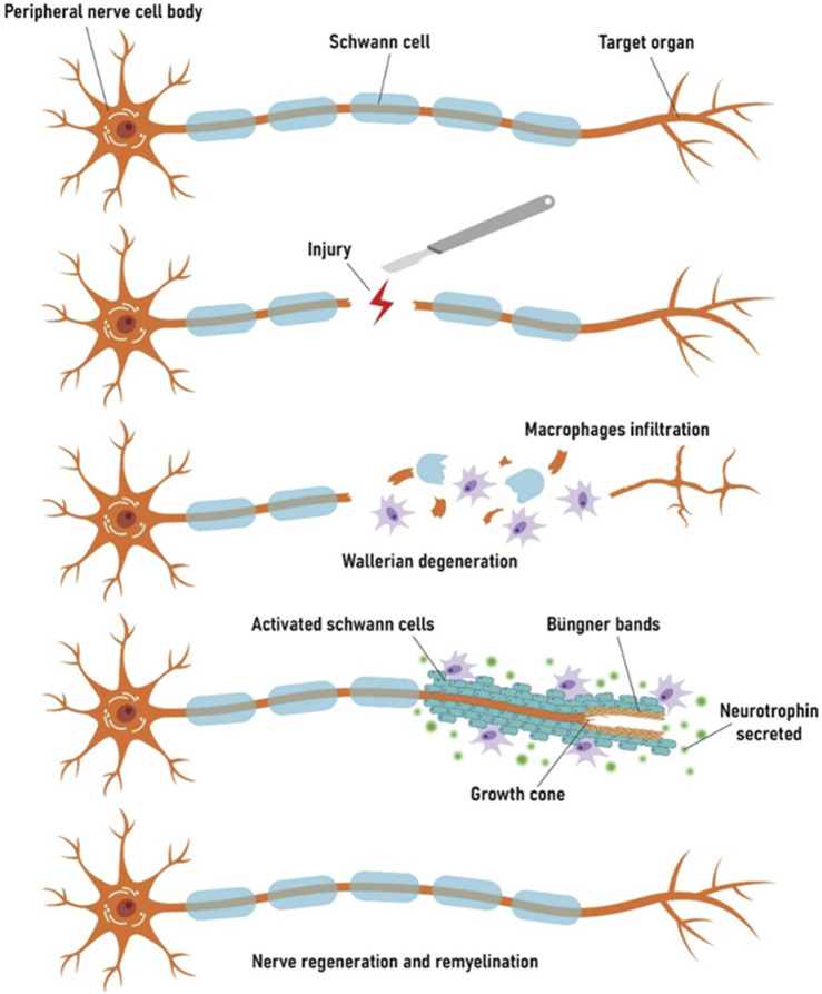
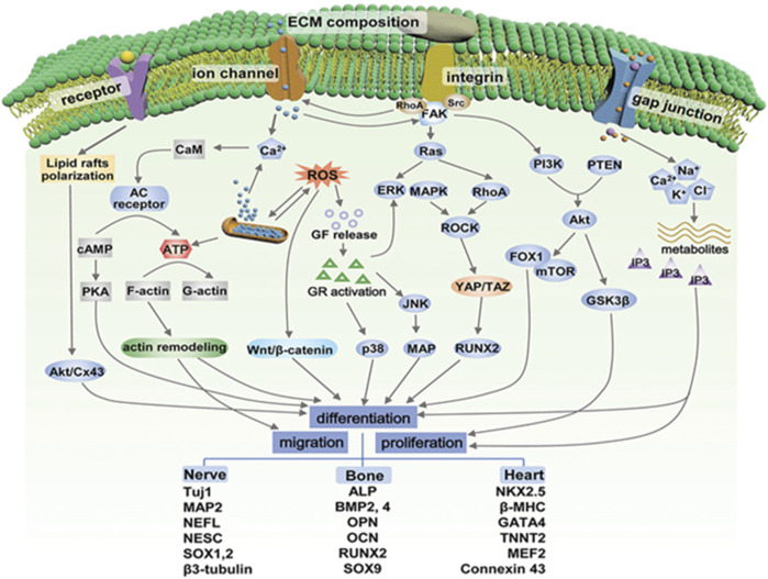
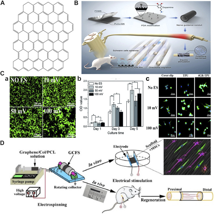
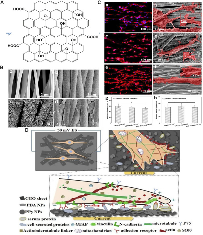
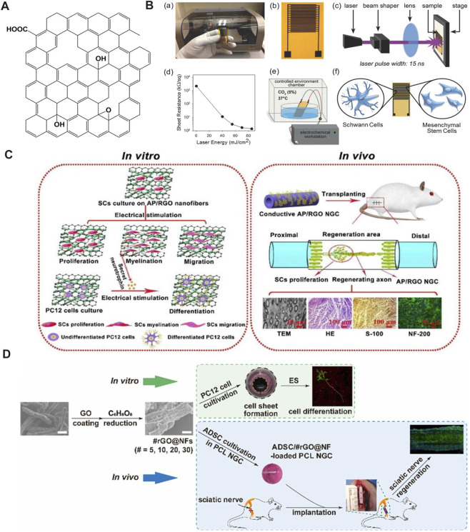
Peripheral nerve injury (PNI) is a common clinical problem, which due to poor recovery often leads to limb dysfunction and sensory abnormalities in patients. Tissue-engineered nerve guidance conduits (NGCs) that are designed and fabricated from different materials are the potential alternative to nerve autografts. However, translation of these NGCs from lab to commercial scale has not been well achieved. Complete functional recovery with the aid of NGCs in PNI becomes a topic of general interest in tissue engineering and regeneration medicine. Electrical stimulation (ES) has been widely used for many years as an effective physical method to promote nerve repair in both pre-clinical and clinical settings. Similarly, ES of conductive and electroactive materials with a broad range of electrical properties has been shown to facilitate the guidance of axons and enhance the regeneration. Graphene and its derivatives possess unique physicochemical and biological properties, which make them a promising outlook for the development of synthetic scaffolds or NGCs for PNI repair, especially in combination with ES. Considering the discussion regarding ES for the treatment of PNI must continue into further detail, herein, we focus on the role of ES in PNI repair and the molecular mechanism behind the ES therapy for PNI, providing a summary of recent advances in context of graphene-based scaffolds (GBSs) in combination with ES. Future perspectives and some challenges faced in developing GBSs are also highlighted with the aim of promoting their clinical applications.
Keywords: electrical stimulation; graphene-based scaffolds; nerve regeneration; neural tissue engineering; peripheral nerve injury.
Copyright © 2024 Zhao, Liu, Kang, Sun, Liu, Wang and Lu.
Conflict of interest statement
The authors declare that the research was conducted in the absence of any commercial or financial relationships that could be construed as a potential conflict of interest.
Figures
Similar articles
-
Reduced Graphene Oxide Fibers Combined with Electrical Stimulation Promote Peripheral Nerve Regeneration.Int J Nanomedicine. 2024 Mar 7;19:2341-2357. doi: 10.2147/IJN.S449160. eCollection 2024. Int J Nanomedicine. 2024. PMID: 38469057 Free PMC article.
-
From innovation to clinic: Emerging strategies harnessing electrically conductive polymers to enhance electrically stimulated peripheral nerve repair.Mater Today Bio. 2024 Dec 19;30:101415. doi: 10.1016/j.mtbio.2024.101415. eCollection 2025 Feb. Mater Today Bio. 2024. PMID: 39816667 Free PMC article.
-
In vitro and in vivo studies of electroactive reduced graphene oxide-modified nanofiber scaffolds for peripheral nerve regeneration.Acta Biomater. 2019 Jan 15;84:98-113. doi: 10.1016/j.actbio.2018.11.032. Epub 2018 Nov 22. Acta Biomater. 2019. PMID: 30471474
-
Nerve guide conduits for peripheral nerve injury repair: A review on design, materials and fabrication methods.Acta Biomater. 2020 Apr 1;106:54-69. doi: 10.1016/j.actbio.2020.02.003. Epub 2020 Feb 8. Acta Biomater. 2020. PMID: 32044456 Review.
-
Advances in nerve guidance conduits for peripheral nerve repair and regeneration.Am J Stem Cells. 2023 Dec 15;12(5):112-123. eCollection 2023. Am J Stem Cells. 2023. PMID: 38213640 Free PMC article. Review.
Cited by
-
Advanced surface modification techniques for titanium implants: a review of osteogenic and antibacterial strategies.Front Bioeng Biotechnol. 2025 Mar 19;13:1549439. doi: 10.3389/fbioe.2025.1549439. eCollection 2025. Front Bioeng Biotechnol. 2025. PMID: 40177619 Free PMC article. Review.
-
Neuromorphic algorithms for brain implants: a review.Front Neurosci. 2025 Apr 11;19:1570104. doi: 10.3389/fnins.2025.1570104. eCollection 2025. Front Neurosci. 2025. PMID: 40292025 Free PMC article. Review.
-
Comparison of Two Synthesis Methods for 3D PLA-Ibuprofen Nanofibrillar Scaffolds.Pharmaceutics. 2025 Jan 14;17(1):106. doi: 10.3390/pharmaceutics17010106. Pharmaceutics. 2025. PMID: 39861754 Free PMC article.
-
The stability and self-assembly of tri-calcium silicate and hydroxyapatite scaffolds in bone tissue engineering applications.J Biol Eng. 2025 Feb 17;19(1):16. doi: 10.1186/s13036-025-00481-4. J Biol Eng. 2025. PMID: 39962588 Free PMC article.
-
Biomaterial-Based Additive Manufactured Composite/Scaffolds for Tissue Engineering and Regenerative Medicine: A Comprehensive Review.Polymers (Basel). 2025 Apr 17;17(8):1090. doi: 10.3390/polym17081090. Polymers (Basel). 2025. PMID: 40284355 Free PMC article. Review.
References
-
- Amani H., Mostafavi E., Arzaghi H., Davaran S., Akbarzadeh A., Akhavan O., et al. (2019). Three-dimensional graphene foams: synthesis, properties, biocompatibility, biodegradability, and applications in tissue engineering. Acs Biomaterials Sci. Eng. 5, 193–214. 10.1021/acsbiomaterials.8b00658 - DOI - PubMed
-
- Aznar-Cervantes S., Pagan A., Martinez J. G., Bernabeu-Esclapez A., Otero T. F., Meseguer-Olmo L., et al. (2017). Electrospun silk fibroin scaffolds coated with reduced graphene promote neurite outgrowth of PC-12 cells under electrical stimulation. Mater. Sci. Eng. C-Materials Biol. Appl. 79, 315–325. 10.1016/j.msec.2017.05.055 - DOI - PubMed
-
- Bai R. G., Ninan N., Muthoosamy K., Manickam S. (2018). Graphene: a versatile platform for nanotheranostics and tissue engineering. Prog. Mater. Sci. 91, 24–69. 10.1016/j.pmatsci.2017.08.004 - DOI
Publication types
LinkOut - more resources
Full Text Sources
Miscellaneous