. 2024 Feb 16:15:1355945.
doi: 10.3389/fimmu.2024.1355945. eCollection 2024.

Sudocetaxel Zendusortide (TH1902) triggers the cGAS/STING pathway and potentiates anti-PD-L1 immune-mediated tumor cell killing

Affiliations

Sudocetaxel Zendusortide (TH1902) triggers the cGAS/STING pathway and potentiates anti-PD-L1 immune-mediated tumor cell killing

Michel Demeule et al. Front Immunol. .

Abstract

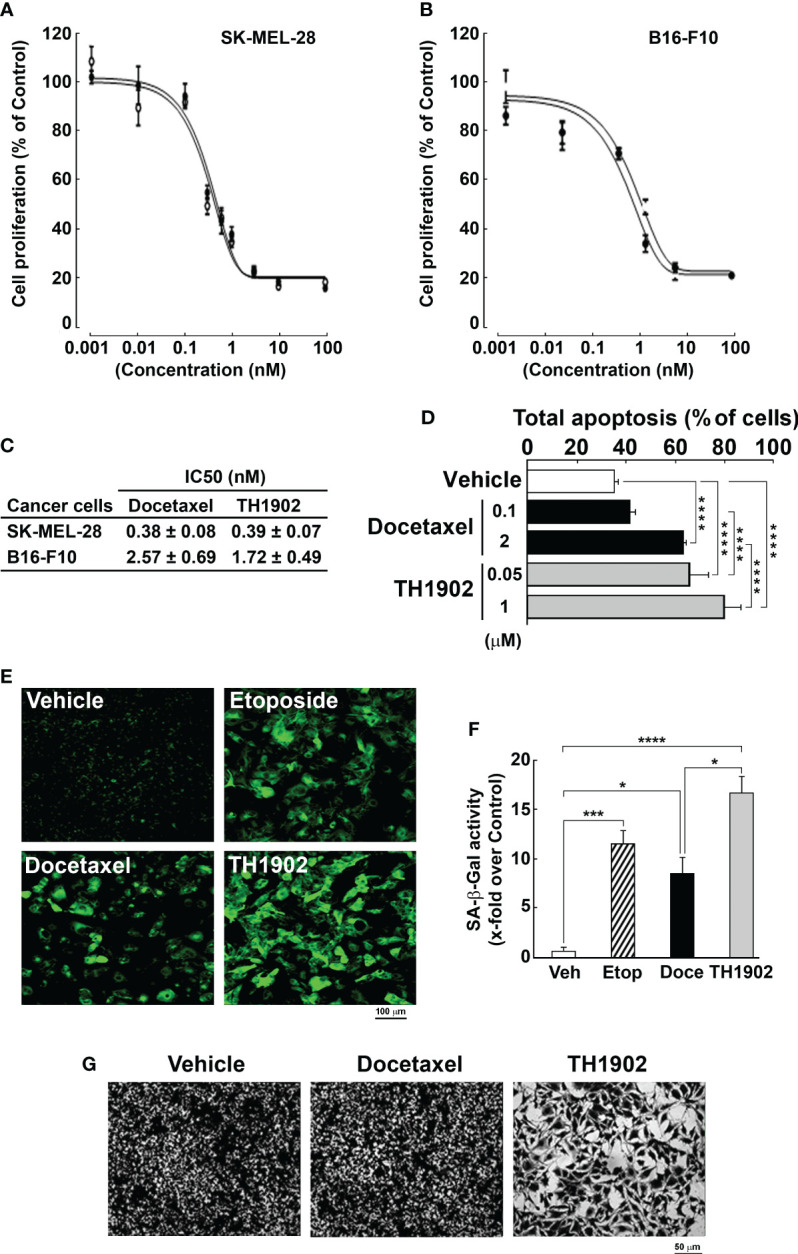
The anticancer efficacy of Sudocetaxel Zendusortide (TH1902), a peptide-drug conjugate internalized through a sortilin-mediated process, was assessed in a triple-negative breast cancer-derived MDA-MB-231 immunocompromised xenograft tumor model where complete tumor regression was observed for more than 40 days after the last treatment. Surprisingly, immunohistochemistry analysis revealed high staining of STING, a master regulator in the cancer-immunity cycle. A weekly administration of TH1902 as a single agent in a murine B16-F10 melanoma syngeneic tumor model demonstrated superior tumor growth inhibition than did docetaxel. A net increase in CD45 leukocyte infiltration within TH1902-treated tumors, especially for tumor-infiltrating lymphocytes and tumor-associated macrophages was observed. Increased staining of perforin, granzyme B, and caspase-3 was suggestive of elevated cytotoxic T and natural killer cell activities. Combined TH1902/anti-PD-L1 treatment led to increases in tumor growth inhibition and median animal survival. TH1902 inhibited cell proliferation and triggered apoptosis and senescence in B16-F10 cells in vitro, while inducing several downstream effectors of the cGAS/STING pathway and the expression of MHC-I and PD-L1. This is the first evidence that TH1902 exerts its antitumor activity, in part, through modulation of the immune tumor microenvironment and that the combination of TH1902 with checkpoint inhibitors (anti-PD-L1) could lead to improved clinical outcomes.

Keywords: PD-L1; STING; checkpoint inhibitor; docetaxel; immune tumor microenvironment; peptide-drug conjugate; sortilin.


Conflict of interest statement

This study received funding from Theratechnologies. The funder had the following involvement with the study : MD, JC-C, CC, AZ, RB, CM, and BA are listed as inventors on patent applications and were involved in study design, data collection and analysis, decision to publish, and preparation of the manuscript. B.A. received research support from Theratechnologies. All authors declare no other competing interests.

Figures

Figure 1
Figure 1
Sustained and prolonged antitumor activity of TH1902 in an immunosuppressed MDA-MB-231 TNBC-derived xenograft model. An in vivo MDA-MB-231 TNBC xenograft model was generated as previously described (5) in immunocompromised nude mice. Mice treatments were performed with intravenous injections of either docetaxel at the MTD of 15 mg/kg/wk, or with TH1902 at 35 mg/kg/wk and halted four days after either (i) three cycles, or for TH1902 after (ii) six cycles. An additional group was treated with TH1902 for (iii) six cycles on followed by three cycles off. (A) Tumor growth was monitored at the indicated days as described in the Methods section. Data are represented as mean ± SEM (three mice/group). All tumors were analyzed by IHC and representative sections of the tumors which were (B) H&E stained, or (C) Stained for SORT1, Ki67, STING, and CD45 as described in the Methods section are shown. (D) The quantity of CD45-stained area was quantified (* p < 0.05, ** p < 0.001). (E) Vasculogenic mimicry was assessed by monitoring CD31-/PAS+ staining (red arrows), whereas normal vasculature was accounted for by CD31+/PAS+ staining (yellow arrows).
Figure 2
Figure 2
Increased SORT1 expression in melanoma tissues and cell line models. (A) Tissue microarrays of healthy tissues and of clinically annotated melanomas from stages II, III, and IV were assessed for SORT1 expression by immunohistochemistry. (B) IHS scoring was performed as described in the Methods section (Normal n=2; Stage II-IV n=10) (** p < 0.01). (C) Cell lysates (20 μg) from different cancer cell lines were assessed for their SORT1 expression levels by Western blot analysis. B16-F10, murine B16-F10 melanoma cells; SKMEL, human SK-MEL-28 melanoma cells; A375, human A375 melanoma cells; MDA, human TNBC-derived MDA-MB-231 cells. GAPDH served as a loading control.
Figure 3
Figure 3
TH1902 exerts in vitro anti-proliferative and apoptotic activities and induces senescence in SORT1-positive melanoma cells. Cell proliferation in response to docetaxel or TH1902 was assessed in (A) SK-MEL-28 and (B) B16-F10 melanoma cells as described in the Methods section, and (C) IC50 values extracted for each test article. (D) Cell apoptosis was assessed in B16-F10 melanoma cells following docetaxel (black bars) and TH1902 (grey bars) treatments at the indicated concentrations. (E) Fluorescence microscopy was used to assess cellular senescence in B16-F10 melanoma cells following docetaxel, TH1902, or etoposide (as a positive control for senescence induction) treatment and representative pictures are shown. (F) Senescence-associated β-galactosidase activity was quantified as described in the Methods section (n=3). (G) Changes in cell morphology were assessed using crystal violet staining of cells upon vehicle (DMSO), 100 nM docetaxel, or 50 nM TH1902 treatment (equivalent docetaxel content) (* p < 0.05, *** p < 0.001, **** p < 0.0001).
Figure 4
Figure 4
Infiltration of leukocytes within B16-F10 tumors treated with vehicle, docetaxel, or TH1902. (A) Tumor growth in syngeneic mice treated with vehicle, 15 mg/kg/wk docetaxel (MTD), or 35 mg/kg/wk TH1902 (equivalent docetaxel content). Data are represented as mean ± SEM (9 mice/group for vehicle and docetaxel, 10 mice/group for TH1902). (B) B16-F10 melanoma tumors were excised, and pictures taken. (C) Excised tumors were fixed with formalin and processed for immunohistochemistry analysis. The top layer of images was stained with haematoxylin and eosin; the bottom layer was assessed immunohistochemically using a monoclonal antibody against CD45 (pan-immune cells). Representative images of whole and magnified tumors are shown for both stainings (black scale bar = 2 mm, white scale bar = 100 µm). (D) The quantities of CD45 stained area were compared for the three groups of mice by one-way ANOVA, followed by Tukey’s multiple comparisons test. The area staining positive for leukocytes was significantly greater in TH1902-treated animals than in animals treated with vehicle or docetaxel. Data are represented as mean ± SEM (* p <0.05, n = 4 tumors analyzed per group).
Figure 5
Figure 5
Effects of docetaxel and TH1902 on the levels of tumor-infiltrating lymphocytes within syngeneic tumors. (A) Each row of photomicrographs depicts cells from B16-F10 tumors in animals treated with either vehicle, docetaxel, or TH1902 (same samples as Figure 4 ). Each column includes representative IHC images of immune cell markers CD3 (T-cells), CD8 (cytotoxic T-cells), CD4 (helper T-cells), FoxP3 (regulatory T-cells), and CD161c (NK cells) performed on paraffin sections from primary tumors (white scale bar = 100 µm). (B) Quantification of IHC stainings. Expression was compared using one-way ANOVA with Tukey’s multiple comparisons test. Data are represented as mean ± SEM (* p <0.05, ** p <0.01, n = 4 tumors analyzed per group).
Figure 6
Figure 6
Effects of docetaxel and TH1902 on the infiltration levels of tissue-associated macrophages within syngeneic tumors. (A) Each row of photomicrographs depicts cells from B16-F10 tumors in animals treated with either vehicle, docetaxel, or TH1902 (same samples as in Figure 4 ). Each column includes representative IHC images of immune cell markers F4/80 (mature macrophage), CD68 (M1 macrophage), CD206 (M2 macrophage), performed on paraffin sections from primary tumors (white scale bar = 100 µm). (B) Quantification of IHC stainings. Expression was compared using one-way ANOVA with Tukey’s multiple comparisons test. Data are represented as mean ± SEM (** p <0.01, *** p <0.001, n = 4 tumors analysed per group).
Figure 7
Figure 7
Effects of docetaxel and TH1902 on expression of tumor markers for immune-stimulated apoptosis. (A) Each row of photomicrographs depicts cells from B16-F10 tumors in animals treated with either vehicle, docetaxel, or TH1902 (same samples as Figure 4 ). Each column includes representative IHC images of markers involved in immune-stimulated apoptosis cleaved caspase-3, perforin, and granzyme B (white scale bar = 100 µm). (B) Quantification of IHC stainings. Expression was compared using one-way ANOVA with Tukey’s multiple comparisons test. Data are represented as mean ± SEM (* p <0.05, ** p <0.01, n = 4 tumors analysed per group).
Figure 8
Figure 8
Effects on tumor growth and mice survival of TH1902 in combination with a checkpoint inhibitor on tumor growth and mice survival. B16-F10 cells were subcutaneously implanted in immunocompetent C57BL/6 mice. (A) Effect on B16-F10 tumor growth of docetaxel, TH1902, and anti-PD-L1 alone or in combination. Mice were treated weekly via intravenous administration of either vehicle, docetaxel (7.5 mg/kg), or TH1902 (17.5 mg/kg; equivalent dose of docetaxel) or bi-weekly via intraperitoneal administration of either anti-PD-L1 (9 mg/kg) and control isotype antibody (9 mg/kg) alone or in combination (docetaxel/anti-PD-L1 or TH1902/anti-PD-L1) for two cycles of treatment. Data are represented as mean ± SEM (n=8 mice/group). (B) Mice were treated weekly via intravenous administration with either vehicle and TH1902 (4.37, 8.75, 17.5 mg/kg) or bi-weekly via intraperitoneal administration of either anti-PD-L1 (9 mg/kg) and control isotype antibody (9 mg/kg) alone or in combination for continuous cycles of treatment until one of the defined endpoints was reached as described in the Methods section. Tumor growth curves were plotted until one individual within a given group reached the tumor size endpoint (tumor >2,000 mm3). Data are represented as mean ± SEM (n=6 mice/group). (C) Effect on survival of increased doses of TH1902 and anti-PD-L1 alone or in combination in B16-F10 tumor-bearing mice. Kaplan-Meier curves were plotted to estimate mice survival and presented as probability of survival in percentage.
Figure 9
Figure 9
TH1902 induces downstream effectors of the STING pathway and the cell surface expression of PD-L1 and MHC-I in B16-F10 melanoma cells. Cells were treated with increasing docetaxel or TH1902 concentrations for 5 minutes, followed by 96 hours of incubation in fresh complete medium. Cell lysates were harvested as described in the Methods section and (A) immunoblotting was performed with anti-STING and anti-GAPDH antibodies, and (B) protein expression was quantified using densitometry. (C) Lysates were further processed for the indicated expression of STING downstream effectors at 100 nM docetaxel or TH1902, and (D) densitometric quantification was performed. (E) Total RNA was extracted and gene expression of IL-6 and TNFα assessed using RT-qPCR in cells treated or not with 100 nM docetaxel or TH1902 for 5 minutes, followed by 24 hours of incubation in fresh complete medium. (F) Immunophenotyping of MHC-I and PD-L1 cell surface expression was performed by flow cytometry as described in the Methods section. Data are represented as mean ± SEM (n=4) (* p < 0.05, ** p < 0.01, *** p < 0.001).
Figure 10
Figure 10
TNFα, but not IL-6, triggers PD-L1 and MHC-1 gene expression in B16-F10 melanoma cells. Cells were treated with the indicated TNFα or IL-6 concentrations for 96 hours. Levels of PD-L1 and MHC-I expression were estimated by flow cytometry as described in the Methods section. PD-L1 and MHC-I cell surface expression upon (A) TNFα treatment, or upon (B) IL-6 treatment. Data are represented as means ± SEM (n = 3). (** p < 0.01, *** p < 0.001).

Similar articles

Cited by

References

    1. Mansoori B, Mohammadi A, Davudian S, Shirjang S, Baradaran B. The different mechanisms of cancer drug resistance: a brief review. Adv Pharm Bull (2017) 7:339–48. doi: 10.15171/apb.2017.041 - DOI - PMC - PubMed
    1. Heh E, Allen J, Ramirez F, Lovasz D, Fernandez L, Hogg T, et al. . Peptide drug conjugates and their role in cancer therapy. Int J Mol Sci (2023) 24:829. doi: 10.3390/ijms24010829 - DOI - PMC - PubMed
    1. Marcusson EG, Horazdovsky BF, Cereghino JL, Gharakhanian E, Emr SD. The sorting receptor for yeast vacuolar carboxypeptidase Y is encoded by the VPS10 gene. Cell (1994) 77:579–86. doi: 10.1016/0092-8674(94)90219-4 - DOI - PubMed
    1. Charfi C, Demeule M, Currie JC, Larocque A, Zgheib A, Danalache BA, et al. . New peptide-drug conjugates for precise targeting of SORT1-mediated vasculogenic mimicry in the tumor microenvironment of TNBC-derived MDA-MB-231 breast and ovarian ES-2 clear cell carcinoma cells. Front Oncol (2021) 11:760787. doi: 10.3389/fonc.2021.760787 - DOI - PMC - PubMed
    1. Demeule M, Charfi C, Currie JC, Larocque A, Zgheib A, Kozelko S, et al. . TH1902, a new docetaxel-peptide conjugate for the treatment of sortilin-positive triple-negative breast cancer. Cancer Sci (2021) 112:4317–34. doi: 10.1111/cas.15086 - DOI - PMC - PubMed

Publication types