

Longitudinal observations of the effects of ischemic stroke on binaural perception
- PMID: 38482140
- PMCID: PMC10936579
- DOI: 10.3389/fnins.2024.1322762
Longitudinal observations of the effects of ischemic stroke on binaural perception
Abstract
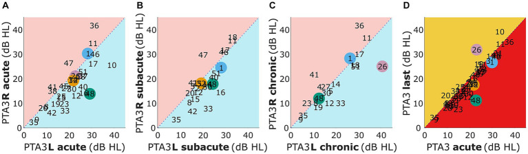
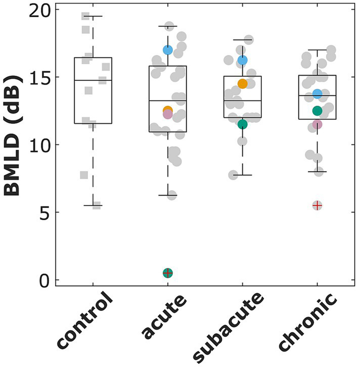
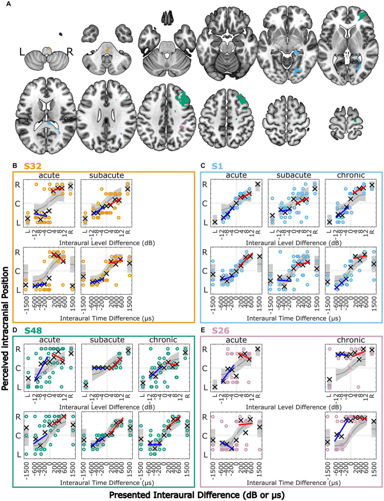
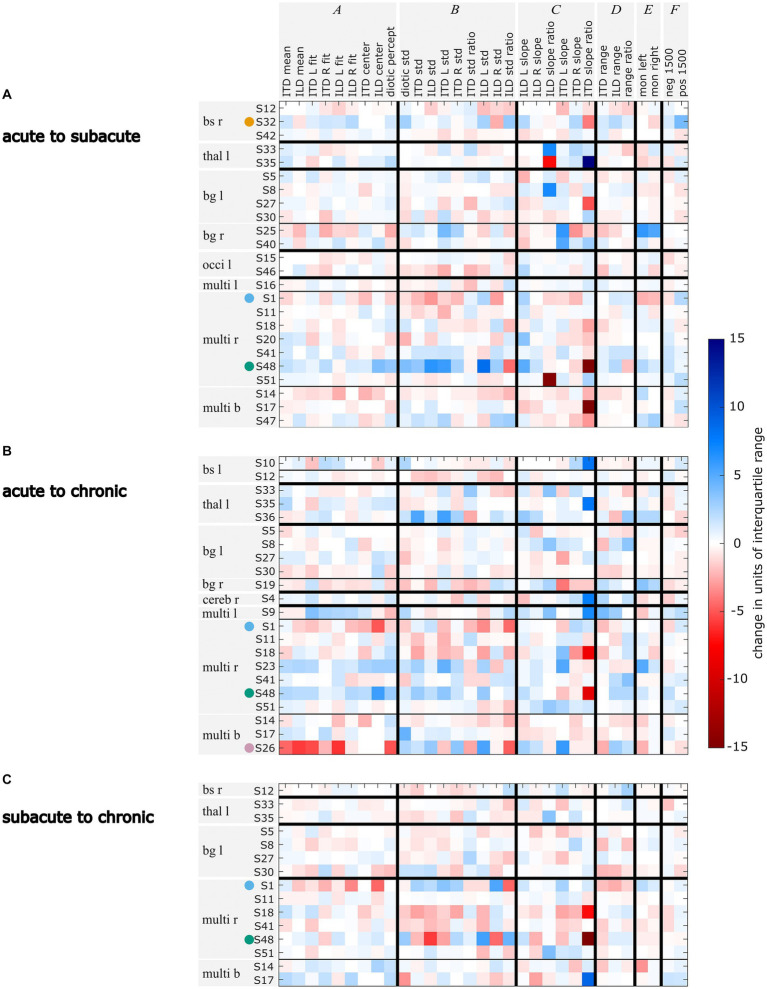
Acute ischemic stroke, characterized by a localized reduction in blood flow to specific areas of the brain, has been shown to affect binaural auditory perception. In a previous study conducted during the acute phase of ischemic stroke, two tasks of binaural hearing were performed: binaural tone-in-noise detection, and lateralization of stimuli with interaural time- or level differences. Various lesion-specific, as well as individual, differences in binaural performance between patients in the acute phase of stroke and a control group were demonstrated. For the current study, we re-invited the same group of patients, whereupon a subgroup repeated the experiments during the subacute and chronic phases of stroke. Similar to the initial study, this subgroup consisted of patients with lesions in different locations, including cortical and subcortical areas. At the group level, the results from the tone-in-noise detection experiment remained consistent across the three measurement phases, as did the number of deviations from normal performance in the lateralization task. However, the performance in the lateralization task exhibited variations over time among individual patients. Some patients demonstrated improvements in their lateralization abilities, indicating recovery, whereas others' lateralization performance deteriorated during the later stages of stroke. Notably, our analyses did not reveal consistent patterns for patients with similar lesion locations. These findings suggest that recovery processes are more individual than the acute effects of stroke on binaural perception. Individual impairments in binaural hearing abilities after the acute phase of ischemic stroke have been demonstrated and should therefore also be targeted in rehabilitation programs.
Keywords: binaural hearing; binaural masking level difference; brain lesions; lateralization; magnetic resonance imaging; psychoacoustics; stroke.
Copyright © 2024 Dietze, Sörös, Pöntynen, Witt and Dietz.
Conflict of interest statement
The authors declare that the research was conducted in the absence of any commercial or financial relationships that could be construed as a potential conflict of interest.
Figures

Similar articles
-
Effects of acute ischemic stroke on binaural perception.Front Neurosci. 2022 Dec 22;16:1022354. doi: 10.3389/fnins.2022.1022354. eCollection 2022. Front Neurosci. 2022. PMID: 36620448 Free PMC article.
-
Effects of reference interaural time and intensity differences on binaural performance in listeners with normal and impaired hearing.Ear Hear. 1995 Aug;16(4):331-53. doi: 10.1097/00003446-199508000-00001. Ear Hear. 1995. PMID: 8549890
-
Binaural sensitivity to temporal fine structure and lateralization ability in children with suspected (central) auditory processing disorder.Auris Nasus Larynx. 2019 Feb;46(1):64-69. doi: 10.1016/j.anl.2018.06.005. Epub 2018 Jun 25. Auris Nasus Larynx. 2019. PMID: 29954636
-
Binaural frequency selectivity in humans.Eur J Neurosci. 2020 Mar;51(5):1179-1190. doi: 10.1111/ejn.13837. Epub 2018 Feb 13. Eur J Neurosci. 2020. PMID: 29359360 Review.
-
Masking level difference: a measure of auditory processing capability.Audiology. 1975;14(4):354-67. doi: 10.3109/00206097509071749. Audiology. 1975. PMID: 1096867 Review.
References
-
- Beck A. T., Steer R. A., Brown G. K. (2013). Beck depressions-inventar – Fs (Bdi-Fs): Deutsche Bearbeitung. Frankfurt: Pearson Assessment & Information Gmbh.
LinkOut - more resources
Full Text Sources