Anatomical variations of the atlas arches: prevalence assessment, systematic review and proposition for an updated classification system
- PMID: 38482143
- PMCID: PMC10932953
- DOI: 10.3389/fnins.2024.1348066
Anatomical variations of the atlas arches: prevalence assessment, systematic review and proposition for an updated classification system
Erratum in
-
Corrigendum: Anatomical variations of the atlas arches: prevalence assessment, systematic review and proposition for an updated classification system.Front Neurosci. 2024 May 13;18:1418734. doi: 10.3389/fnins.2024.1418734. eCollection 2024. Front Neurosci. 2024. PMID: 38803688 Free PMC article.
Abstract
Objective and background: This study focuses on the atlas, a pivotal component of the craniovertebral junction, bridging the cranium and spinal column. Notably, variations in its arches are documented globally, necessitating a thorough assessment and categorization due to their significant implications in clinical, diagnostic, functional, and therapeutic contexts. The primary objective is to ascertain the frequency of these anatomical deviations in the atlas arches among a Colombian cohort using cone-beam computed tomography (CBCT).
Methodology: Employing a descriptive, cross-sectional approach, this research scrutinizes the structural intricacies of the atlas arches in CBCT scans. Analytical parameters included sex distribution and the nature of anatomical deviations as per Currarino's classification. Statistical analyses were conducted to identify significant differences, including descriptive statistics and Chi-square tests. A systematic review of the literature was conducted in order to enhance the current Currarino's classification.
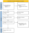
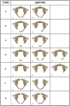
Results: The study examined 839 CBCT images, with a nearly equal sex distribution (49.7% female, 50.3% male). Anatomical variations were identified in 26 instances (3%), displaying a higher incidence in females (X2 [(1, N = 839) = 4.0933, p = 0.0430]). The most prevalent variation was Type A (2.5%), followed by Type B (0.4%), and Type G (0.2%) without documenting any other variation. The systematic review yielded 7 studies. A novel classification system for these variations is proposed, considering global prevalence data in the cervical region.
Conclusion: The study highlights a statistically significant predominance of Type A variations in the female subset. Given the critical nature of the craniovertebral junction and supporting evidence, it recommends an amendment to Currarino's classification to better reflect these clinical observations. A thorough study of anatomical variations of the upper cervical spine is relevant as they can impact important functional aspects such as mobility as well as stability. Considering the intricate anatomy of this area and the pivotal function of the atlas, accurately categorizing the variations of its arches is crucial for clinical practice. This classification aids in diagnosis, surgical planning, preventing iatrogenic incidents, and designing rehabilitation strategies.
Keywords: anatomical variation; atlas (C1 vertebra); cervical instability; cone-beam computed tomography (CBCT); congenital abnormalities; vertebral arch.
Copyright © 2024 Baena-Caldas, Mier-García, Griswold, Herrera-Rubio and Peckham.
Conflict of interest statement
The authors declare that the research was conducted in the absence of any commercial or financial relationships that could be construed as a potential conflict of interest.
Figures


Similar articles
-
Corrigendum: Anatomical variations of the atlas arches: prevalence assessment, systematic review and proposition for an updated classification system.Front Neurosci. 2024 May 13;18:1418734. doi: 10.3389/fnins.2024.1418734. eCollection 2024. Front Neurosci. 2024. PMID: 38803688 Free PMC article.
-
Cervical vertebral column morphology in patients with obstructive sleep apnoea assessed using lateral cephalograms and cone beam CT. A comparative study.Dentomaxillofac Radiol. 2013;42(6):20130060. doi: 10.1259/dmfr.20130060. Epub 2013 Mar 15. Dentomaxillofac Radiol. 2013. PMID: 23503808 Free PMC article.
-
Three-Dimensional Evaluation and Classification of the Anatomy Variations of Vertebral Artery at the Craniovertebral Junction in 120 Patients of Basilar Invagination and Atlas Occipitalization.Oper Neurosurg. 2019 Dec 1;17(6):594-602. doi: 10.1093/ons/opz076. Oper Neurosurg. 2019. PMID: 31127851
-
Fusions at the craniovertebral junction.Childs Nerv Syst. 2008 Oct;24(10):1209-24. doi: 10.1007/s00381-008-0607-7. Epub 2008 Apr 4. Childs Nerv Syst. 2008. PMID: 18389260 Review.
-
Pediatric cervical kyphosis in the MRI era (1984-2008) with long-term follow up: literature review.Childs Nerv Syst. 2022 Feb;38(2):361-377. doi: 10.1007/s00381-021-05409-z. Epub 2021 Nov 22. Childs Nerv Syst. 2022. PMID: 34806157 Review.
Cited by
-
Anatomical variability, morphofunctional characteristics, and clinical relevance of accessory ossicles of the back: implications for spinal pathophysiology and differential diagnosis.J Orthop Surg Res. 2025 Mar 6;20(1):240. doi: 10.1186/s13018-024-05407-2. J Orthop Surg Res. 2025. PMID: 40050963 Free PMC article. Review.
References
-
- Cattrysse E., Buzzatti L., Provyn S., Barbero M., Van Roy P. (2016). Variability of upper cervical anatomy: a reflection on its clinical relevance. J. Funct. Morphol. Kinesiol. 1, 126–139. doi: 10.3390/jfmk1010126 - DOI
-
- Chau A. M., Wong J. H., Mobbs R. J. (2009). Cervical myelopathy associated with congenital C2/3 canal stenosis and deficiencies of the posterior arch of the atlas and laminae of the axis: case report and review of the literature. Spine 34, E886–E891. doi: 10.1097/BRS.0b013e3181b64f0a, PMID: - DOI - PubMed
LinkOut - more resources
Full Text Sources

