. 2024 Feb 28:11:1338563.
doi: 10.3389/fvets.2024.1338563. eCollection 2024.

Comparative pathogenicity of CA1737/04 and Mass infectious bronchitis virus genotypes in laying chickens

Affiliations

Comparative pathogenicity of CA1737/04 and Mass infectious bronchitis virus genotypes in laying chickens

Ahmed Ali et al. Front Vet Sci. .

Abstract

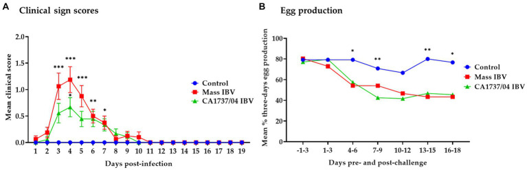
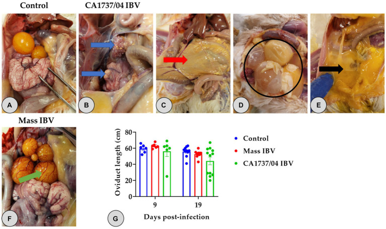
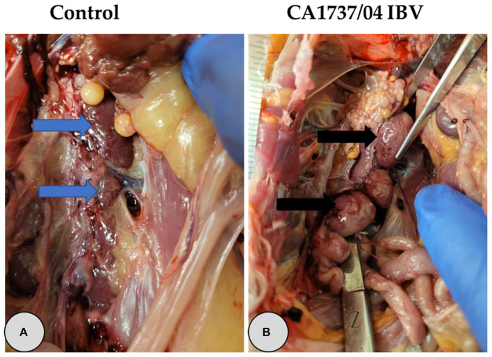
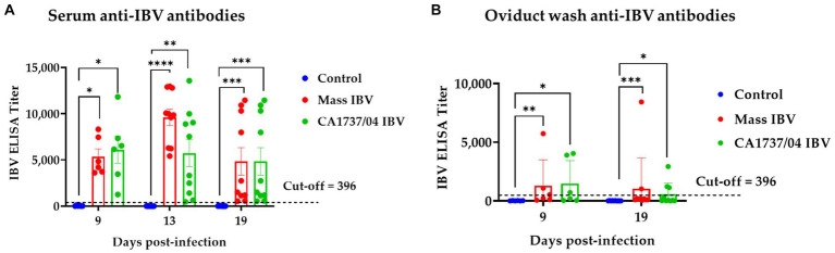
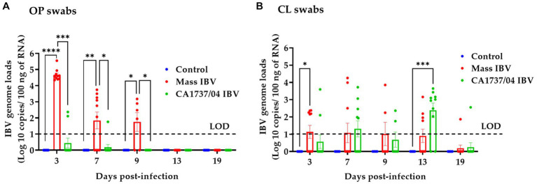
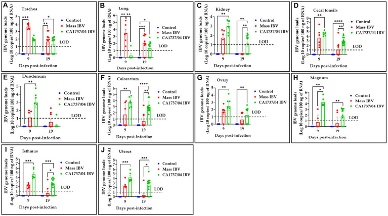
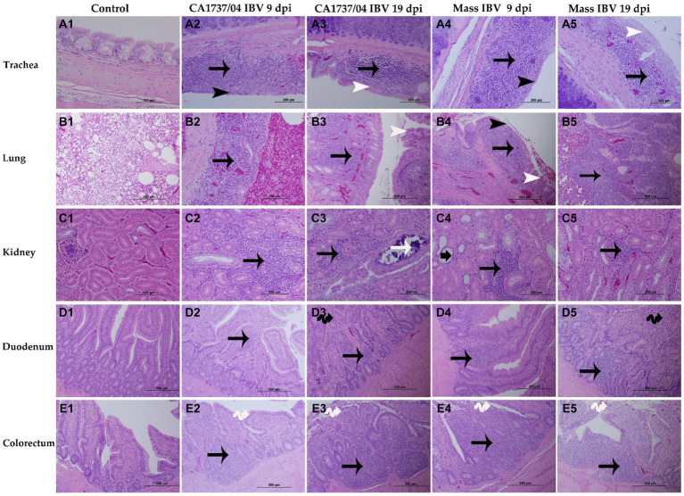
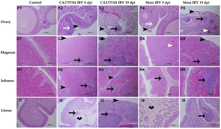
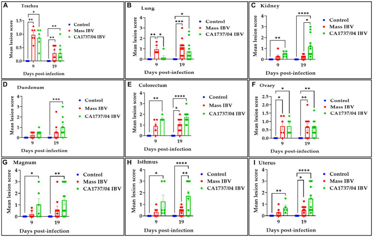
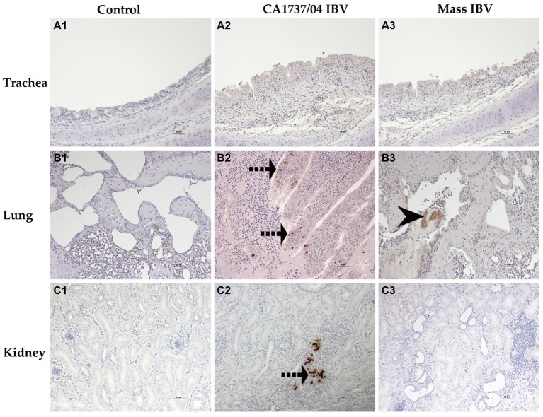
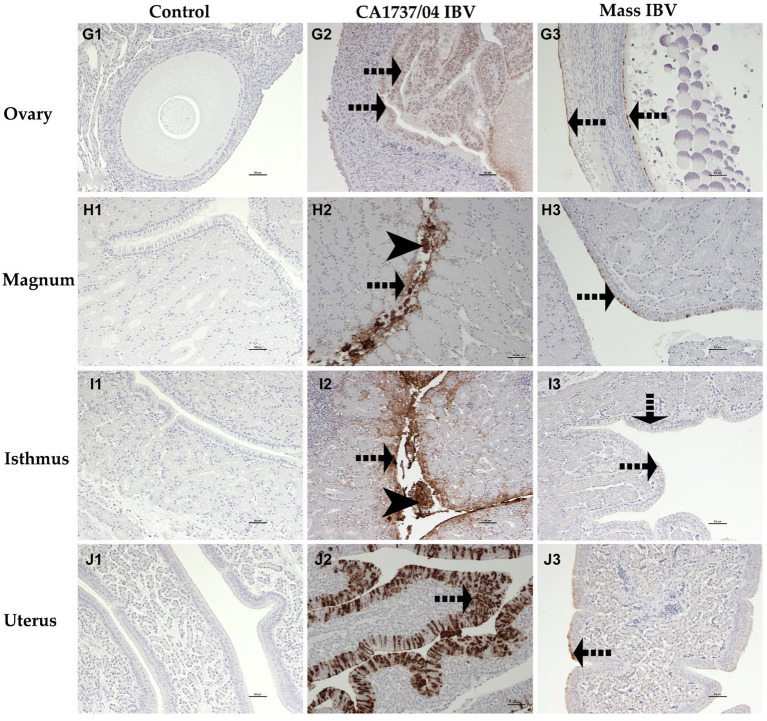
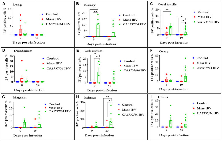
Infectious bronchitis virus (IBV) is a respiratory virus causing atropism in multiple body systems of chickens. Recently, the California 1737/04 (CA1737/04) IBV strain was identified as one of the circulating IBV variants among poultry operations in North America. Here, the pathogenicity and tissue tropism of CA1737/04 IBV strain in specific-pathogen-free (SPF) hens were characterized in comparison to Massachusetts (Mass) IBV. In 30 weeks-old SPF hens, Mass or CA1737/04 IBV infections were carried out, while the third group was maintained as a control group. Following infection, we evaluated clinical signs, egg production, viral shedding, serology, necropsy examination, and histopathology during a period of 19 days. Also, certain tissue affinity parameters were investigated, which involved the localization of viral antigens and the detection of viral RNA copies in designated tissues. Our findings indicate that infection with CA1737/04 or Mass IBV strain could induce significant clinical signs, reduced egg production, and anti-IBV antibodies locally in oviduct wash and systemically in serum. Both IBV strains showed detectable levels of viral RNA copies and induced pathology in respiratory, renal, enteric, and reproductive tissues. However, the CA1737/04 IBV strain had higher pathogenicity, higher tissue tropism, and higher replication in the kidney, large intestine, and different segments of the oviduct compared to the Mass IBV strain. Both IBV strains shed viral genome from the cloacal route, however, the Mass IBV infected hens shed higher IBV genome loads via the oropharyngeal route compared to CA1737/04 IBV-infected hens. Overall, the current findings could contribute to a better understanding of CA1737/04 IBV pathogenicity in laying hens.

Keywords: Canada; hen; infectious bronchitis virus; pathogenicity; tissue tropism.


Conflict of interest statement

The authors declare that the research was conducted in the absence of any commercial or financial relationships that could be construed as a potential conflict of interest.

Figures

Figure 1
Figure 1
Daily mean clinical signs scores (A) and mean percentages of 3 days interval of egg production (B) following infection with Mass (15AB-01) and CA1737/04 IBV strains. The group differences in the mean clinical signs scores were identified by the Kruskal–Wallis test followed by Dunn’s multiple comparison test. Error bars represent the standard error of the mean (SEM). Fisher’s exact test was employed to compare the proportion of 3 days intervals of egg production. Asterisks indicate significant differences (*p < 0.05, **p < 0.01, and ***p < 0.001).
Figure 2
Figure 2
Reproductive gross lesions (A–F) and oviduct lengths (G) at 9 and 19 days following infection with Mass (15AB-01) and CA1737/04 IBV strains. (A) Refers to the control group without gross lesions. (B–E) Shows gross lesions observed in the CA1737/04 IBV-infected group; blue arrows reveal regressed ovary and oviduct; red arrow indicates yellowish peritoneum; black circle and arrow refer to eggs and ruptured yolk inside the abdominal cavity, respectively. (F) Shows congested ovarian follicles in the Mass IBV-infected group (green arrow). (G) Represents oviduct lengths means which were compared between the groups using the Kruskal–Wallis test followed by Dunn’s multiple comparison test. Error bars represent SEM.
Figure 3
Figure 3
Representative gross lesions observed in the kidney at 9 and 19 days following infection with CA1737/04 IBV strain. (A) Refers to the control group without gross lesions (indicated with blue arrows). (B) Reveals swollen kidney (shown by black arrows).
Figure 4
Figure 4
Anti-IBV antibodies in serum (A) and oviduct washes (B) collected at different time points following infection with Mass IBV (15AB-01) and CA1737/04 IBV strains. The mean titers were compared between the experimental groups using the Kruskal–Wallis test and Dunn’s multiple comparison test. Error bars represent SEM. Asterisks indicate significant differences (*p < 0.05, **p < 0.01, ***p < 0.001, and ****p < 0.0001).
Figure 5
Figure 5
IBV genome loads were quantified from OP (A) and CL (B) swabs collected at 3, 7, 9, 13, and 19 days following infection with Mass (15AB-01) and CA1737/04 IBV strains. The Kruskal–Wallis test followed by Dunn’s multiple comparison test was used to analyze the group differences. Error bars represent SEM. Asterisks indicate significant differences (*p < 0.05, **p < 0.01, ***p < 0.001, and ****p < 0.0001). The dotted lines indicate the limit of detection (LOD) of RT-qPCR assay.
Figure 6
Figure 6
IBV genome loads were determined in trachea (A), lung (B), kidney (C), cecal tonsils (D), duodenum (E), colorectum (F), ovary (G), magnum (H), isthmus (I), and uterus (J) collected at 9 and 19 days following infection with Mass (15AB-01) and CA1737/04 IBV strains. The group difference was identified using the Kruskal–Wallis test followed by Dunn’s multiple comparison test. Error bars represent SEM. Asterisks indicate significant differences (*p < 0.05, **p < 0.01, ***p < 0.001, and ****p < 0.0001). The dotted lines show the limit of detection (LOD) of RT-qPCR assay.
Figure 7
Figure 7
Microscopic changes observed in the trachea (A1–A5), lung (B1–B5), kidney (C1–C5), duodenum (D1–D5), and colorectum (E1–E5) at 9 and 19 days following infection with Mass (15AB-01) and CA1737/04 IBV strains. A1–E1 are controls. A2–E2 and A3–E3 show lesions in the CA1737/04 IBV-infected group at 9 and 19 dpi, respectively. While A4–E4 and A5–E5 refer to lesions in the Mass IBV-infected group at 9 and 19 dpi, respectively. Black arrows indicate inflammatory cell infiltrations; black head arrows show epithelial thinning; white head arrows refer to epithelial hyperplasia; white arrow shows intra-luminal dark blue deposits; short-thick black arrow refers to tubular dilatation; double black arrows indicate villus atrophy; double white arrows show necrosis of epithelial cells from intestinal villi.
Figure 8
Figure 8
Microscopic changes observed in the ovary (F1–F5), magnum (G1–G5), isthmus (H1–H5), and uterus (I1–I5) at 9 and 19 days following infection with CA1737/04 and Mass IBV strains. F1–I1 are controls. F2–I2 and F3–I3 represent lesions detected in the CA1737/04 IBV-infected group at 9 and 19 dpi, respectively. Whereas, F4–I4 and F5–I5 show lesions observed in the Mass group at 9 and 19 dpi, respectively. Black-headed arrows refer to epithelial and ciliary losses; white-headed arrows show congested blood capillaries; black arrows indicate mononuclear cell infiltrations; white arrows refer to heterophils; double black arrows show edema.
Figure 9
Figure 9
Histopathological lesions were scored in the trachea (A), lung (B), kidney (C), duodenum (D), colorectum (E), ovary (F), magnum (G), isthmus (H), and uterus (I) at 9 and 19 days following infection with Mass (15AB-01) and CA1737/04 IBV strains. The mean scores were compared among the groups using the Kruskal–Wallis test followed by Dunn’s multiple comparison test. Error bars represent SEM. Asterisks indicate significant differences (*p < 0.05, **p < 0.01, ***p < 0.001, and ****p < 0.0001).
Figure 10
Figure 10
Immunohistochemical detection of IBV antigen in trachea (A1–A3), lung (B1–B3), and kidney (C1–C3) collected at 9 and 19 dpi. A1–C1 are controls. A2,A3 represent no expression of IBV antigen in trachea of both IBV-infected groups. B2,C2 reveal expression of IBV antigen in the CA1737/04-IBV infected group. B3 shows expression of IBV antigen in the Mass IBV-infected group. Dotted black arrows and black head arrow indicate viral antigen immunolabeling in intact and sloughed epithelia, respectively. Scale bar = 50 μm.
Figure 11
Figure 11
Immunohistochemical detection of IBV antigen in cecal tonsils (D1–D3), duodenum (E1–E3), and colorectum (F1–F3) collected at 9 and 19 dpi. D1–F1 are controls. D2–F2 represents the expression of IBV antigen in the CA1737/04 IBV-infected group. D3,E3 show expression of IBV antigen in the Mass-infected group. The dotted black arrows refer to viral antigen immunolabeling in the epithelial cells. Scale bar = 50 μm.
Figure 12
Figure 12
Immunohistochemical detection of IBV antigen in the ovary (G1–G3), magnum (H1–H3), isthmus (I1–I3), and uterus (J1–J3) collected at 9 and 19 dpi. G1–J1 are controls. G2–J2 represent expression of IBV antigen in the CA1737/04 IBV-infected group. G3–J3 show expression of IBV antigen in the Mass IBV-infected group. Dotted black arrows and black head arrows indicate viral antigen immunolabeling in intact and sloughed epithelia, respectively. Scale bar = 50 μm.
Figure 13
Figure 13
Percentage of IBV-positive cells detected in the lung (A), kidney (B), cecal tonsils (C), duodenum (D), colorectum (E), ovary (F), magnum (G), isthmus (H), and uterus (I) at 9 and 19 days following infection with CA1737/04 and Mass IBV strains. The Kruskal–Wallis test followed by Dunn’s multiple comparison test was used to identify the group differences. Error bars represent SEM. Asterisks indicate significant differences (*p < 0.05 and **p < 0.01).

Similar articles

Cited by

References

    1. Cook JK, Jackwood M, Jones RC. The long view: 40 years of infectious bronchitis research. Avian Pathol. (2012) 41:239–50. doi: 10.1080/03079457.2012.680432, PMID: - DOI - PubMed
    1. Cavanagh D. Coronavirus avian infectious bronchitis virus. Vet Res. (2007) 38:281–97. doi: 10.1051/vetres:2006055 - DOI - PubMed
    1. Amarasinghe A, Popowich S, De Silva Senapathi U, Abdul-Cader MS, Marshall F, van der Meer F, et al. . Shell-less egg syndrome (SES) widespread in western Canadian layer operations is linked to a Massachusetts (Mass) type infectious bronchitis virus (IBV) isolate. Viruses. (2018) 10:437. doi: 10.3390/v10080437, PMID: - DOI - PMC - PubMed
    1. Reddy VR, Trus I, Desmarets LM, Li Y, Theuns S, Nauwynck HJ. Productive replication of nephropathogenic infectious bronchitis virus in peripheral blood monocytic cells, a strategy for viral dissemination and kidney infection in chickens. Vet Res. (2016) 47:70. doi: 10.1186/s13567-016-0354-9, PMID: - DOI - PMC - PubMed
    1. Lin SY, Chen HW. Infectious bronchitis virus variants: molecular analysis and pathogenicity investigation. Int J Mol Sci. (2017) 18:2030. doi: 10.3390/ijms18102030 - DOI - PMC - PubMed