. 2024 May 1;326(5):F704-F726.
doi: 10.1152/ajprenal.00259.2023. Epub 2024 Mar 14.

Mice with a Pax2 missense variant display impaired glomerular repair

Affiliations

Mice with a Pax2 missense variant display impaired glomerular repair

Joanna Cunanan et al. Am J Physiol Renal Physiol. .

Abstract

PAX2 regulates kidney development, and its expression persists in parietal epithelial cells (PECs), potentially serving as a podocyte reserve. We hypothesized that mice with a Pax2 pathogenic missense variant (Pax2A220G/+) have impaired PEC-mediated podocyte regeneration. Embryonic wild-type mouse kidneys showed overlapping expression of PAX2/Wilms' tumor-1 (WT-1) until PEC and podocyte differentiation, reflecting a close lineage relationship. Embryonic and adult Pax2A220G/+ mice have reduced nephron number but demonstrated no glomerular disease under baseline conditions. Pax2A220G/+ mice compared with wild-type mice were more susceptible to glomerular disease after adriamycin (ADR)-induced podocyte injury, as demonstrated by worsened glomerular scarring, increased podocyte foot process effacement, and podocyte loss. There was a decrease in PAX2-expressing PECs in wild-type mice after adriamycin injury accompanied by the occurrence of PAX2/WT-1-coexpressing glomerular tuft cells. In contrast, Pax2A220G/+ mice showed no changes in the numbers of PAX2-expressing PECs after adriamycin injury, associated with fewer PAX2/WT-1-coexpressing glomerular tuft cells compared with injured wild-type mice. A subset of PAX2-expressing glomerular tuft cells after adriamycin injury was increased in Pax2A220G/+ mice, suggesting a pathological process given the worse outcomes observed in this group. Finally, Pax2A220G/+ mice have increased numbers of glomerular tuft cells expressing Ki-67 and cleaved caspase-3 compared with wild-type mice after adriamycin injury, consistent with maladaptive responses to podocyte loss. Collectively, our results suggest that decreased glomerular numbers in Pax2A220G/+ mice are likely compounded with the inability of their mutated PECs to regenerate podocyte loss, and together these two mechanisms drive the worsened focal segmental glomerular sclerosis phenotype in these mice.NEW & NOTEWORTHY Congenital anomalies of the kidney and urinary tract comprise some of the leading causes of kidney failure in children, but our previous study showed that one of its genetic causes, PAX2, is also associated with adult-onset focal segmental glomerular sclerosis. Using a clinically relevant model, our present study demonstrated that after podocyte injury, parietal epithelial cells expressing PAX2 are deployed into the glomerular tuft to assist in repair in wild-type mice, but this mechanism is impaired in Pax2A220G/+ mice.

Keywords: PAX2; glomerular repair; parietal epithelial cells; podocyte; regeneration.


Conflict of interest statement

No conflicts of interest, financial or otherwise, are declared by the authors.

Figures

None
Graphical abstract
Figure 1.
Figure 1.
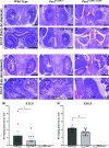
Embryonic mice with the c.A220G variant in Pax2 had congenital anomalies of the kidney and urinary tract (CAKUT) phenotypes characterized by decreased glomerular numbers. A–L: representative images of hematoxylin and eosin-stained kidney sections from embryonic day (E)13.5 and E16.5 wild-type, Pax2A220G/+, and Pax2A220G/A220G embryos. Wild-type E13.5 (A and B) and E16.5 (C and D) embryonic kidneys demonstrated normal development of early nephrogenic structures and formation of maturing glomeruli (dashed circles). Pax2A220G/+ E13.5 (E and F) and E16.5 (G and H) embryonic kidneys showed decreased numbers of early nephrogenic structures and maturing glomeruli (dashed circles) compared with wild-type kidneys. The majority (90.9%) of E13.5 Pax2A220G/+ embryonic kidneys showed renal hypoplasia (congenitally smaller kidneys with decreased glomerular numbers) (E), whereas few (9.1%) of E13.5 Pax2A220G/+ embryonic kidneys showed unilateral dysplastic kidney formation characterized by abnormal patterning of the renal parenchyma (F). Pax2A220G/A220G E13.5 (I and J) and E16.5 (K and L) embryonic kidneys showed a complete absence of glomerular development. Fifty percent of Pax2A220G/A220G mice had a small severely underdeveloped kidney and unilateral renal aplasia (absence of one kidney) (I), whereas the other 50% showed bilateral renal aplasia (absence of both kidneys) (J). Red asterisks indicate absence of kidneys (I–K). Quantification of developing glomeruli per mm2 kidney tissue in E13.5 (M) and E16.5 (N) embryonic kidneys showed similar numbers of developing glomeruli at E13.5 between wild-type kidneys (n = 8 embryos from 3 separate litters) and Pax2A220G/+ kidneys (n = 11 embryos from 3 separate litters) [not statistically significant (ns)], but at E16.5 Pax2A220G/+ embryonic kidneys (n = 13 embryos from 2 separate litters) showed significantly fewer developing glomeruli compared with wild-type kidneys (n = 8 embryos from 2 separate litters) (*P < 0.05). Scale bars = 250 µm in A, B, E, F, I, and J and 500 µm in C, D, G, H, K, and L.
Figure 2.
Figure 2.
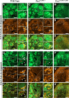
Early glomerular precursor cells displayed overlapping expression of PAX2 and Wilms’ tumor-1 (WT-1) until parietal epithelial cell (PEC) and podocyte differentiation, supporting a close developmental relationship between the two cell types. A–R: representative images of immunofluorescence staining for PAX2 and WT-1 on embryonic day (E)13.5 and E16.5 wild-type, Pax2A220G/+, and Pax2A220G/A220G embryonic kidneys. PAX2-expressing cells are shown in green, WT-1-expressing cells are shown in orange, and overlapping PAX2 and WT-1 expression in cells is shown in yellow (merge). In both E13.5 wild-type and Pax2A220G/+ kidneys, PAX2 expression was more predominant than WT-1 in condensed mesenchyme cells (A–C and G–I insets and dashed lines), whereas overlapping PAX2 and WT-1 expression was detected in PEC precursors (A–C and G–I insets and block arrows) and podocyte precursors (A–C and G–I insets and arrowheads) within early glomerular structures. In both E16.5 wild-type and Pax2A220G/+ kidneys, WT-1 expression but no PAX2 expression was detected in podocyte precursor cells within the glomerular tuft of maturing glomeruli (D–F and J–L insets and arrowheads). Predominant WT-1 expression and limited PAX2 coexpression was detected in few PEC precursors that line Bowman’s capsule (D–F and J–L insets and block arrows). Red blood cells that produce green autofluorescence within the glomerular tuft are indicated by red arrows (D and F and J and L, red arrows). In Pax2A220G/A220G E13.5 and E16.5 kidneys, PAX2 and WT-1 expression in disorganized mesenchyme-like cells was observed (M–O and P–R, dashed lines), but there was a complete absence of any PAX2 and WT-1-positive developing glomeruli. Scale bars = 60 µm in A–R.
Figure 3.
Figure 3.
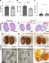
Postnatal and adult mice with the Pax2 c.A220G variant had reduced nephron numbers but did not display any significant differences in glomerular function at baseline. Body weight measurements were not different between Pax2A220G/+ mice (n = 18 mice) and wild-type mice (n = 20 mice) [not statistically significant (ns)] (A), but quantification of glomeruli isolated using Dynabeads (J and K) showed significantly decreased glomerular numbers in 3-mo-old Pax2A220G/+ kidneys (n = 8 mice) compared with wild-type kidneys (n = 7 mice) (**P < 0.005) (B). Urinary albumin-to-creatinine ratios (ACRs) revealed no changes in urinary albumin excretion in Pax2A220G/+ mice (n = 35 mice) compared with wild-type mice (n = 44 mice) (ns) (C). Representative images of hematoxylin and eosin-stained kidney sections from newborn postnatal day 3 (postnatal D3) wild-type and Pax2A220G/+ kidneys showed that the majority (80%) of Pax2A220G/+ kidneys had histology comparable with wild-type kidneys (D and E) while few (20%) Pax2A220G/+ kidneys showed unilateral cystic formation (asterisks) (F). Representative images of gross histology observations also showed that the majority (67%) of 3-mo-old Pax2A220G/+ kidneys were comparable with wild-type kidneys (G and H), with some Pax2A220G/+ kidneys showing unilateral renal hypoplasia (22%) (I) and unilateral cystic formation (11%) (L). Scale bars = 1 mm in A–C and 2 cm in D–F and I.
Figure 4.
Figure 4.
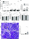
Adult Pax2A220G/+ mice compared with wild-type mice had a worsened focal segmental glomerulosclerosis (FSGS) phenotype after adriamycin (ADR)-induced glomerular injury, including increased glomerulosclerosis and mortality. Urinary albumin-to-creatinine ratio (uACR) measurements showed significant albuminuria in both wild-type and Pax2A220G/+ mice beginning at day 14 after ADR injury (****P < 0.0001). However, there were no statistically significant differences in urinary albumin excretion between Pax2A220G/+ mice (n = 35 mice) and wild-type mice (n = 34 mice) at baseline before either vehicle or ADR injections, and day 7 (n = 26 wild-type mice and n = 27 Pax2A220G/+ mice), day 14 (n = 26 wild-type mice and n = 25 Pax2A220G/+ mice), day 21 (n = 25 wild-type mice and n = 22 Pax2A220G/+ mice), and day 28 (n = 26 wild-type mice and n = 29 Pax2A220G/+ mice) after ADR injury [not statistically significant (ns)] (A). Pax2A220G/+ mice had higher mortality rates compared with wild-type mice over a period of 28 days after ADR injury (n = 26 ADR-injured wild-type mice and n = 29 ADR-injured Pax2A220G/+ mice) (B). Glomerular measurements demonstrated no significant differences in glomerular area (C), total number of cells per glomerular section (D), and area of nucleus per glomerular cell (E) (n = 310 mice/group) (ns). F–I: representative images of periodic acid-Schiff staining of vehicle-treated and ADR-injured wild-type and Pax2A220G/+ glomeruli. At baseline, vehicle-treated Pax2A220G/+ mice showed similar glomerular histology as wild-type mice (F and G). After ADR injury, both wild-type and Pax2A220G/+ mice showed segmental scars (red dashed circles), but this was more pronounced in Pax2A220G/+ mice (H and I). Quantification of the percentage of sclerotic glomeruli relative to the total number of glomeruli showed significantly increased numbers of sclerotic glomeruli in ADR-injured Pax2A220G/+ mice (n = 11 mice) compared with wild-type mice (n = 11 mice) (*P < 0.05) (J). Scale bars = 40 µm in C–F.
Figure 5.
Figure 5.
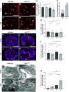
Adult Pax2A220G/+ compared with wild-type mice had a worsened focal segmental glomerulosclerosis (FSGS) phenotype after adriamycin (ADR)-induced glomerular injury, including increased podocyte loss and decreased podocyte slit diaphragm integrity. A–D: representative images of immunofluorescence staining for the podocyte marker Wilms’ tumor-1 (WT-1) on vehicle-treated and ADR-injured wild-type and Pax2A220G/+ glomeruli. Quantification of WT-1-positive glomerular tuft cells per glomerular section (A–D, arrowheads) showed no difference in podocyte numbers between vehicle-treated Pax2A220G/+ mice (n = 4 mice) and wild-type mice (n = 3 mice) [not statistically significant (ns)], but there were significantly decreased podocyte numbers in ADR-injured Pax2A220G/+ mice (n = 6 mice) compared with injured wild-type mice (n = 7 mice) (*P < 0.05) (E). E: decreased podocyte numbers were also found in ADR-injured Pax2A220G/+ mice compared with vehicle-treated Pax2A220G/+ mice (**P < 0.005), whereas there was no significant change in podocyte numbers in ADR-injured wild-type mice compared with vehicle-treated wild-type controls. F: Pax2A220G/+ mice compared with wild-type mice showed a higher percentage of podocyte loss after ADR. Quantification of WT-1-positive parietal epithelial cells (PECs) per glomerular section (A–D, block arrows) showed no statistically significant difference in any of the four groups (n = 37 mice per group) (ns) (G). H–K: representative images of immunostaining for the podocyte slit diaphragm protein nephrin on vehicle-treated and ADR-injured wild-type and Pax2A220G/+ glomeruli. At baseline, vehicle-treated wild-type and Pax2A220G/+ glomeruli showed a linear-like, continuous pattern of nephrin staining (H and I, arrowheads). ADR-injured Pax2A220G/+ mice showed an exacerbated discontinuous pattern with sporadic areas of dense and granular-like nephrin staining (J and K, arrowheads). L: quantification of the percentage of glomerular area with nephrin staining demonstrated no statistically significant difference between vehicle-treated wild-type mice (n = 3 mice) and Pax2A220G/+ (n = 3 mice) or in ADR-injured wild-type mice (n = 7 mice) and Pax2A220G/+ mice (n = 5 mice) (ns). M–P: representative transmission electron microscopy images from vehicle-treated and ADR-injured wild-type and Pax2A220G/+ glomeruli. Vehicle-treated Pax2A220G/+ mice showed normal podocyte foot process morphology similar to wild-type mice (M and N, black arrowheads). ADR-injured wild-type mice showed scattered areas of mild podocyte foot process effacement (O, red arrowheads) whereas other areas were normal (O, black arrowheads). In contrast, ADR-injured Pax2A220G/+ mice showed more severe foot process effacement spanning larger areas of the glomerular basement membrane (P, red arrowheads). Q: quantifications showed significantly increased foot process effacement in ADR-injured Pax2A220G/+ mice (n = 5 mice) compared with injured wild-type mice (n = 4 mice) (**P <0.005) as well as in ADR-injured Pax2A220G/+ mice compared with vehicle-treated Pax2A220G/+ controls (***P < 0.0005). Q: in addition, there was no significant difference in foot process effacement in ADR-injured wild-type mice compared with vehicle-treated wild-type controls as well as between vehicle-treated Pax2A220G/+ mice (n = 5 mice) and wild-type mice (n = 5 mice) (ns). Scale bars = 25 µm in A–J and 2 µm in L–O.
Figure 6.
Figure 6.
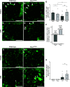
PAX2-positive parietal epithelial cells (PECs) significantly decreased in wild-type mice after adriamycin (ADR) injury but did not change in Pax2A220G/+ mice after injury. This was accompanied by the emergence of PAX2-expressing cells in the glomerular tuft in both groups but was more marked in Pax2A220G/+mice. A–J: representative images and quantifications of immunofluorescence staining for PAX2 on vehicle-treated and ADR-injured wild-type and Pax2A220G/+ glomeruli. At baseline, vehicle-treated wild-type mice (n = 5 mice) and Pax2A220G/+ mice (n = 6 mice) showed PAX2 expression limited to PECs in Bowman’s capsule, with no significant difference between these groups in the number of PAX2-expressing PECs [not statistically significant (ns)] (A and B, block arrows, and E). After ADR, wild-type mice (n = 12 mice) demonstrated significantly decreased numbers of PAX2-expressing PECs compared with vehicle-treated wild-type mice (n = 5) (*P < 0.05) (A and C, block arrows, and E), whereas there was no change in PAX2-expressing PECs in Pax2A220G/+ mice (n = 13 mice) after injury compared with vehicle-treated Pax2A220G/+ mice (n = 6 mice) (ns) (B and D, block arrows, and E). There were also significantly increased numbers of PAX2-expressing PECs in ADR-injured Pax2A220G/+ mice (n = 13 mice) compared with ADR-injured wild-type mice (n = 12 mice) (**P < 0.005) (C and D, block arrows, and E). F: wild-type mice showed a decrease whereas Pax2A220G/+ mice showed an increase in the proportion of PAX2+ve PECs within Bowman’s capsule after ADR relative to their baseline numbers. Next, there were no PAX2-expressing glomerular tuft cells observed in both wild-type mice (n = 5 mice) and Pax2A220G/+ mice (n = 6 mice) at baseline (G, H, and K). However, PAX2-positive glomerular tuft cells were observed in both types of mice after ADR injury (I and J, arrowheads). Quantification of PAX2-positive glomerular tuft cells per glomerular section revealed there were significantly more in ADR-injured Pax2A220G/+ mice (n = 13 mice) compared with ADR-injured wild type mice (n = 12 mice) (*P < 0.05) (I and J, arrowheads, and K). Scale bars = 40 µm in A–D and F–I.
Figure 7.
Figure 7.
PAX2-expressing glomerular tuft cells were also observed in kidney tissue from patients with PAX2-associated focal segmental glomerulosclerosis (FSGS). Representative images of periodic acid Schiff (PAS) staining and immunohistochemistry for PAX2 on serial sections from a normal human glomerulus (A) and from two patients [IDs: FG-EQ IV (8) and FG-EQ III (8)] with PAX2-associated FSGS (B and C) are shown. In healthy human glomeruli, PAX2 expression was limited to parietal epithelial cells (A, block arrows) and was absent within the glomerular tuft (A). In patients with FSGS associated with PAX2 missense variants, PAX2 expression was detected in parietal epithelial cells (PECs) (B and C, block arrows) but was also detected in cells within the glomerular tuft (B and C, arrowheads) and overlapping with areas of sclerosis [C, asterisk (*)], suggestive of a pathogenic role. The PAS image in C from patient FG-EQ III (8) has been previously published in Barua et al. (18) and was reused in this study with copyright transfer permissions granted by the Journal of the American Society of Nephrology (License No. 5670261395616, date issued: November 15, 2023). Scale bars = 20 µm in A–C.
Figure 8.
Figure 8.
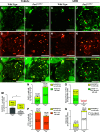
Rare glomerular tuft cells expressing PAX2 with coexpression of Wilms’ tumor-1 (WT-1) were not observed at baseline but emerged after adriamycin (ADR) injury and were significantly increased in wild-type mice compared with Pax2A220G/+ mice. A–M: representative images and quantifications of immunofluorescence costaining for PAX2 and WT-1 in vehicle-treated and ADR-injured wild-type and Pax2A220G/+ glomeruli. A–F: at baseline, vehicle-treated wild-type mice (n = 3 mice) and Pax2A220G/+ mice (n = 4 mice) did not show any PAX2/WT-1 coexpression within glomerular tuft cells. Glomerular tuft cells with PAX2/WT-1 coexpression were shown in injured wild-type mice (n = 8 mice) as demonstrated by yellow cells in the “merge” panel shown by white arrowheads (G–I), although these cells were relatively rare and their emergence was an infrequent event overall, as shown by the scale of the y-axis in M. There was less overlap of PAX2/WT-1 costaining in glomerular tuft cells in injured Pax2A220G/+ mice (n = 9 mice) as shown in J–L, supported by quantifications (*P < 0.05) (M). Red arrowheads point to glomerular tuft cells with PAX2 staining without overlap of WT-1, which were seen more frequently in ADR-injured Pax2A220G/+ glomeruli (J–L) and were rarely observed in injured wild-type glomeruli (G–I). Bar graphs demonstrating the proportion of PAX2/WT-1 glomerular tuft cells relative to the post-ADR average numbers of PAX2-positive glomerular tuft cells (N), migrated PAX2-positive parietal epithelial cells (PECs) (O), total WT-1+ve tuft cells (P), and podocytes lost after injury (Q). N–Q: in all comparisons, wild-type mice showed a higher proportion of PAX2/WT-1-coexpressing glomerular tuft cells compared to Pax2A220G/+ mice. Scale bars = 40 µm in A–L.
Figure 9.
Figure 9.
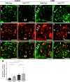
Parietal epithelial cells (PECs) with PAX2/Wilms’ tumor-1 (WT-1) coexpression showed no significant change after adriamycin (ADR) injury in both wild-type and Pax2A220G/+ mice. A–M: representative images and quantifications of immunofluorescence costaining for PAX2 and WT-1 on vehicle-treated and ADR-injured wild-type and Pax2A220G/+ glomeruli. Overlapping areas of PAX2/WT-1 coexpression are shown by block arrows. At baseline, vehicle-treated wild-type mice (n = 4 mice) and Pax2A220G/+ mice (n = 5 mice) showed PECs with PAX2/WT-1 coexpression; however, there was no significant difference in the numbers of these cells between the two groups [not statistically significant (ns)] (A–F, block arrows, and M). After ADR injury, there was also no significant difference between wild-type mice (n = 8 mice) and Pax2A220G/+ mice (n = 7 mice) in the numbers of PAX2/WT-1-coexpressing PECs (G–L, block arrows, and M). Scale bars = 40 µm in A–L.
Figure 10.
Figure 10.
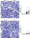
Adult Pax2A220G/+ mice compared with wild-type mice had significantly increased glomerular tuft cell division and cell death after adriamycin (ADR)-induced glomerular injury. A–J: representative images and quantifications of immunohistochemistry for Ki-67 and cleaved caspase-3 (CC3) in vehicle-treated and ADR-injured wild-type and Pax2A220G/+ glomeruli. At baseline, vehicle-treated wild-type mice (n = 3 mice) and Pax2A220G/+ mice (n = 3 mice) did not show any Ki-67-positive glomerular tuft cells (A, B, and E). ADR-injured Pax2A220G/+ mice (n = 10 mice) showed significantly increased numbers of Ki-67-positive glomerular tuft cells compared with wild-type mice (n = 12 mice) (*P < 0.05) (C and D, arrowheads, and E). At baseline, vehicle-treated wild-type mice (n = 3 mice) and Pax2A220G/+ mice (n = 3 mice) did not show any CC3-positive glomerular tuft cells (F, G, and J). ADR-injured Pax2A220G/+ mice (n = 13 mice) showed significantly increased numbers of CC3-positive glomerular tuft cells compared with wild-type mice (n = 12 mice) (*P < 0.05) (H and I, arrowheads, and J). Scale bars = 40 µm in A–D and F–I.
Figure 11.
Figure 11.
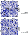
Adult Pax2A220G/+ compared with wild-type mice showed no significant differences in parietal epithelial cell (PEC) division and cell death after adriamycin (ADR)-induced glomerular injury. A–J: representative images and quantifications of immunohistochemistry for Ki-67 and cleaved caspase-3 (CC3) in vehicle-treated and ADR-injured wild-type and Pax2A220G/+ glomeruli. At baseline, vehicle-treated wild-type mice (n = 3 mice) and Pax2A220G/+ mice (n = 3 mice) did not show any Ki-67-positive PECs (A, B, and E). ADR-injured Pax2A220G/+ mice (n = 13 mice) and wild-type mice (n = 11 mice) showed Ki-67-expressing PECs; however, the numbers of these cells were not significantly different between these two groups [not statistically significant (ns)] (C and D, block arrows, and E). At baseline, vehicle-treated wild-type mice (n = 3 mice) and Pax2A220G/+ mice (n = 3 mice) did not show any CC3-positive PECs (F, G, and J). Some ADR-injured Pax2A220G/+ mice (n = 12 mice) and wild-type mice (n = 11 mice) showed few CC3-expressing PECs, although overall these cells were rare as shown by the scale of the y-axis in J; however, the numbers of these cells were not significantly different between these two groups (H and I, block arrows, and J).

References

    1. Jha V, Garcia-Garcia G, Iseki K, Li Z, Naicker S, Plattner B, Saran R, Wang AY-M, Yang C-W. Chronic kidney disease: global dimension and perspectives. Lancet 382: 260–272, 2013. [Erratum in Lancet 382: 208, 2013]. doi:10.1016/S0140-6736(13)60687-X. - DOI - PubMed
    1. Verberne WR, Das-Gupta Z, Allegretti AS, Bart HAJ, van Biesen W, García-García G, Gibbons E, Parra E, Hemmelder MH, Jager KJ, Ketteler M, Roberts C, Al Rohani M, Salt MJ, Stopper A, Terkivatan T, Tuttle KR, Yang C-W, Wheeler DC, Bos WJW. Development of an international standard set of value-based outcome measures for patients with chronic kidney disease: a report of the International Consortium for Health Outcomes Measurement (ICHOM) CKD Working Group. Am J Kidney Dis 73: 372–384, 2019. doi:10.1053/j.ajkd.2018.10.007. - DOI - PubMed
    1. Jager KJ, Kovesdy C, Langham R, Rosenberg M, Jha V, Zoccali C. A single number for advocacy and communication-worldwide more than 850 million individuals have kidney diseases. Kidney Int 96: 1048–1050, 2019. doi:10.1016/j.kint.2019.07.012. - DOI - PubMed
    1. Reidy K, Kaskel FJ. Pathophysiology of focal segmental glomerulosclerosis. Pediatr Nephrol 22: 350–354, 2007. doi:10.1007/s00467-006-0357-2. - DOI - PMC - PubMed
    1. Song B, Smink AM, Jones CV, Callaghan JM, Firth SD, Bernard CA, Laslett AL, Kerr PG, Ricardo SD. The directed differentiation of human iPS cells into kidney podocytes. PLoS One 7: e46453, 2012. doi:10.1371/journal.pone.0046453. - DOI - PMC - PubMed

Publication types

MeSH terms

LinkOut - more resources