. 2024 Mar 14;19(3):e0298323.
doi: 10.1371/journal.pone.0298323. eCollection 2024.

The polyglutamine domain is the primary driver of seeding in huntingtin aggregation

Affiliations

The polyglutamine domain is the primary driver of seeding in huntingtin aggregation

Adam Skeens et al. PLoS One. .

Abstract

Huntington's Disease (HD) is a fatal, neurodegenerative disease caused by aggregation of the huntingtin protein (htt) with an expanded polyglutamine (polyQ) domain into amyloid fibrils. Htt aggregation is modified by flanking sequences surrounding the polyQ domain as well as the binding of htt to lipid membranes. Upon fibrillization, htt fibrils are able to template the aggregation of monomers into fibrils in a phenomenon known as seeding, and this process appears to play a critical role in cell-to-cell spread of HD. Here, exposure of C. elegans expressing a nonpathogenic N-terminal htt fragment (15-repeat glutamine residues) to preformed htt-exon1 fibrils induced inclusion formation and resulted in decreased viability in a dose dependent manner, demonstrating that seeding can induce toxic aggregation of nonpathogenic forms of htt. To better understand this seeding process, the impact of flanking sequences adjacent to the polyQ stretch, polyQ length, and the presence of model lipid membranes on htt seeding was investigated. Htt seeding readily occurred across polyQ lengths and was independent of flanking sequence, suggesting that the structured polyQ domain within fibrils is the key contributor to the seeding phenomenon. However, the addition of lipid vesicles modified seeding efficiency in a manner suggesting that seeding primarily occurs in bulk solution and not at the membrane interface. In addition, fibrils formed in the presence of lipid membranes displayed similar seeding efficiencies. Collectively, this suggests that the polyQ domain that forms the amyloid fibril core is the main driver of seeding in htt aggregation.


Conflict of interest statement

The authors have declared that no competing interests exist.

Figures

Fig 1
Fig 1. Htt-exon1(46Q) fibrils seed aggregation in a C. elegans strain (EAK102) expressing a non-pathogenic N-terminal htt fragment [htt-513(Q15)].
Representative fluorescence microscopy images of the (A) EAK102 and (B) EAK103 C. elegans strains that express htt513(15Q) and htt513(128Q), respectively. (C) Worm viability of EAK102 and a control strain (N2) 48 h after exposure to various concentrations of htt-exon1(46Q) seeds. Error bars represents standard error of the mean. * represents p < 0.01 based on a T-test comparing N2 and EAK102 worm viability at a given dose (n = 3 for both conditions). (D) Representative fluorescence microscopy images and (E) quantification of visible inclusion bodies per worm comparing control EAK102 worms to EAK102 worms exposed to 10 μM htt-exon1(46Q) seeds. Error bars represent standard error of the mean. Error bars represents standard error of the mean. * represents p < 0.01 based on a T-test comparing the number of inclusions observed in EAK102 worms to those treated with 10 μM htt seeds (n = 5 for the no seeds condition and n = 8 for the seeded condition).
Fig 2
Fig 2. Fibrils formed by htt-mimicking peptides.
(A) ThT assay demonstrating the different rate of fibril formation for the Nt17-Q35-KK, Nt17-Q35-P10, KK-Q35-KK, and KK-Q35-P10 peptides. Error bars represent standard error of the mean (n = 3 for all conditions). (B) Representative ex situ AFM images of fibrils formed from Nt17-Q35-KK, Nt17-Q35-P10, KK-Q35-KK, and KK-Q35-P10. (C) Histograms of the average height along the contour of fibrils corresponding to each htt peptide.
Fig 3
Fig 3. The impact of peptide seeds on htt-exon1(46Q) aggregation.
(A) ThT assays tracking the impact of seeds comprised of Nt17-Q35-KK, Nt17-Q35-P10, KK-Q35-KK, or KK-Q35-P10 on htt-exon1(46Q) aggregation. The fluorescence signals were normalized to the control htt-exon1(46Q) alone aggregation reaction. Error bars represent standard error of the mean (n = 3 for all conditions). (B) Representative ex situ AFM images comparing fibrils of htt-exon1(46Q) formed in the absence and presence of seeds comprised of Nt17-Q35-KK, Nt17-Q35-P10, KK-Q35-KK, or KK-Q35-P10. (C) Histograms of the average height along the contour of htt-exon1(46Q) fibril formed in the absence or presence of the indicated peptide seeds. (D) Representative ex situ AFM images of htt-exon1(46Q) fibril bundles aggregated in the absence and presence of seeds comprised of Nt17-Q35-KK, Nt17-Q35-P10, KK-Q35-KK, or KK-Q35-P10.
Fig 4
Fig 4. CD analysis of resulting fibril structure.
(A) CD spectra of htt-exon1(46Q) aggregates formed in the absence (htt-exon1(46Q) 8h) or presence of seeds derived from Nt17-Q35-KK, Nt17-Q35-P10, KK-Q35-KK, or KK-Q35-P10 peptides. For comparison, a spectra of htt-exon1(46Q) after 3 h of incubation without seeds is provided, which is a condition comprised predominately of oligomers. Due to the presence of GST, a GST spectra is also provided. (B) CD spectra of htt-exon1(46Q) with the subtraction of the GST spectra.
Fig 5
Fig 5. The impact of peptide seeds on htt-exon1(20Q) aggregation.
(A) ThT assays tracking the impact of seeds comprised of Nt17-Q35-KK, Nt17-Q35-P10, KK-Q35-KK, or KK-Q35-P10 on htt-exon1(20Q) aggregation. The fluorescence signals were normalized to the control htt-exon1(20Q) alone aggregation reaction. Error bars represent standard deviation (n = 3 for all conditions). (B) Representative ex situ AFM images comparing fibrils of htt-exon1(20Q) formed in the absence and presence of seeds comprised of Nt17-Q35-KK, Nt17-Q35-P10, KK-Q35-KK, or KK-Q35-P10. Colored lines in each image correspond to the height profiles. (C) Histograms of the average height along the contour of htt-exon1(20Q) fibril formed in the presence of the indicated peptide seeds. A distribution for htt-exon1(20Q) fibrils formed in the absence of seeds is not provided because no fibrils were observed under that condition.
Fig 6
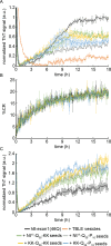
Fig 6. The impact of lipids on the seeding of htt-exon1(46Q) by peptides.
(A) ThT assays tracking the impact of TBLE vesicles (20:1 lipid:protein molar ratio) on the ability of seeds comprised of Nt17-Q35-KK, Nt17-Q35-P10, KK-Q35-KK, or KK-Q35-P10 to alter htt-exon1(46Q) aggregation. (B) PDA/TBLE assay measuring the impact of seeds comprised of Nt17-Q35-KK, Nt17-Q35-P10, KK-Q35-KK, or KK-Q35-P10 on the ability of htt-exon1(46Q) to bind TBLE vesicles. (C) Htt-exon1(46Q) aggregation tracked by ThT assay in the presence of Nt17-Q35-KK, Nt17-Q35-P10, KK-Q35-KK, or KK-Q35-P10 seeds produced in the presence of POPG vesicles. All ThT plots were normalized to the control htt-exon1(46Q) alone aggregation reaction. Error bars represent standard deviation for each plot (n = 3 for all conditions).
Fig 7
Fig 7. Peptide-derived fibrils seed aggregation in a C. elegans strain (EAK102) expressing a non-pathogenic N-terminal htt fragment.
(A) The viability of N2 (not expressing htt) and EAK102 [expressing htt-513(Q15)] worms after 48 h of exposure to seeds derived from the peptides KK-Q35-KK, Nt17-Q35-KK, KK-Q35-P10-KK, and Nt17-Q35-P10-KK was determined. The control condition represents worms that were not exposed to any seeds. * indicates p < 0.05 based on a T-test (n = 3 for all conditions). Error bars represent the standard deviation. (B) Representative fluorescence microscopy images of an EAK102 worm that was not exposed to any peptide-derived seeds and EAK102 worms that were exposed to the various peptide-derived seeds (5 μM) for 48 h. (C) Quantification of the number of visual inclusion observed in EAK102 worms that have been exposed to peptide-derived seeds. * indicates p < 0.05 based on a T-test (n = 12 for no seeds and n = 5 for all other conditions). Error bars represent the standard deviation.

Similar articles

References

    1. The Collaborative Huntingtons Disease Research Group. A novel gene containing a trinucleotide repeat that is exanded and unstable on Huntingtons-disease chromosomes Cell. 1993;72(6):971–83. - PubMed
    1. Chen SM, Ferrone FA, Wetzel R. Huntington’s disease age-of-onset linked to polyglutamine aggregation nucleation. P. Natl. Acad. Sci. USA. 2002;99(18):11884–9. doi: 10.1073/pnas.182276099 - DOI - PMC - PubMed
    1. Legleiter J, Mitchell E, Lotz GP, Sapp E, Ng C, DiFiglia M, et al.. Mutant Huntingtin Fragments Form Oligomers in a Polyglutamine Length-dependent Manner in Vitro and in Vivo. J. Biol. Chem. 2010;285(19):14777–90. doi: 10.1074/jbc.M109.093708 - DOI - PMC - PubMed
    1. Lajoie P, Snapp EL. Formation and Toxicity of Soluble Polyglutamine Oligomers in Living Cells. PLoS One. 2010;5(12). doi: 10.1371/journal.pone.0015245 - DOI - PMC - PubMed
    1. Nucifora LG, Burke KA, Feng X, Arbez N, Zhu S, Miller J, et al.. Identification of Novel Potentially Toxic Oligomers Formed in Vitro from Mammalian-derived Expanded huntingtin Exon-1 Protein. J. Biol. Chem. 2012;287(19):16017–28. doi: 10.1074/jbc.M111.252577 - DOI - PMC - PubMed