. 2024 Mar 19;121(12):e2319582121.
doi: 10.1073/pnas.2319582121. Epub 2024 Mar 14.

Identification of a negative-strand RNA virus with natural plant and fungal hosts

Affiliations

Identification of a negative-strand RNA virus with natural plant and fungal hosts

Ruoyin Dai et al. Proc Natl Acad Sci U S A. .

Abstract

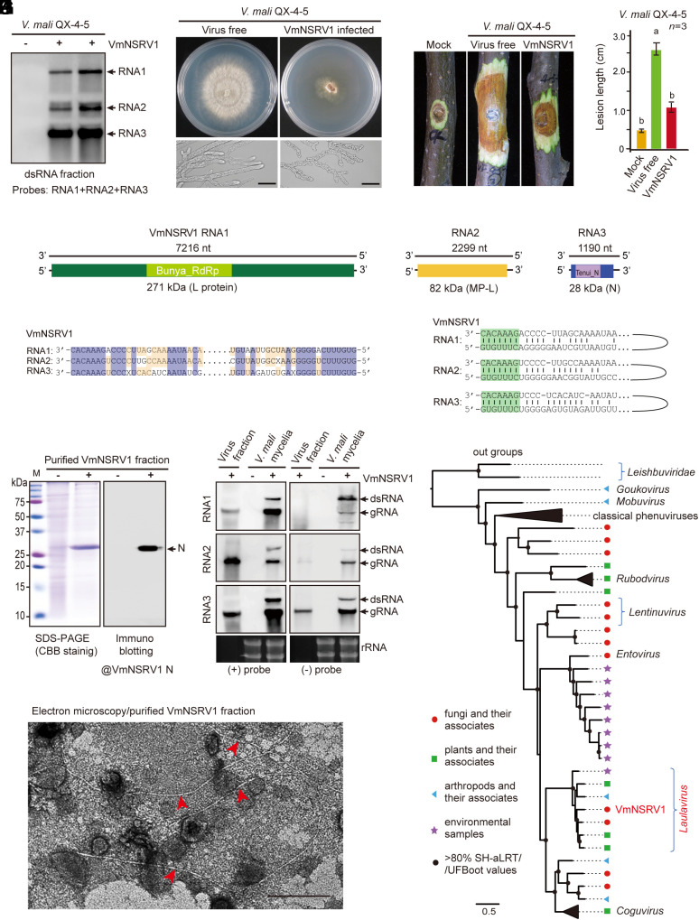
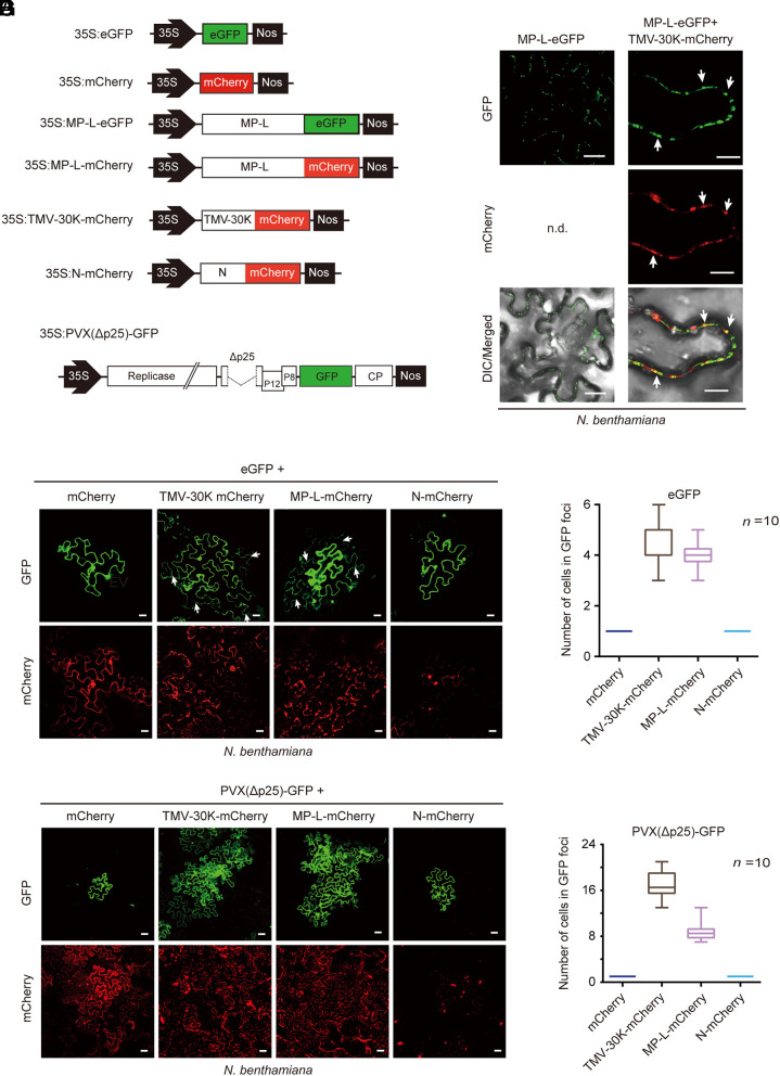
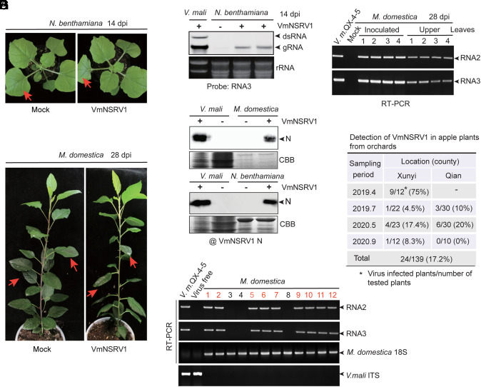
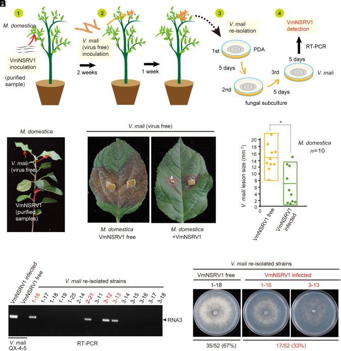
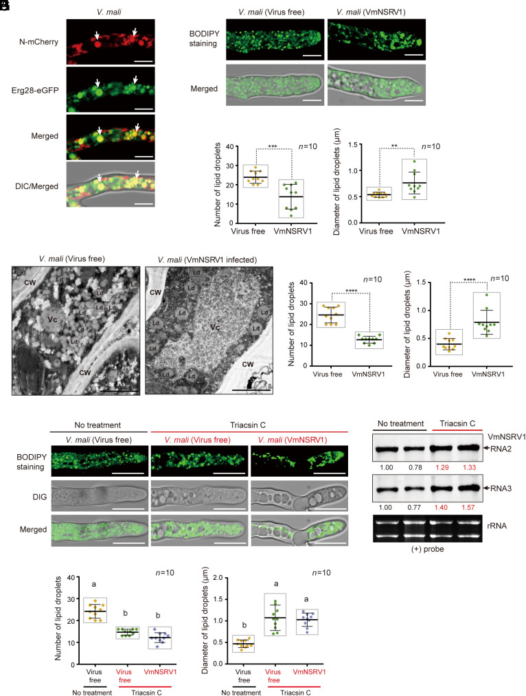
The presence of viruses that spread to both plant and fungal populations in nature has posed intriguingly scientific question. We found a negative-strand RNA virus related to members of the family Phenuiviridae, named Valsa mali negative-strand RNA virus 1 (VmNSRV1), which induced strong hypovirulence and was prevalent in a population of the phytopathogenic fungus of apple Valsa canker (Valsa mali) infecting apple orchards in the Shaanxi Province of China. Intriguingly, VmNSRV1 encodes a protein with a viral cell-to-cell movement function in plant tissue. Mechanical leaf inoculation showed that VmNSRV1 could systemically infect plants. Moreover, VmNSRV1 was detected in 24 out of 139 apple trees tested in orchards in Shaanxi Province. Fungal inoculation experiments showed that VmNSRV1 could be bidirectionally transmitted between apple plants and V. mali, and VmNSRV1 infection in plants reduced the development of fungal lesions on leaves. Additionally, the nucleocapsid protein encoded by VmNSRV1 is associated with and rearranged lipid droplets in both fungal and plant cells. VmNSRV1 represents a virus that has adapted and spread to both plant and fungal hosts and shuttles between these two organisms in nature (phyto-mycovirus) and is potential to be utilized for the biocontrol method against plant fungal diseases. This finding presents further insights into the virus evolution and adaptation encompassing both plant and fungal hosts.

Keywords: cross-kingdom infection; hypovirulence; movement protein; mycovirus; negative-strand RNA virus.


Conflict of interest statement

Competing interests statement:The authors declare no competing interest.

Figures

Fig. 1.
Fig. 1.
Identification of a hypovirulence-inducing negative-strand RNA virus from V. mali. (A) Detection of three RNA segments corresponding to the RNA1, 2, and 3 genomes of VmNSRV1 in a V. mali strain by RNA blotting with dsRNA fractions. (B) Phenotypic growth and mycelial morphology of a V. mali strain (VmNSRV1-free and -infected, QX-4-5) on PDA medium. The fungi were photographed at 3 d after culturing. (Scale bar, 20 µm.) (C) Virulence assay of the V. mali (QX-4-5) strain on apple twigs. Fungal disease lesions (peeled twigs) were photographed at 5 d after inoculation. (D) Measurement of fungal disease lesions observed in the experiment described in (C). Different letters indicate significant differences (P < 0.05, one-way ANOVA). (E) A schematic genome structure of VmNSRV1. Black lines represent negative-strand genomic RNAs. Colored boxes represent open reading frames (ORFs) encoded by positive-strand genomic RNAs. The estimated molecular weight of each encoded protein (L[RdRp], MP-L, and N) is indicated. The relative position of conserved domains in the encoded RdRp and N proteins is presented (small colored boxes). (F) Sequence similarities of the 5′- and 3′-terminal regions of VmNSRV1 RNA1, 2, and 3 segments. (G) Sequence complementarity of the 5′- and 3′-terminal regions of each VmNSRV1 RNA1, 2, and 3 segment. (H) SDS-PAGE and immunoblot analyses of the protein associated with VmNSRV1 purified fractions. Immunoblotting was carried out using an antibody specific to the VmNSRV1 N protein. (I) Electron microscopy of a VmNSRV1 purified fraction from a virus-infected V. mali (QX-4-5) strain. Red arrowheads mark the potential VmNSRV1 particles. (Scale bar, 200 nm.) (J) Detection of positive- and negative-strand RNA of VmNSRV1 in purified virus fraction and mycelia of a virus-infected V. mali (QX-4-5) strain by RNA blotting. (K) Phylogenetic relationships of VmNSRV1 with phenuiviruses or other selected phenui-like viruses. The maximum likelihood tree was based on multiple sequence alignment of the RNA1-encoded L protein (RdRp). For the virus names and details of the tree, see SI Appendix, Fig. S6 and its legend.
Fig. 2.
Fig. 2.
Viral plant MP function of the MP-L protein encoded by RNA2 of VmNSRV1. (A) A schematic diagram of the constructs used to assess the ability of MP-L to increase the SEL of plasmodesmata and complement the cell-to-cell movement of movement-defective potato virus X expressing GFP [PVX(Δp25)-GFP]. TMV 30K fused to mCherry (TMV-30K-mCherry) was used as a plasmodesmatal marker. 35S and Nos represent the cauliflower mosaic virus (CaMV) 35S promoter and nopaline synthase terminator sequences, respectively. (B) Subcellular localization of MP-L protein fused to eGFP (MP-L-eGFP) in the epidermal cells of N. benthamiana plants transiently expressed by Agrobacterium infiltration. Fluorescence was observed by CLSM 3 d after inoculation. (Scale bar, 25 µm.) (C) Coexpression of MP-L-eGFP with TMV-30K-mCherry in epidermal cells. Arrows indicate the representative overlapping fluorescence signals. (Scale bar, 10 µm.) (D) Coexpression of the tag-free eGFP with TMV-30K-mCherry, MP-L-mCherry, N-mCherry, or unfused mCherry in epidermal cells. An Agrobacterium culture harboring 35S: eGFP (OD600 = 1) was diluted 10,000-fold and mixed (1:1) with an Agrobacterium culture harboring one of the mCherry expression constructs. Arrows indicate cells with diffused eGFP. (Scale bar, 20 µm.) (E) Quantification of the GFP-expressing cells in GFP foci observed in the experiment described in (D). (F) Coexpression of PVX(Δp25)-GFP with TMV-30K-mCherry, MP-L-mCherry, N-mCherry, or unfused mCherry in epidermal cells. (Scale bar, 40 µm.) (G) Quantification of the GFP-expressing cells in GFP foci observed in the experiment described in (F).
Fig. 3.
Fig. 3.
Infectivity of VmNSRV1 in plants. (A) N. benthamiana plant mechanically inoculated with purified VmNSRV1 fractions. Plants were photographed at 14 dpi. Arrows indicate the inoculated leaves. (B) RNA blot detection of VmNSRV1 RNA3 accumulation in the upper leaves of N. benthamiana plants described in (A). Leaves were harvested at 14 dpi. (C) Apple plant (Malus domestica) mechanically inoculated with purified VmNSRV1 fraction. Plants were photographed at 28 dpi. Arrows indicate the inoculated leaves. (D) RT-PCR detection of VmNSRV1 RNA2 and RNA3 accumulation in the upper leaves of apple plants described in (C). Leaves were harvested at 28 dpi. (E) Immunoblot analysis of the protein fractions from noninoculated upper leaves of virus-free and VmNSRV1-infected N. benthamiana and M. domestica. Immunoblotting was carried out using an antibody specific to the VmNSRV1 N protein. (F) RT-PCR detection of VmNSRV1 RNA2 and RNA3 accumulation in the upper leaves of field-grown apple trees. Primers specific for M. domestica 18S rRNA and V. mali ITS were used as a plant reference gene or to confirm the absence of V. mali in leaf samples, respectively. (G) Detection rate of VmNSRV1 in apple trees grown in orchards located in two counties of Shaanxi Province, China. VmNSRV1 detection was performed as described in (F).
Fig. 4.
Fig. 4.
Transmission of VmNSRV1 from the plant to fungus. (A) Diagram describing the experimental procedure used to study VmNSRV1 acquisition by V. mali. (1) Mechanical inoculation of purified VmNSRV1 fractions on apple plants. (2) Inoculation of upper leaves with mycelia of a virus-free V. mali (QX-4-5) strain 2 wk after VmNSRV1 inoculation. (3) Reisolation of V. mali from fungal lesions on leaves 1 wk after inoculation with V. mali mycelia. (4) RT-PCR detection of VmNSRV1 in reisolated V. mali strains after at least three subcultures. (B) A young apple plant inoculated with purified VmNSRV1 fractions on the lower leaves and a virus-free V. mali (QX-4-5) strain on the noninoculated upper leaves. Arrows indicate the inoculated leaves. (C) Representative V. mali lesions on the leaves of virus-free and VmNSRV1-infected apple plants. Fungal disease lesions were photographed at 5 d after inoculation. (D) Lesion areas of V. mali measured on leaves described in (C). “*” indicates significant differences (P < 0.05, Student’s t test). (E) RT-PCR detection of VmNSRV1 in V. mali strains reisolated from lesions on leaves described in (C). (F) Phenotypic growth of representative reisolated V. mali strains with VmNSRV1 infection or without virus infection. The fungi were photographed at 3 d after culturing. The numbers of VmNSRV1-free and VmNSRV1-infected V. mali strains are shown below the images.
Fig. 5.
Fig. 5.
Association of the VmNSRV1 N protein with lipid droplets. (A) Coexpression of N protein fused to mCherry (N-mCherry) with Erg28-eGFP (a lipid droplet marker) in V. mali (QX-4-5) strain. Fluorescence in mycelial cells was observed by CLSM. Arrows indicate representative overlapping fluorescence signals. (Scale bar, 10 µm.) (B) Staining of lipid droplets with BODIPY 493/503. (Scale bar, 10 µm.) (C) Quantification and measurement of the number and size of lipid droplets in the area of 10 µm2 per cell observed in the staining experiment described in (B). “**” or “***” indicates significant differences (P < 0.01 or 0.001, Student’s t test). (D) Electron microscopy observations of ultrathin sections prepared from the mycelia of virus-free and VmNSRV1-infected V. mali (QX-4-5) strains. CW, Vc, and Ld indicate cell wall, vacuolar, and lipid droplet, respectively. (Scale bar, 2.0 μm.) (E) Quantification and measurement of the number and size of lipid droplets in the area of 10 µm2 per cell observed in the staining experiment described in (D). “****” indicates significant differences (P < 0.0001, Student’s t test). (F) Treatment of virus-free and virus-infected V. mali (QX-4-5) strains with triacsin C (acetyl-CoA synthetase inhibitor). Lipid droplets were stained with BODIPY 493/503. (Scale bar, 10 µm.) (G) Quantification and measurement of the number and size of lipid droplets in the area of 10 µm2 per fungal cell observed in the staining experiment described in (F). Different letters indicate significant differences (P < 0.01, one-way ANOVA). (H) RNA blot analysis of VmNSRV1 negative-strand genomic RNA in triacsin C-treated and non-treated V. mali (QX-4-5) strains obtained from the experiment described in (F).

References

    1. Pommerville J. C., Alcamo’s Fundamentals of Microbiology: Body Systems (Jones & Bartlett Publishers, 2012).
    1. Sun L., Kondo H., Andika I. B., “Cross-kingdom virus infection” in Encyclopedia of Virology, Bamford D. H., Zuckerman M., Eds. (Elsevier, Amsterdam, 2020), pp. 443–449.
    1. Navaratnarajah C., Warrier R., Kuhn R. J., “Assembly of viruses: Enveloped particles” in Encyclopedia of Virology, van Regenmortel M. H. V., Mahy B. W. J., Eds. (Elsevier, Amsterdam, 2008), pp. 193–200, 10.1016/B978-012374410-4.00667-1. - DOI
    1. Buchmann J. P., Holmes E. C., Cell walls and the convergent evolution of the viral envelope. Microbiol. Mol. Biol. Rev. 79, 403–418 (2015). - PMC - PubMed
    1. Whitfield A. E., Falk B. W., Rotenberg D., Insect vector-mediated transmission of plant viruses. Virology 479, 278–289 (2015). - PubMed

Supplementary concepts

LinkOut - more resources