. 2024 Apr 5;384(6691):119-124.
doi: 10.1126/science.adf0224. Epub 2024 Mar 14.

Sister chromatid cohesion establishment during DNA replication termination

Affiliations

Sister chromatid cohesion establishment during DNA replication termination

George Cameron et al. Science. .

Abstract

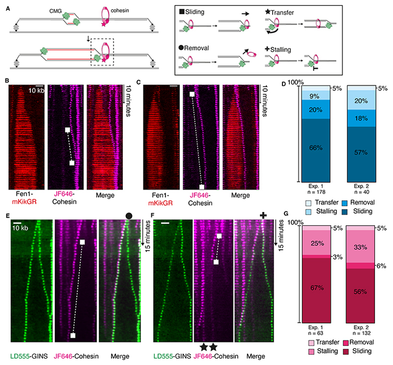
Newly copied sister chromatids are tethered together by the cohesin complex, but how sister chromatid cohesion coordinates with DNA replication is poorly understood. Prevailing models suggest that cohesin complexes, bound to DNA before replication, remain behind the advancing replication fork to keep sister chromatids together. By visualizing single replication forks colliding with preloaded cohesin complexes, we find that the replisome instead pushes cohesin to where a converging replisome is met. Whereas the converging replisomes are removed during DNA replication termination, cohesin remains on nascent DNA and provides cohesion. Additionally, we show that CMG (CDC45-MCM2-7-GINS) helicase disassembly during replication termination is vital for proper cohesion in budding yeast. Together, our results support a model wherein sister chromatid cohesion is established during DNA replication termination.


Conflict of interest statement

Competing interests

Authors declare that they have no competing interests.

Figures

Fig. 1
Fig. 1. Replisomes push cohesin during DNA replication.
(A) Cartoon showing DNA replication from a single origin on surface-tethered λ DNA. Replication is performed in Xenopus laevis egg extracts while collisions between replisomes and cohesins are visualized. (B and C) Collisions between replication forks, labelled with Fen1-mKiKGR (red), and pre-loaded JF646-cohesin (magenta) are visualized. Examples showing cohesin sliding ahead of a Fen1-mKikGR-labelled replication fork. (D) Comparison of primary cohesin fate after collision with replication forks in extracts. Two independent experiments are shown. (E and F) Representative kymograms showing LD555-GINS collision with JF646-cohesin. Examples of different cohesin fates (sliding, removal, transfer and fork stalling) are marked with symbols. (G) Proportions of cohesin fates after collision by labelled replisomes. Two independent experiments are shown.
Fig. 2
Fig. 2. Cohesin is pushed to positions of DNA replication termination.
(A and B) Kymograms showing replication forks colliding with JF646-cohesin complexes under conditions of high origin firing. After a period of replication, a high salt wash (HSW) was performed and the same DNAs were imaged. (C) Quantification of cohesin fates at converging replication forks. The fate of cohesin that was pushed to a converging replication fork was measured. Two independent experiments are shown. (D to F) Kymogram examples showing replisome (LD555-GINS) progression on DNA from multiple origins and colliding with JF646-cohesin. (G) Quantification of JF646-cohesin fate at sites where converging replisomes (LD555-GINS) are removed, with two independent experiments shown.
Fig. 3
Fig. 3. Cohesin holds together newly replicated surface-tethered sister DNAs.
(A) Diagram showing sister DNA collapse experiments. Replication forks are paused at DNA ends by LacI. Upon IPTG addition replication forks reach DNA ends and collapsing sister DNAs are visualized. (B) Example kymograms where both sister DNA strands collapse (see cartoon). The time that collapsed DNA strands colocalized before separating is indicated. (C) Time that collapsed DNA strands colocalized under different extract depletion conditions. n=3 independent experiments were performed, with the following number of individual molecules: Mock (n=32, 36, 59), Depletion (n=13, 40, 53), and Rescue (n=16, 41, 53). Data are mean ± SEM, compared with a two-sided t-test. (D) Percentage of sister DNAs that have separated from one another within 2 minutes of collapsing from both ends under different extract depletion conditions. (E) Example kymogram showing JF549-cohesin associating with both collapsed sister DNAs. The red arrow indicates cohesin at the end of DNA tethers, which is excluded from the analysis. (F) Quantification of cohesin position after sister DNA collapse of either one strand or both strands, with data from two independent experiments shown.
Fig. 4
Fig. 4. Cohesin dynamics during replication termination when replisome disassembly is inhibited.
(A) Replisomes were labeled directly using LD555-GINS and replisome disassembly upon fork convergence was inhibited with 200 μM p97i (NMS-873). In this example, converging replisomes bypass one another and one replisome continues pushing a labelled cohesin (white square). (B) Quantification of cohesin and replisome fates observed during replication termination when replisome disassembly is inhibited. (C) Plots showing timings of sister chromatid colocalization ±p97i. Data were collected from two experiments for each condition (DMSO, n = 124 and p97i, n = 140). (D) Model of cohesion establishment during replication termination. Possible scenarios describing termination-coupled cohesion establishment are shown in fig. S15.

References

    1. Yatskevich S, Rhodes J, Nasmyth K. Organization of Chromosomal DNA by SMC Complexes. Annu Rev Genet. 2019;53:445–482. - PubMed
    1. Davidson IF, Bauer B, Goetz D, Tang W, Wutz G, Peters JM. DNA loop extrusion by human cohesin. Science. 2019;366:1338–1345. - PubMed
    1. Kim Y, Shi Z, Zhang H, Finkelstein IJ, Yu H. Human cohesin compacts DNA by loop extrusion. Science. 2019;366:1345–1349. - PMC - PubMed
    1. Ciosk R, Shirayama M, Shevchenko A, Tanaka T, Toth A, Shevchenko A, Nasmyth K. Cohesin’s binding to chromosomes depends on a separate complex consisting of Scc2 and Scc4 proteins. Mol Cell. 2000;5:243–254. - PubMed
    1. Takahashi TS, Yiu P, Chou MF, Gygi S, Walter JC. Recruitment of Xenopus Scc2 and cohesin to chromatin requires the pre-replication complex. Nat Cell Biol. 2004;6:991–996. - PubMed

Publication types

MeSH terms

LinkOut - more resources