. 2024 Mar 15;383(6688):eadk4422.
doi: 10.1126/science.adk4422. Epub 2024 Mar 15.

Continuous evolution of compact protein degradation tags regulated by selective molecular glues

Affiliations

Continuous evolution of compact protein degradation tags regulated by selective molecular glues

Jaron A M Mercer et al. Science. .

Abstract

Conditional protein degradation tags (degrons) are usually >100 amino acids long or are triggered by small molecules with substantial off-target effects, thwarting their use as specific modulators of endogenous protein levels. We developed a phage-assisted continuous evolution platform for molecular glue complexes (MG-PACE) and evolved a 36-amino acid zinc finger (ZF) degron (SD40) that binds the ubiquitin ligase substrate receptor cereblon in complex with PT-179, an orthogonal thalidomide derivative. Endogenous proteins tagged in-frame with SD40 using prime editing are degraded by otherwise inert PT-179. Cryo-electron microscopy structures of SD40 in complex with ligand-bound cereblon revealed mechanistic insights into the molecular basis of SD40's activity and specificity. Our efforts establish a system for continuous evolution of molecular glue complexes and provide ZF tags that overcome shortcomings associated with existing degrons.


Figures

Figure 1.
Figure 1.
Phage-assisted continuous evolution circuit for molecular glue complexes (MG-PACE). (A) In PACE, the gene for a protein of interest (POI) is placed on a selection phage (SP) in place of the phage gene gIII, which encodes the phage coat protein pIII. Host cells harbor an accessory plasmid (AP) encoding a selection circuit that provides pIII and a mutagenesis plasmid (MP) that increases the incidence of mutation during SP replication. (B) Rapamycin-sensitive MG-PACE circuit. (C) Rapamycin-induced activation of the MG-PACE circuit (1 hr). (D) Pomalidomide and IMiD derivatives PT-179 and PK-1016. (E) Whole-transcriptome RNA-Seq of MM1.S cells treated with pomalidomide (1 μM, 48 hrs, left) or PT-179 (1 μM, 48 hrs, right). Values and error bars in (C) represent the mean and standard deviation of three replicates, each normalized to a control treatment with DMSO only. Values in (E) represent the mean of five replicates; p-values for each gene were calculated using the Wald test and adjusted using the Benjamini-Hochberg method (84).
Figure 2.
Figure 2.
Phage-assisted continuous evolution of new ZF degrons. (A) Full-length CRBN MG-PACE circuit. (B) CRBNCTD MG-PACE circuit. (C) Pomalidomide-induced activation of both CRBN MG-PACE circuits (1 hr). (D) PACE in CRBNCTD circuit. (E) PT-179-induced CRBNCTD circuit activation with evolved variants (1 hr). (F) PANCE in the full-length CRBN circuit. (G) PACE in the full-length CRBN circuit. (H) PT-179-induced full-length CRBN circuit activation with evolved variants (1 hr). (I) Mutations in PANCE- and PACE-evolved degron variants. SD17 and SD20 contain a frame-shift mutation recoding residues 57–60 and extending the reading frame by two residues. Values and error bars in (C), (E), and (H) represent the mean and standard deviation of three replicates, each normalized to a control treatment with DMSO only. Dashed vertical lines in (D), (F), and (G) represent neutral drift passages.
Figure 3.
Figure 3.
SD40 degrades exogenously- and endogenously-expressed tagged proteins in human HEK293T and K562 cells. (A) Cartoon representation of PT-179 recruiting a degron-tagged protein to the CUL4•RBX1•DDB1•CRBN E3 ligase complex. (B) PT-179-induced eGFP degradation (≥20 hrs) with evolved degrons in HEK293T cells. (C) Degradation of proteins fused to SD40 (24 hrs). The corresponding genes were genomically integrated in HEK293T cells using a lentiviral vector. (D) Time course of PT-179-induced degradation of genomically expressed SD40-PRKRA in HEK293T cells. (E) Insertion of a 108-bp sequence encoding SD40 in-frame in endogenous genomic target genes using twin prime editing. SD40 was precisely inserted before the endogenous PLK1 stop codon in HEK293T cells and after the endogenous BRD4 start codon in K562 cells. (F) Degradation of endogenous SD40-tagged PLK1 (2 hrs) and BRD4 (24 hrs) in cell lines generated by prime editing. (G) Thermodynamic and kinetic binding parameters for SD0 and SD40 binding to CRBN•pomalidomide or CRBN•PT-179. Values and error bars in (B) represent the mean and standard deviation of three replicates, normalized to control treatment with DMSO only. Values and error bars in (E) represent the mean and standard deviation of three replicates. Thermodynamic and kinetic binding parameters in (G) reflect the mean and standard error of three replicates.
Figure 4.
Figure 4.
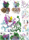
Cryo-EM structure of the ternary complex of SD40 with DDB1•CRBN bound to IMiD derivatives. (A) EM map of DDB1ΔBPB•CRBN•PT-179•SD40, colored by domains. Map obtained from local refinement on CRBN and sharpened using deepEMhancer (98). (B) Cartoon model of DDB1ΔBPB•CRBN•PT-179•SD40 with PT-179 highlighted in space-filling representation. (C) Comparison of the structures of SD40 bound to CRBN•PT-179 and to CRBN•pomalidomide. Backbone trace of DDB1ΔBPB•CRBN•pomalidomide•SD40 is shown in translucent gray superimposed on DDB1ΔBPB•CRBN•PT-179•SD40 in color aligned on the CRBN residues around the ligand. (D) Closeup of the cartoon model in (B) near the CRBN•PT-179•SD40 interface with key residues of SD40 shown in stick representation. (E) Interaction of F18 and PT-179 though stacking of the side chain against the morpholine ring of the compound. (F) A constellation of hydrophobic residues (P20, I36, F45, and L49) shown in space-filling representation. These residues pack tightly against each other and H353 of CRBN. (G) Additional interactions made by the C-terminal tail of SD40 with CRBNNTD. CRBN is shown in surface representation and SD40 is shown as a transparent surface and cartoon. SD40 residues contributing most to the interaction are shown in stick representation. (H) Interaction of Y47 and H48 of SD40 with CRBN residues. π–π stacking interactions with H103 in CRBNNTD and Y355 in CRBNCTD are highlighted. In panels E–H, figure insets show the map orientation, depth cue, and cropping applied to arrive at that panel.
Figure 5.
Figure 5.
MG-PACE evolves degrons that engage PT-179-bound mouse CRBN. (A) The mmCRBN MG-PACE circuit. Stringency is increased by expression of a competing ZF degron fused to maltose binding protein (MBP). (B) PACE in the mmCRBN MG-PACE circuit. (C) Mutations in evolved PT-179•mmCRBN-binding degrons. (D) PT-179-induced mmCRBN circuit activation (1 hr) by SD0 and evolved variants. (E) PT-179-induced degradation (≥20 hrs) of degron-tagged eGFP in mouse 3T3 cells. Values and error bars in (D) and (E) represent the mean and standard deviation of three replicates, each normalized to control treatment with DMSO only, except for the highest PT-179 concentration in (E), which used two replicates.

References

    1. Schreiber SL, The Rise of Molecular Glues. Cell 184, 3–9 (2021). - PubMed
    1. Raina K, Crews CM, Chemical Inducers of Targeted Protein Degradation. J. Biol. Chem 285, 11057–11060 (2010). - PMC - PubMed
    1. Fischer ES, Park E, Eck MJ, Thomä NH, SPLINTS: small-molecule protein ligand interface stabilizers. Curr. Opin. Struct. Biol 37, 115–122 (2016). - PMC - PubMed
    1. Bier D, Thiel P, Briels J, Ottmann C, Stabilization of Protein–Protein Interactions in chemical biology and drug discovery. Prog. Biophys. Mol. Biol 119, 10–19 (2015). - PubMed
    1. Krönke J, Udeshi ND, Narla A, Grauman P, Hurst SN, McConkey M, Svinkina T, Heckl D, Comer E, Li X, Ciarlo C, Hartman E, Munshi N, Schenone M, Schreiber SL, Carr SA, Ebert BL, Lenalidomide Causes Selective Degradation of IKZF1 and IKZF3 in Multiple Myeloma Cells. Science 343, 301–305 (2014). - PMC - PubMed

Publication types