. 2024 May 9;45(5):655-661.
doi: 10.3174/ajnr.A8165.

Optic Nerve Sheath MR Imaging Measurements in Patients with Orthostatic Headaches and Normal Findings on Conventional Imaging Predict the Presence of an Underlying CSF-Venous Fistula

Affiliations

Optic Nerve Sheath MR Imaging Measurements in Patients with Orthostatic Headaches and Normal Findings on Conventional Imaging Predict the Presence of an Underlying CSF-Venous Fistula

Wouter I Schievink et al. AJNR Am J Neuroradiol. .

Abstract

Background and purpose: Spontaneous spinal CSF leaks typically cause orthostatic headache, but their detection may require specialized and invasive spinal imaging. We undertook a study to determine the value of simple optic nerve sheath MR imaging measurements in predicting the likelihood of finding a CSF-venous fistula, a type of leak that cannot be detected with routine spine MR imaging or CT myelography, among patients with orthostatic headache and normal conventional brain and spine imaging findings.

Materials and methods: This cohort study included a consecutive group of patients with orthostatic headache and normal conventional brain and spine imaging findings who underwent digital subtraction myelography under general anesthesia to look for spinal CSF-venous fistulas.

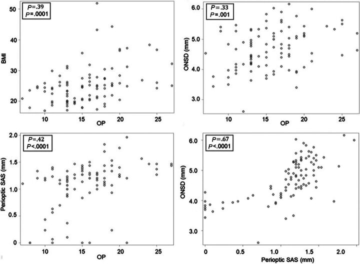
Results: The study group consisted of 93 patients (71 women and 22 men; mean age, 47.5 years; range, 17-84 years). Digital subtraction myelography demonstrated a CSF-venous fistula in 15 patients. The mean age of these 8 women and 7 men was 56 years (range, 23-83 years). The mean optic nerve sheath diameter was 4.0 mm, and the mean perioptic subarachnoid space was 0.5 mm in patients with a CSF-venous fistula compared with 4.9 and 1.2 mm, respectively, in patients without a fistula (P < .001). Optimal cutoff values were found at 4.4 mm for optic nerve sheath diameter and 1.0 mm for the perioptic subarachnoid space. Fistulas were detected in about 50% of patients with optic nerve sheath diameter or perioptic subarachnoid space measurements below these cutoff values compared with <2% of patients with optic nerve sheath diameter or perioptic subarachnoid space measurements above these cutoff values. Following surgical ligation of the fistula, optic nerve sheath diameter increased from 4.0 to 5.3 mm and the perioptic subarachnoid space increased from 0.5 to 1.2 mm (P < .001).

Conclusions: Concerns about a spinal CSF leak should not be dismissed in patients with orthostatic headache when conventional imaging findings are normal, and simple optic nerve sheath MR imaging measurements can help decide if more imaging needs to be performed in this patient population.


Figures

FIG 1.
FIG 1.
How to measure the ONSD and perioptic subarachnoid space. Illustration (A) depicting the measurements for the ONSD and the perioptic subarachnoid space. Pre- (B) and post- (C) operative coronal fat-suppressed T2-weighted MR imaging shows restoration of the perioptic subarachnoid space (arrows) following ligation of a spinal CSF-venous fistula.
FIG 2.
FIG 2.
ONSD and perioptic subarachnoid space measurements in patients with and without spinal CSF-venous fistulas. Box and whisker plots of the ONSD and perioptic SAS in patients without and with a spinal CSF-venous fistula. The box represents the upper and lower quartiles with the line splitting the box representing the median. The diamond represents the mean. The whiskers represent the upper and lower values of the data, up to 1.5 times the interquartile range. The single points represent the outliers.
FIG 3.
FIG 3.
Predicted probability of finding a spinal CSF-venous fistula based on the ONSD and perioptic subarachnoid space measurements. The graphs depict the predicted probability (and 80% confidence intervals) of identifying a CSF-venous fistula according to ONSD and perioptic SAS.
FIG 4.
FIG 4.
The relationship among the BMI, CSF opening pressure, ONSD, and perioptic subarachnoid space. Scatterplots depict the relationships among BMI, CSF OP (in centimeters CSF), ONSD, and the perioptic SAS. By ranking from the weakest to the strongest correlation, positive correlations are found between the ONSD and OP, BMI and OP, perioptic SAS and OP, and ONSD and perioptic SAS.
FIG 5.
FIG 5.
Normalization of the ONSD and perioptic subarachnoid space following ligation of spinal CSF-venous fistulas. Individual measurements of the pre- and postoperative ONSD and perioptic SAS following ligation of the spinal CSF-venous fistula.

References

    1. Schievink WI. Spontaneous intracranial hypotension. N Engl J Med 2021;385:2173–78 10.1056/NEJMra2101561 - DOI - PubMed
    1. Mehta D, Cheema S, Davagnanam I, et al. . Diagnosis and treatment evaluation in patients with spontaneous intracranial hypotension. Front Neurol 2023;14:1145949 10.3389/fneur.2023.1145949 - DOI - PMC - PubMed
    1. Schievink WI. Spontaneous spinal cerebrospinal fluid leaks and intracranial hypotension. JAMA 2006;295:2286–96 10.1001/jama.295.19.2286 - DOI - PubMed
    1. D’Antona L, Jaime Merchan MA, Vassiliou A, et al. . Clinical presentation, investigation findings, and treatment outcomes of spontaneous intracranial hypotension syndrome: a systematic review and meta-analysis. JAMA Neurol 2021;78:329–37 10.1001/jamaneurol.2020.4799 - DOI - PMC - PubMed
    1. Schievink WI, Maya MM, Moser FG, et al. . Lateral decubitus digital subtraction myelography to identify spinal CSF-venous fistulas in spontaneous intracranial hypotension. J Neurosurg Spine 2019;31:1–4 10.3171/2019.6.SPINE19487 - DOI - PubMed

MeSH terms

LinkOut - more resources