

Germline HAVCR2/TIM-3 Checkpoint Inhibitor Receptor Deficiency in Recurrent Autoinflammatory Myocarditis
- PMID: 38485795
- PMCID: PMC10940375
- DOI: 10.1007/s10875-024-01685-x
Germline HAVCR2/TIM-3 Checkpoint Inhibitor Receptor Deficiency in Recurrent Autoinflammatory Myocarditis
Abstract
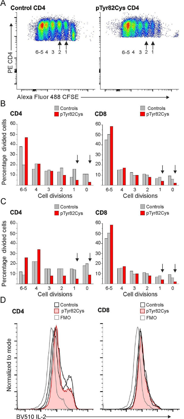
Myocarditis can be caused by viral infection, drug reaction or general inflammatory condition. To provide understanding on inflammatory myocarditis, we describe clinical, genetic, and immunological properties of a young male patient who suffered from recurrent myocarditis episodes since the age of four years. Electrocardiography, troponin I/T, echocardiography, myocardial magnetic resonance imaging and histological findings were consistent with recurrent myocarditis episodes. Homozygous c.245 A > G p.Tyr82Cys pathogenic variant in Hepatitis A Virus Cellular Receptor 2 (HAVCR2) gene encoding T cell immunoglobulin and mucin domain-containing protein 3 (TIM-3) receptor was found. Peripheral blood mononuclear cells were collected when the patient was asymptomatic; CD4+ and CD8+ T lymphoblasts, CD56+ natural killer cells and CD14+ monocytes were negative for surface TIM-3 expression. In vitro, TLR4 mediated interleukin-1β (IL-1β) response was high after LPS/ATP stimulation. Clinical symptoms responded to IL-1 receptor antagonist anakinra. TIM-3 p.Tyr82Cys CD4+ and CD8+ T cell proliferation in vitro was unrestrained. Findings on IL-2, interferon gamma, regulatory T cells, signal transducer and activator of transcription (STAT) 1, 3 and 4 phosphorylation, and PD-1 and LAG-3 checkpoint inhibitor receptor analyses were comparable to controls. We conclude that TIM-3 deficiency due to homozygous HAVCR2 c.245 A > G p.Tyr82Cys pathogenic variant in the patient described here is associated with autoinflammatory symptoms limited to early onset recurrent febrile myocarditis. Excessive IL-1β production and defective regulation of T cell proliferation may contribute to this clinical condition responsive to anakinra treatment.
Keywords: Autoinflammation; Inborn error in immunity; Interleukin-1; Myocarditis; T-lymphocytes.
© 2024. The Author(s).
Conflict of interest statement
The authors declare no competing interests.
Figures


References
-
- Frisancho-Kiss S, Nyland JF, Davis SE, Barrett MA, Gatewood SJL, Njoku DB et al. Cutting Edge: T Cell Ig Mucin-3 Reduces Inflammatory Heart Disease by Increasing CTLA-4 during Innate Immunity [Internet]. The Journal of Immunology. 2006. p. 6411–5. 10.4049/jimmunol.176.11.6411. - PubMed
-
- Gayden T, Sepulveda FE, Khuong-Quang D-A, Pratt J, Valera ET, Garrigue A, et al. Germline HAVCR2 mutations altering TIM-3 characterize subcutaneous panniculitis-like T cell lymphomas with hemophagocytic lymphohistiocytic syndrome. Nat Genet. 2018;50:1650–7. doi: 10.1038/s41588-018-0251-4. - DOI - PubMed
Publication types
MeSH terms
Substances
LinkOut - more resources
Full Text Sources
Molecular Biology Databases
Research Materials
