. 2024 Mar 14;15(1):1876.
doi: 10.1038/s41467-024-46194-y.

Harnessing whole human liver ex situ normothermic perfusion for preclinical AAV vector evaluation

Affiliations

Harnessing whole human liver ex situ normothermic perfusion for preclinical AAV vector evaluation

Marti Cabanes-Creus et al. Nat Commun. .

Abstract

Developing clinically predictive model systems for evaluating gene transfer and gene editing technologies has become increasingly important in the era of personalized medicine. Liver-directed gene therapies present a unique challenge due to the complexity of the human liver. In this work, we describe the application of whole human liver explants in an ex situ normothermic perfusion system to evaluate a set of fourteen natural and bioengineered adeno-associated viral (AAV) vectors directly in human liver, in the presence and absence of neutralizing human sera. Under non-neutralizing conditions, the recently developed AAV variants, AAV-SYD12 and AAV-LK03, emerged as the most functional variants in terms of cellular uptake and transgene expression. However, when assessed in the presence of human plasma containing anti-AAV neutralizing antibodies (NAbs), vectors of human origin, specifically those derived from AAV2/AAV3b, were extensively neutralized, whereas AAV8- derived variants performed efficiently. This study demonstrates the potential of using normothermic liver perfusion as a model for early-stage testing of liver-focused gene therapies. The results offer preliminary insights that could help inform the development of more effective translational strategies.


Conflict of interest statement

L.L. and I.E.A. are co-founders of Exigen Biotherapeutics, a company that utilizes similar technologies broadly discussed in this paper. M.C.-C., I.E.A., and L.L. are inventors on patent applications filed by Children’s Medical Research Institute related to AAV capsid sequences and in vivo function of novel AAV variants (WO2021168509). The remaining authors declare no competing interests.

Figures
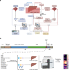
Fig. 1
Fig. 1. Study design.
a Graphical flow chart of the liver perfusion machine. b Transgene composition and study design. CMV Cytomegalovirus, ITR Inverted Terminal Repeat.
Fig. 2
Fig. 2. Performance parameters during ex situ normothermic perfusion.
ac Course of perfusate flows, lactate concentration and bile flow throughout the perfusion of Donor 1. d Liver histology of representative core biopsies from Donor 1 at the indicated time points (n = 2 core biopsies per time point). Scale: 100 μm. eg Course of perfusate flows, lactate concentration and bile flow throughout the perfusion of Donor 2. h Liver histology of representative core biopsies from Donor 2 at the indicated time points (n = 2 core biopsies per time point). Scale: 100 μm. PV Portal Vein, HA Hepatic Artery, LLSG Left Lateral Sector Graft, ERG Extended Right Graft.
Fig. 3
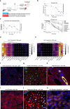
Fig. 3. Functional evaluation of AAV vectors in the ex situ human liver perfused with non-neutralizing human plasma.
a Graphical representation of the study. b Concentration of total (top panel) and de-multiplexed AAV vector genomes in the perfusate (bottom panel). Concentration of individual vectors is expressed as the estimated percentage of the initial concentration as studied with NGS. Vectors are ranked from most to least abundant (top to bottom). c Total vector genomes per haploid cell found in biopsies from both liver grafts extracted at the indicated perfusion times. d Percentage of NGS reads mapped to each barcoded AAV capsid variant. The transgene DNA, indicating vector uptake, was extracted from biopsies taken from both grafts at the indicated time points. e Similar analysis performed on transgenes recovered from RNA, which indicate functional transduction. Percentages are normalized to the pre-injection mix. f Immunofluorescence analysis of the net eGFP signal from collective AAV transduction in the left graft. g Similar immunofluorescence image for the right graft. The vector-encoded eGFP was also counterstained with an anti-eGFP antibody (red). Blue: DAPI (nuclei). Scale = 100 μm. h Immunofluorescence analysis of the bile ducts and hepatocytes of the left graft. Yellow: Cytokeratin 7 (bile ducts); red: albumin; blue: DAPI (nuclei). Scale: 50 μm. i Immunofluorescence analysis of liver zonation. Red: Histidine ammonia-lyase (HAL); blue: DAPI (nuclei). Scale: 500 μm. j Immunofluorescence analysis of liver integrity. Red: albumin; Purple: Hepatocyte nuclear factor 4 alpha (HNF4α); blue: DAPI (non-hepatocyte nuclei). Scale: 20 μm. k Immunofluorescence analysis of AAV Receptor (AAVR) in the human liver. Orange: AAVR; Blue: DAPI (nuclei). Scale: 100 μm.
Fig. 4
Fig. 4. Functional evaluation of AAV vectors in the ex situ human liver perfused with neutralizing human plasma.
a Graphical representation of the study. b Concentration of total (top panel) and de-multiplexed AAV vector genomes in the perfusate (bottom panel). Concentration of individual vectors is expressed as the estimated percentage of the initial concentration as studied with NGS. Vectors are ranked from most to least abundant (top to bottom). c Total vector genomes per haploid cell found in biopsies the left graft. d Percentage of NGS reads mapped to each barcoded AAV capsid variant. The transgene DNA, indicating vector uptake, was extracted from biopsies taken from both grafts at the indicated time points. e Similar analysis performed on transgenes recovered from RNA, which indicate functional transduction. Percentages are normalized to the pre-injection mix. f Immunofluorescence analysis of the net eGFP signal from collective AAV transduction in the left graft. The vector-encoded eGFP was also counterstained with an anti-eGFP antibody (red). Blue: DAPI (nuclei). Scale = 100 μm. g Immunofluorescence analysis of the presence of Kupffer cells in the left graft. Green: vector-encoded eGFP; Red: CD68 (Human macrophage marker); blue: DAPI (nuclei). Scale 100 μm. h Immunofluorescence analysis of the bile ducts and hepatocytes of the left graft. Yellow: Cytokeratin 7 (bile ducts); red: albumin; blue: DAPI (nuclei). Scale: 50 μm. i Immunofluorescence analysis of liver zonation. Red: Histidine ammonia-lyase (HAL); blue: DAPI (nuclei). Scale: 500 μm. j Immunofluorescence analysis of liver integrity. Red: albumin; Purple: Hepatocyte nuclear factor 4 alpha (HNF4α); blue: DAPI (non-hepatocyte nuclei). Scale: 20 μm. k Immunofluorescence analysis of AAV Receptor (AAVR) in the human liver. Orange: AAVR; Blue: DAPI (nuclei). Scale: 100 μm.

References

    1. Au HKE, Isalan M, Mielcarek M. Gene therapy advances: a meta-analysis of AAV usage in clinical settings. Front Med (Lausanne) 2021;8:809118. doi: 10.3389/fmed.2021.809118. - DOI - PMC - PubMed
    1. Kishimoto TK, Samulski RJ. Addressing high dose AAV toxicity - ‘one and done’ or ‘slower and lower’? Expert Opin. Biol. Ther. 2022;22:1067–1071. doi: 10.1080/14712598.2022.2060737. - DOI - PubMed
    1. Mergental H, et al. Transplantation of discarded livers following viability testing with normothermic machine perfusion. Nat. Commun. 2020;11:2939. doi: 10.1038/s41467-020-16251-3. - DOI - PMC - PubMed
    1. Lau NS, et al. Prolonged ex vivo normothermic perfusion of a split liver: an innovative approach to increase the number of available grafts. Transpl. Direct. 2021;7:e763. doi: 10.1097/TXD.0000000000001216. - DOI - PMC - PubMed
    1. Lau NS, et al. Long-term normothermic perfusion of human livers for longer than 12 days. Artif. Organs. 2022;46:2504–2510. doi: 10.1111/aor.14372. - DOI - PMC - PubMed

Substances