. 2024 Mar 14;10(1):140.
doi: 10.1038/s41420-024-01910-x.

MCT4-driven CAF-mediated metabolic reprogramming in breast cancer microenvironment is a vulnerability targetable by miR-425-5p

Affiliations

MCT4-driven CAF-mediated metabolic reprogramming in breast cancer microenvironment is a vulnerability targetable by miR-425-5p

Alessandra Affinito et al. Cell Death Discov. .

Abstract

Multiple oncogenic alterations contribute to breast cancer development. Metabolic reprogramming, deeply contributing to tumor microenvironment (TME) education, is now widely recognized as a hallmark of cancer. The reverse Warburg effect induces cancer-associated fibroblasts (CAFs) to produce and secrete L-lactate, enhancing malignant characteristics such as neoangiogenesis, metastatic dissemination, and treatment resistance. Monocarboxylate transporter (MCT) 4 is involved in lactate efflux from CAFs into stromal and epithelial cells. Here, we first assess the expression of miR-425-5p and its target MCT4 in breast cancer CAFs and normal fibroblasts. We analyzed the metabolic changes induced by miR-425-5p in CAFs and its role in the education of breast cancer epithelial cells. We show that miR-425-5p-induced MCT4 knockdown decreased lactate extrusion from CAFs and its availability in the TME. miR-425-5p overexpression induced profound metabolic transformation in CAFs, ultimately influencing breast cancer metabolism. Furthermore, miR-425-5p impaired the capacity of CAFs to sustain vessel formation and breast cancer cell migration, viability, and proliferation. These findings emphasize the key role of miR-425-5p in breast cancer metabolism and aggressiveness, and its possible importance for breast cancer therapy and monitoring.


Conflict of interest statement

All authors have read and approved the manuscript, its contents, and its submission and disclose no potential conflict of interest.

Figures
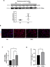
Fig. 1
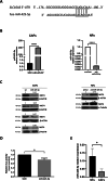
Fig. 1. MCT4 is highly expressed in Cancer-Associated Fibroblasts.
Representative Western blot analysis of MCT4 performed on plasmamembrane fractions of Cancer-Associated Fibroblasts (CAFs) (n = 4) and Normal Fibroblasts (NFs) (n = 3). Na + /K+ ATPase was used for data normalization of the fractionation in the sample preparation. Densitometry data are expressed after normalization on Na + /K+ ATPase band intensity A. A representative confocal microscopy image of MCT4 expression in CAFs (n = 3) (DAPI blue, MCT4 red) B. Three different subpopulations of two primary fibroblasts were collected at different growing distances from the tumor (intratumoral, IT; peritumoral, PT; normal, NF) and MCT4 gene expression was evaluated by quantitative real-time PCR. Values are expressed as a mean of two biological replicates of technical duplicates C. Culture media from NFs (n = 2) and CAFs (n = 2) were collected and assessed by a colorimetric L-lactate assay. Values are expressed as mean of two technical replicates D. Graph shows mean ± SD. * P values < 0.05; ***p < 0.001. Bar = 100 μm.
Fig. 2
Fig. 2. MCT4 is a direct target of miR-425-5p.
Bioinformatics predictions (TargetScan and miRANDA) indicated miR-425-5p as a putative miRNA targeting MCT4 mRNA A. Representative Real Time PCR for miR-425-5p upon miR-425-5p and anti miR-425-5p transfection in Cancer-Associated Fibroblasts (CAF n = 1) and Normal Fibroblasts (NF n = 1) respectively. Data are expressed as mean of two technical replicates B. Representative Western Blot analysis of MCT4 upon miR-425-5p and anti-miR-425 transfection in CAFs (n = 4) and NFs (n = 2) respectively C. Dual luciferase assay of the predicted binding site for miR-425-5p on the 3’UTR region of SLC16A3. Data are expressed as mean of two technical replicates D. Quantitative Real Time PCR for miR-425-5p performed on CAFs (n = 3) and NFs (n = 2), to evaluate miR-425-5p levels. Values are expressed as mean of two technical replicates E. Graph shows mean ± SD over control. * P values < 0.05.
Fig. 3
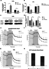
Fig. 3. miR425-5p-induced metabolic changes in Cancer-Associated Fibroblasts.
L-lactate assessed with a colorimetric assay in Cancer-Associated Fibroblasts (CAFs n = 2) and culture media 72 h after miR-425-5p transfection. Values are expressed as a mean of two biological replicates of technical duplicates A. L-lactate assessed in Normal Fibroblasts (NFs n = 2) and culture media 48 h after antimiR-425-5p transfection. Values are expressed as a mean of two biological replicates of technical duplicates B. Representative Western blot C and real-time analysis D of GLUT-1 expression in CAFs (n = 2) upon miR-425-5p transfection. Data are expressed as mean of two technical replicates. Metabolic changes in CAFs transfected with miR-425-5p, as assessed by glycolytic proton efflux rate (glycoPER) kinetics E, extracellular acidification rate (ECAR) F, proton efflux rate (PER) G, and ATP production H. Data are presented as a mean of two biological replicates of eight technical replicates. Data are mean percentage ± SD over control. * P values < 0.05; ** P values < 0.01.
Fig. 4
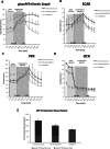
Fig. 4. miR-425-5p reverts MCT4-mediated metabolic shift resulting in reprogramming of Cancer-Associated Fibroblasts.
Metabolic changes in MCT4-transfected MS-5 cells with or without miR-425-5p overexpression, as assessed by glycolytic proton efflux rate (glycoPER) kinetics A, proton efflux rate (PER) B, extracellular acidification rate (ECAR) C, oxygen consumption rate OCR D, and ATP production E. Data are presented as a mean of biological duplicates of eight technical replicates ± SD over control.
Fig. 5
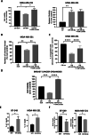
Fig. 5. Metabolic reprogramming of breast cancer cells by conditioned medium (CM) from miR-425-5p-overexpressing Cancer-Associated Fibroblasts.
Cell viability and caspase 3-activation A, colony formation assay B, and migration assay C of MDA-MB-231 cells treated with CM from Cancer-Associated Fibroblasts (CAFs) overexpressing miR-425-5p or a scrambled sequence. 1 mM of lactate was used in rescue experiments. Cell viability in luminal B-type 3D BC organoids upon treatment with CM from CAFs overexpressing miR-425-5p or a scrambled sequence. Data are expressed as mean of two biological replicates D. Cell death E and cell viability F of CMDFA-labelled BT-549 or MDA-MB-231 is shown upon co-culture with CAFs overexpressing miR-425-5p/scrambled sequence. Data are Values are expressed as biological means (n = 2) of technical triplicates. * P values < 0.05; ** P values < 0.01; *** P values < 0.001, **** P values < 0.0001.
Fig. 6
Fig. 6. miR-425-5p overexpression in stromal fibroblasts modulates angiogenesis in the tumor microenvironment.
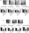
Representative images and quantifications of tube formation by HUVECs after culture in conditioned medium (CM) from miR-425-5p-overexpressing Cancer-Associated Fibroblasts (CAFs), compared to the control. 1 mM of lactate was used in rescue experiments A. Representative images and quantifications of tube formation by HUVECs cultured in CM from Normal Fibroblasts (NFs) with downregulated miR-425-5p, compared to the control. 1 mM of lactate was used in rescue experiments B. Data are presented as mean of six different images for each experimental point. * P values < 0.05; ** P values < 0.01; *** P values < 0.001. Bar = 500 μm.
Fig. 7
Fig. 7. Schematic representation of miR-425-5p’s mechanisms of action.
MiR-425-5p alters the metabolism of breast Cancer-associated Fibroblasts (CAFs) by downregulating MCT4 and indirectly reducing GLUT1, leading to decreased lactate extrusion. The interaction between miR-425-5p reprogrammed CAFs and breast cancer epithelial cells has a significant impact on breast cancer cell viability, proliferation, and migration. Angiogenesis is also influenced. This graphical abstract was drawn by using and adapting pictures from Servier Medical Art (Smart.Servier.com), provided by Servier, licensed under a Creative Commons Attribution 3.0 unported license (http://creativecommons.org/licenses/by/3.0/).

References

    1. Houthuijzen JM, Jonkers J. Cancer-associated fibroblasts as key regulators of the breast cancer tumor microenvironment. Cancer Metastasis Rev. 2018;37:577–97. doi: 10.1007/s10555-018-9768-3. - DOI - PubMed
    1. Eiro N, Gonzalez L, Martinez-Ordonez A, Fernandez-Garcia B, Gonzalez LO, Cid S, et al. Cancer-associated fibroblasts affect breast cancer cell gene expression, invasion and angiogenesis. Cell Oncol. (Dordr.) 2018;41:369–78. doi: 10.1007/s13402-018-0371-y. - DOI - PubMed
    1. Dou D, Ren X, Han M, Xu X, Ge X, Gu Y, et al. Cancer-associated fibroblasts-derived exosomes suppress immune cell function in breast cancer via the miR-92/PD-L1 pathway. Front Immunol. 2020;11:2026. doi: 10.3389/fimmu.2020.02026. - DOI - PMC - PubMed
    1. Arcucci A, Ruocco MR, Granato G, Sacco AM, Montagnani S. Cancer: an oxidative crosstalk between solid tumor cells and cancer associated fibroblasts. BioMed. Res. Int. 2016;2016:4502846. doi: 10.1155/2016/4502846. - DOI - PMC - PubMed
    1. Xing Y, Zhao S, Zhou BP, Mi J. Metabolic reprogramming of the tumour microenvironment. FEBS J. 2015;282:3892–8. doi: 10.1111/febs.13402. - DOI - PubMed