. 2024 Mar;56(3):700-710.
doi: 10.1038/s12276-024-01166-6. Epub 2024 Mar 14.

Cancer-derived exosomal Alu RNA promotes colorectal cancer progression

Affiliations

Cancer-derived exosomal Alu RNA promotes colorectal cancer progression

Sara Magliacane Trotta et al. Exp Mol Med. 2024 Mar.

Abstract

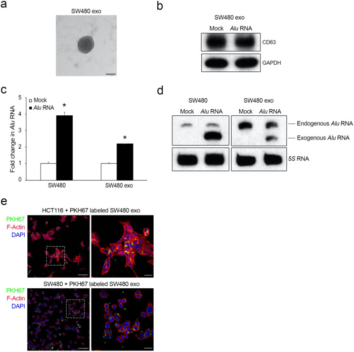
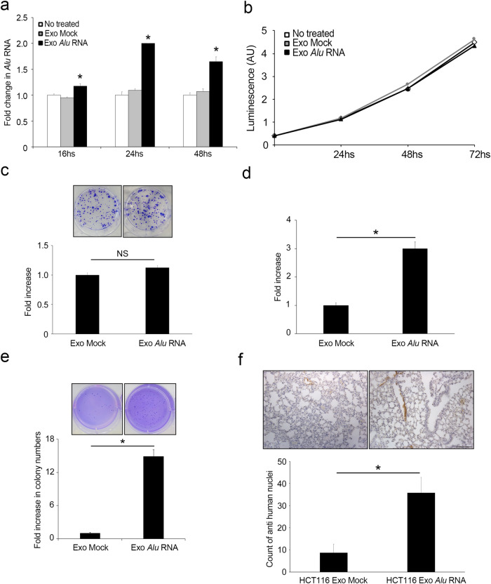
Inflammation plays a crucial role in cancer progression, but the relevance of the inflammasome remains unclear. Alu RNA was the first endogenous nucleic acid shown to activate the NLRP3 (nucleotide-binding domain leucine-rich repeat containing 3) inflammasome. Here, we showed that Alu RNA can induce epithelial-to-mesenchymal transition (EMT) through NLRP3 inflammasome activation and IL-1β release in colorectal cancer (CRC) cells. Alu RNA is stored, transported and transferred to CRC cells by exosomes. Exosomal Alu RNA promotes tumorigenesis by inducing invasion, metastasis and EMT via NLRP3 inflammasome activation. Consistent with these data, we found that significantly increased Alu RNA expression correlates with the induction of NLRP3 priming in human CRC patients. Furthermore, the level of Alu RNA in circulating exosomes correlates with CRC progression in a preclinical model. These findings reveal the direct involvement of Alu RNA in cancer pathogenesis, and its presence in CRC cell-derived exosomes could be used as a noninvasive diagnostic biomarker.


Conflict of interest statement

The authors declare no competing interests.

Figures

Fig. 1
Fig. 1. Alu RNA induces EMT through NLRP3 inflammasome activation in CRC cell lines.
a Alu RNA exerts cytotoxic effects on HCT116 and LS174T cells but does not alter the viability of HT29 and SW480 cells, as determined by the MTT assay (n = 3). b Dose-dependent effects of Alu RNA on HCT116 cell viability, as determined by the MTT assay (n = 3). c Western blotting analysis showing the activation of Caspase-1, increased expression of the mesenchymal marker Fibronectin, decreased expression of E-cadherin and increased expression in the EMT transcription factor ZEB1 in HCT116 cells after transfection with high or low levels of Alu RNA. Densitometric values are normalized to Vinculin and are shown in parentheses. d High and low Alu RNA levels induce the secretion of IL-1β from HCT116 cells, as measured by ELISA. IL-1β protein levels were normalized to the number of adherent cells (n = 3). e Representative images of HCT116 cells transfected with vehicle (Mock, left), high Alu RNA levels (middle), or low Alu RNA levels (right) and stained for F-actin with rhodamine phalloidin (red). Nuclei were counterstained with 4’,6-diamidino-2-phenylindole (DAPI, blue). Scale bar: 100 μm. f Transfection with an siRNA targeting NLRP3 (siNLRP3) reduced the abundance of the target mRNA in HCT116 cells compared with that in cells transfected with a control siRNA (siLuc), as determined by qRT‒PCR (n = 3). siNLRP3 transfection prevented Alu-induced EMT in HCT116 cells, as determined by (g) western blotting and h immunofluorescence analyses. Alu RNA (central lane, g) induces Caspase-1 activation as well as EMT, decreasing E-cadherin expression and increasing Fibronectin and ZEB1 expression compared to those in the Mock group (left lane, g). Transfection of siNLPR3-knockdown cells with Alu RNA prevented Alu-induced EMT and inhibited Alu-induced Caspase-1 activation (right lane, g). Densitometric values are normalized to Vinculin and are shown in parentheses. h Representative images of F-actin (red) staining. Nuclei were counterstained with DAPI (blue). Scale bar: 100 μm. For all panels, *p < 0.05; NS = not statistically significant. The error bars denote the s.e.m.
Fig. 2
Fig. 2. Alu RNA is stored and transported by exosomes by CRC cells.
a TEM image showing vesicles with morphologies and sizes that are characteristic of exosomes. Scale bar: 50 nm. b Western blotting analysis of CD63 levels in exosomes that were purified by ultracentrifugation from the culture supernatants of SW480 cells (SW480 Exo) that were transfected with Alu RNA or vehicle (Mock). GAPDH was used as a loading control. c qRT‒PCR results showing the abundance of Alu RNA in exosomes derived from Alu RNA-transfected SW480 cells (SW480 Exo) compared to that in exosomes derived from vehicle-transfected cells (Mock). On the left, as a control, the same analysis was performed on Alu RNA- or vehicle (Mock)-transfected SW480 cells (n = 3); *p < 0.05. Error bars denote the s.e.m. d Northern blotting analysis shows the abundance of Alu RNA in exosomes from SW480 cells (SW480 Exo) transfected with Alu RNA compared to those from vehicle-transfected cells (Mock). On the left, as a control, the same analysis was performed on Alu RNA- or vehicle (Mock)-transfected SW480 cells. We observed a band corresponding to endogenous Alu RNA (approximately 300 nucleotides in length) and a faster band corresponding to the transfected exogenous Alu RNA (281 nucleotides in length). 5S RNA was used as a loading control (n = 3). e Representative confocal images of PKH67-labeled SW480-derived exosomes (green) in HCT116 and SW480 cells that were stained for F-actin with rhodamine phalloidin (red). Nuclei were counterstained with DAPI (blue). Scale bar: 100 μm. A higher magnification is shown on the right. Scale bar: 25 μm.
Fig. 3
Fig. 3. Cancer-derived exosomal Alu RNA promotes tumorigenesis and metastasis dissemination.
a qRT‒PCR analysis of Alu RNA expression in HCT116 cells treated with exosomes from Alu-transfected SW480 cells (Exo Alu) after 16, 24 and 48 hours compared with that in HCT116 cells treated with control exosomes (Exo Mock) or untreated cells (n = 3); *p < 0.05. Error bars denote the s.e.m. b Exosomal Alu RNA did not alter HCT116 cell viability over time, as determined by luminescence assay (n = 3). c Exosomal Alu RNA does not alter the ability of HCT116 cells to form colonies. The data are expressed as the fold increase compared with HCT116 cells treated with Exo Mock (n = 3). Error bars denote the s.e.m. Representative images of the two conditions are shown. d Exosomal Alu RNA increases HCT116 cell invasion. The data are expressed as the fold increase compared with HCT116 cells treated with Exo Mock (n = 3); *p = 0.019. Error bars denote the s.e.m. e Exosomal Alu RNA increases the ability of HCT116 cells to grow in an anchorage-dependent manner. The bars indicate the average number of colonies in each well (n = 3); *p = 0.001. Error bars denote the s.e.m. Representative images of the two conditions are shown. f Exosomal Alu RNA increases the metastatic potential of HCT116 cells in vivo. The bars indicate the average number of human nuclei-positive cells in lung sections from mice that were injected with Exo Alu-pretreated HCT116 cells via the tail vein compared to that in lung sections from mice that were injected Exo Mock-pretreated HCT116 cells; *p = 0.012. Error bars denote the s.e.m. Representative images of anti-human nuclei-stained lung sections are shown. Scale bar: 100 μm.
Fig. 4
Fig. 4. Exosomal Alu RNA induces tumorigenesis via NLRP3 inflammasome activation.
a Exosomal Alu RNA induces NLRP3 inflammasome priming by increasing the mRNA expression of NLRP3 and IL-1B in HCT116 cells, as shown by qRT‒PCR (n = 3); *p < 0.05. No difference in IL18 gene expression was observed. The error bars denote the s.e.m. b Western blotting analysis showing the activation of Caspase-1 (p20) in HCT116 cells that were treated with Exo Alu. Densitometric values are normalized to β-Tubulin and are shown in parentheses. c Western blotting analysis (above) showing a decrease in E-cadherin expression, an increase in mesenchymal marker expression (Fibronectin and Vimentin), and an increase in EMT-TF ZEB1 expression in HCT116 cells treated with Exo Alu. Densitometric values are normalized to Vinculin and are shown in parentheses. Below, representative images of HCT116 cells that were treated with Exo Mock or Exo Alu and stained for F-actin (red). Nuclei were counterstained with DAPI (blue). Scale bar: 100 μm. d OLT1177 (100 μM) reduces exosomal Alu RNA-induced HCT116 cell invasion. The data are expressed as the fold increase compared with HCT116 cells treated with Exo Mock (n = 3); *p = 0.015; **p = 0.027. Error bars denote the s.e.m. e Anti-IL1β (500 ng/ml) reduces exosomal Alu RNA-induced HCT116 cell invasion. The data are expressed as the fold increase compared with HCT116 cells that were treated with Exo Mock (n = 3); *p = 0.026; **p = 0.047. Error bars denote the s.e.m. f NLRP3 and IL-1B mRNA expression in 13 matched nontumoral (NT) tissues, primary colon tumor (T) tissues and liver metastasis (M) tissues from patients was evaluated by qRT‒PCR and normalized to 18S RNA expression; *p = 0.03; **p = 0.014; ***p = 0.019. The error bars denote the s.e.m.
Fig. 5
Fig. 5. Exosomal Alu RNA abundance is correlated with CRC progression.
a Northern blotting analysis showing the abundance of Alu RNA in exosomes that were purified by ultracentrifugation from serum of xenotransplanted mice bearing large (n = 8; 1.52 ± 0.09) or small CRC tumors (n = 6; 0.88 ± 0.14). U6 RNA was used as a loading control. b qRT‒PCR analysis showing Alu RNA levels in exosomes purified from the serum of xenotransplanted mice bearing small (n = 8; 0.68 g±0.11) or large (n = 6; 1.58 g±0.19) CRC tumors, as evaluated by qRT‒PCR and normalized to human 18S RNA; *p = 0.037. c qRT‒PCR analysis showing Alu RNA levels in exosomes that were purified from the serum of mice that were intravenously injected with Exo Alu- or Exo Mock-pretreated HCT116 cells. The values were normalized to human 18S RNA; *p = 0.025. The error bars denote the s.e.m.

References

    1. Deininger P. Alu elements: know the SINEs. Genome Biol. 2011;12:236. doi: 10.1186/gb-2011-12-12-236. - DOI - PMC - PubMed
    1. Batzer MA, Deininger PL. Alu repeats and human genomic diversity. Nat Rev Genet. 2002;3:370–379. doi: 10.1038/nrg798. - DOI - PubMed
    1. Häsler J, Samuelsson T, Strub K. Useful ‘junk’: Alu RNAs in the human transcriptome. Cell Mol Life Sci. 2007;64:1793–1800. doi: 10.1007/s00018-007-7084-0. - DOI - PMC - PubMed
    1. Burns KH. Repetitive DNA in disease. Science. 2022;376:353–354. doi: 10.1126/science.abl7399. - DOI - PubMed
    1. Kaneko H, et al. DICER1 deficit induces Alu RNA toxicity in age-related macular degeneration. Nature. 2011;471:325–330. doi: 10.1038/nature09830. - DOI - PMC - PubMed

MeSH terms