. 2024 Mar 14;29(1):37.
doi: 10.1186/s11658-024-00547-z.

MRE11A: a novel negative regulator of human DNA mismatch repair

Affiliations

MRE11A: a novel negative regulator of human DNA mismatch repair

Demin Du et al. Cell Mol Biol Lett. .

Abstract

Background: DNA mismatch repair (MMR) is a highly conserved pathway that corrects DNA replication errors, the loss of which is attributed to the development of various types of cancers. Although well characterized, MMR factors remain to be identified. As a 3'-5' exonuclease and endonuclease, meiotic recombination 11 homolog A (MRE11A) is implicated in multiple DNA repair pathways. However, the role of MRE11A in MMR is unclear.

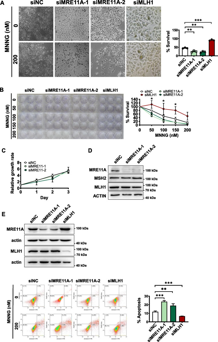
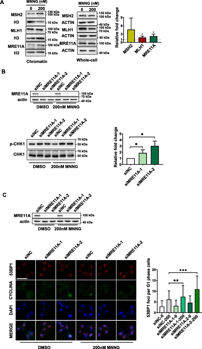
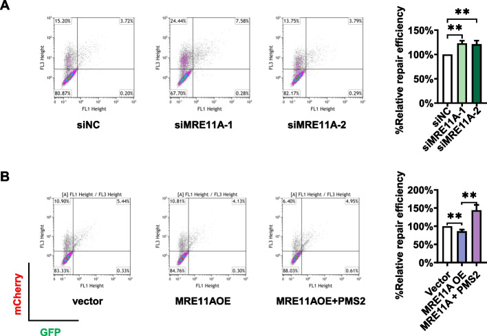
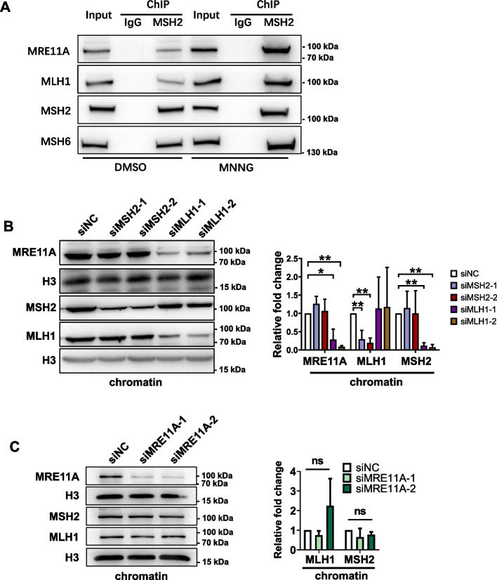
Methods: Initially, short-term and long-term survival assays were used to measure the cells' sensitivity to N-methyl-N'-nitro-N-nitrosoguanidine (MNNG). Meanwhile, the level of apoptosis was also determined by flow cytometry after MNNG treatment. Western blotting and immunofluorescence assays were used to evaluate the DNA damage within one cell cycle after MNNG treatment. Next, a GFP-heteroduplex repair assay and microsatellite stability test were used to measure the MMR activities in cells. To investigate the mechanisms, western blotting, the GFP-heteroduplex repair assay, and chromatin immunoprecipitation were used.

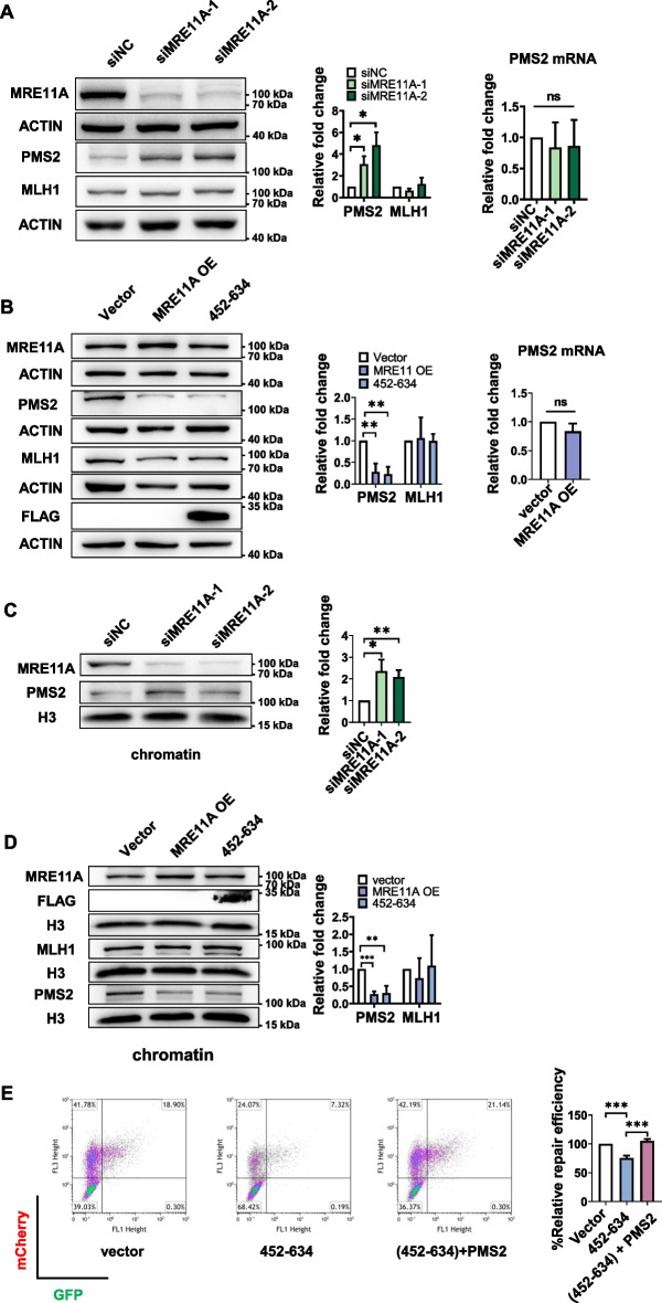
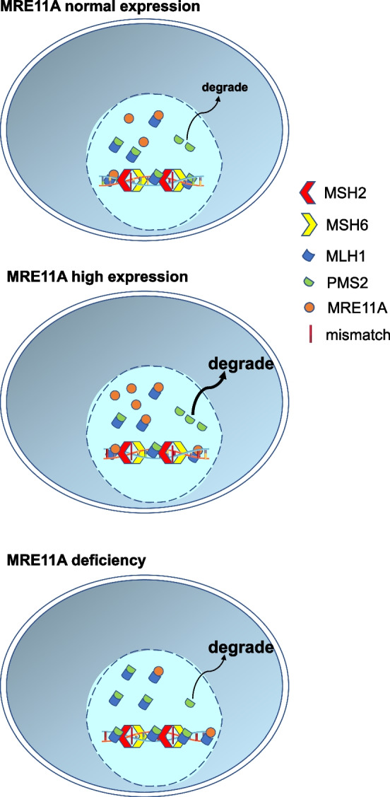
Results: We show that knockdown of MRE11A increased the sensitivity of HeLa cells to MNNG treatment, as well as the MNNG-induced DNA damage and apoptosis, implying a potential role of MRE11 in MMR. Moreover, we found that MRE11A was largely recruited to chromatin and negatively regulated the DNA damage signals within the first cell cycle after MNNG treatment. We also showed that knockdown of MRE11A increased, while overexpressing MRE11A decreased, MMR activity in HeLa cells, suggesting that MRE11A negatively regulates MMR activity. Furthermore, we show that recruitment of MRE11A to chromatin requires MLH1 and that MRE11A competes with PMS2 for binding to MLH1. This decreases PMS2 levels in whole cells and on chromatin, and consequently comprises MMR activity.

Conclusions: Our findings reveal that MRE11A is a negative regulator of human MMR.

Keywords: Alkylating agents; DNA mismatch repair; DNA repair; MRE11A; PMS2.


Conflict of interest statement

The authors declare that they have no competing interests.

Figures

Fig. 1
Fig. 1
MRE11A deficiency sensitizes cells to MNNG treatment. A HeLa cells were transfected with two different siRNAs targeting MRE11A and exhibited abnormal morphology and reduced survival 72 h after 200 nM MNNG treatment. The survival rate was the percentage of surviving cells to parallel cells treated only with O6-benzylguaine and DMSO in each group, and cells with MLH1 deficiency were used as a positive control. B MRE11A knockdown cells were treated with MNNG and seeded in triplicate in six-well plates, and after approximately 2 weeks, the cells were stained with Crystal Violet, and the colonies with ≥ 200 cells were counted. The survival rate was the percentage of surviving clones in parallel wells treated only with DMSO in each group, and cells with MLH1 deficiency were used as a positive control. C The growth rates of control cells and MRE11A knockdown cells were measured in 96-well plates with CCK8 reagents. D Western blotting showed no significant changes in the protein levels of MSH2 and MLH1 in MRE11A knockdown cells. E Representative flow cytometry pictures of scatter plots of PI versus Annexin V staining of the siNC control, MRE11A and MLH1 knockdown cells 72 h after 200 nM MNNG treatment. The right graph shows the statistical analysis of the left flow cytometry data, quantification, and comparison of the proportions of apoptotic cells in each group. The % apoptosis was calculated as the % apoptosis of cells with 200 nM MNNG minus that with only DMOS treatment. All data were analyzed with an unpaired two-tailed Student’s t test. Data are shown as mean ± SD, n = 3, *P < 0.05, **P < 0.01, ***P < 0.001
Fig. 2
Fig. 2
MRE11A deficiency increases DNA damage signals 12 h after MNNG treatment. A Representative western blotting pictures of chromatin binding and whole-cell MSH2, MLH1, and MRE11 proteins 12 h after DMSO or 200 nM MNNG treatment of HeLa cells. The level of histone H3 was set as an internal control. The right graph shows the quantification of the fold change in the ratio of chromatin binding to the whole-cell proteins MSH2, MLH1, and MRE11A after exposure to 200 nM MNNG. B Representative western blotting of the phosphorylation levels of CHK1 12 h after DMSO or 200 nM MNNG treatment. The alteration of phosphorylation level was calculated as p-CHK1 level normalized by total CHK1 protein after 200 nM MNNG treatment minus that with only DMSO treatment. The right graph shows the quantification of protein level changes relative to siNC. C Representative immunofluorescence images of 53BP1 foci in G1 phase 12 h after DMSO or 200 nM MNNG treatment. The right graph shows the quantification of the number of 53BP1 foci per cell in G1 phase (CYCLINA-). At least 250 cells were counted for each group. Data are shown as mean ± SD, *P < 0.05, **P < 0.01, ***P < 0.001, using unpaired two-tailed Student’s t test
Fig. 3
Fig. 3
MRE11A negatively regulates MMR activity in HeLa cells. The left pictures represent the scatter plots of cells cotransfected with GFP-heteroduplex and mCherry plasmids as described in “Materials and Methods” section. The x-axis and y-axis represent the signal intensities of GFP and mCherry, respectively. The MMR repair efficiency was calculated as the ratio of the number of GFP-positive cells to mCherry-positive cells, and the quantification results relative to siNC or empty vector controls are shown in the right graphs. A Cells were transfected with siNC or two MRE11A siRNAs followed by GFP-heteroduplex and mCherry plasmid cotransfection after 2 days. The next day, the cells were subjected to flow cytometry for GFP and mCherry signal analysis. B Cells were transfected with empty vector, MRE11A overexpression plasmid (MRE11AOE), or MRE11A + PMS2 overexpression plasmids (MRE11OE + PMS2) followed by GFP-heteroduplex and m-cherry plasmid cotransfection after 2 days. The next day, the cells were subjected to flow cytometry for GFP and mCherry signal analysis. Data are shown as mean ± SD, *P < 0.05, **P < 0.01, ***P < 0.001, using unpaired two-tailed Student’s t test
Fig. 4
Fig. 4
MRE11A is recruited to chromatin by MMR proteins. A Representative western blotting of chromatin proteins coprecipitated with MSH2 12 h after DMSO or 200 nM MNNG treatment. B, C Representative western blotting of the chromatin binding MSH2, MLH1, and MRE11A proteins after knockdown of MSH2, MLH1, and MRE11A independently. The right graphs show the quantification of the western blotting bands relative to siNC controls. Data shown as mean ± SD, *P < 0.05, **P < 0.01, ***P < 0.001, using unpaired two-tailed Student’s t test
Fig. 5
Fig. 5
MRE11A negatively regulates PMS2 levels. Representative western blotting of the indicated protein levels in whole cells (A, B) or on chromatin (C, D) with MRE11A knockdown/overexpression or expression of flag-tagged 452–634AA of MRE11A. The right graphs show the quantification of the western blotting intensities relative to siNC or empty vector controls. The changes in the mRNA levels of PMS2 after MRE11A knockdown (A) or overexpression (B) were quantified using qPCR. Representative scatter plots of flow cytometry analysis of cells expressing GFP or mCherry, reflecting MMR repair efficiencies of cells expressing 452–634AA of MRE11A with/without PMS2 overexpression (E). The MMR repair efficiency was calculated as the ratio of the number of GFP-positive cells to mCherry-positive cells, and the quantification results relative to siNC or empty vector controls are shown in the right graphs. Data shown as mean ± SD, *P < 0.05, **P < 0.01, ***P < 0.001, using unpaired two-tailed Student’s t test
Fig. 6
Fig. 6
MRE11A levels negatively correlate with PMS2 levels in various cell lines. Representative western blotting of the PMS2 protein levels in whole cell lysates after MRE11A knockdown (A) or overexpression (B) in the indicated cell lines. The blue arrow indicates the PMS2 bands in the context of MRE11A overexpression
Fig. 7
Fig. 7
Schematic summary of the study. In naïve cells, a proportion of MRE11A may interact with MLH1 but does not interfere with the proper interaction between intrinsic PMS2 and MLH1. In MRE11A-overexpressing cells, excess MRE11A occupied the binding site of PMS2 to MLH1, leading to the degradation of unbound PMS2 and decreased MLH1·PMS2 heterodimer on chromatin, consequently compromising MMR activity. In MRE11A-deficient cells, more intrinsic PMS2 binds to MLH1, leading to increased MMR activity and thus increased sensitivity to MNNG treatment

Similar articles

Cited by

References

    1. Liu D, Keijzers G, Rasmussen LJ. DNA mismatch repair and its many roles in eukaryotic cells. Mutat Res Rev Mutat Res. 2017;773:174–187. doi: 10.1016/j.mrrev.2017.07.001. - DOI - PubMed
    1. Fishel R. Mismatch repair. J Biol Chem. 2015;290:26395–26403. doi: 10.1074/jbc.R115.660142. - DOI - PMC - PubMed
    1. Stelloo E, Jansen AML, Osse EM, Nout RA, Creutzberg CL, Ruano D, et al. Practical guidance for mismatch repair-deficiency testing in endometrial cancer. Ann Oncol. 2016;28:96–102. doi: 10.1093/annonc/mdw542. - DOI - PubMed
    1. Lynch HT, Snyder CL, Shaw TG, Heinen CD, Hitchins MP. Milestones of Lynch syndrome: 1895–2015. Nat Rev Cancer. 2015;15:181–194. doi: 10.1038/nrc3878. - DOI - PubMed
    1. Baretti M, Le DT. DNA mismatch repair in cancer. Pharmacol Ther. 2018;189:45–62. doi: 10.1016/j.pharmthera.2018.04.004. - DOI - PubMed