. 2024 Mar 14;23(1):96.
doi: 10.1186/s12933-024-02178-2.

MCT4-dependent lactate transport: a novel mechanism for cardiac energy metabolism injury and inflammation in type 2 diabetes mellitus

Affiliations

MCT4-dependent lactate transport: a novel mechanism for cardiac energy metabolism injury and inflammation in type 2 diabetes mellitus

Xiu Mei Ma et al. Cardiovasc Diabetol. .

Abstract

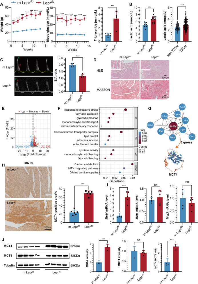
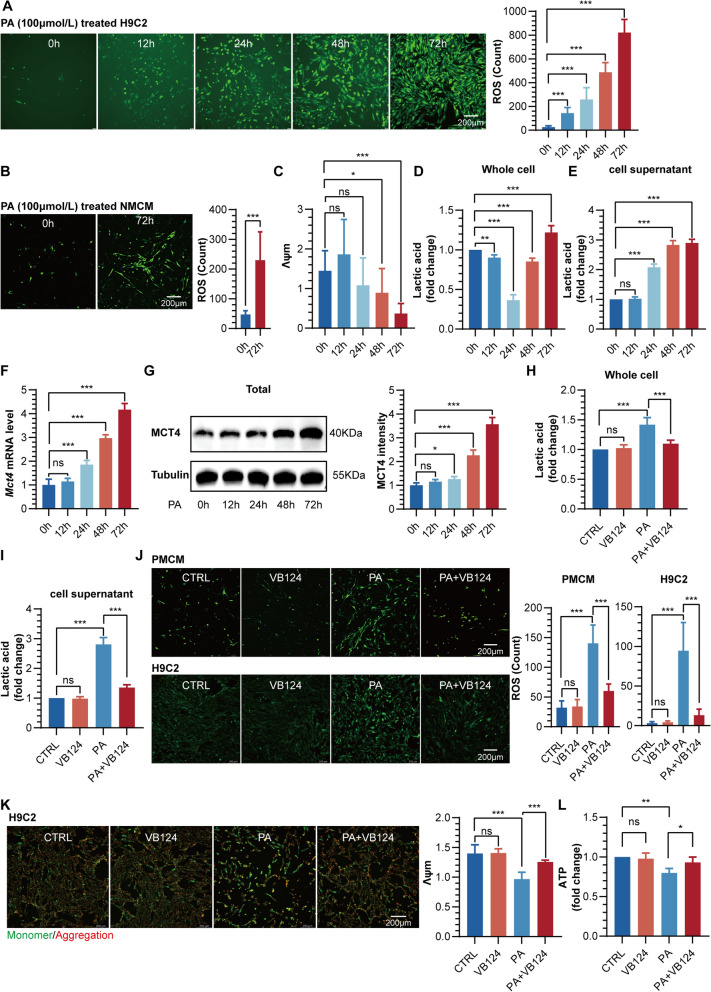
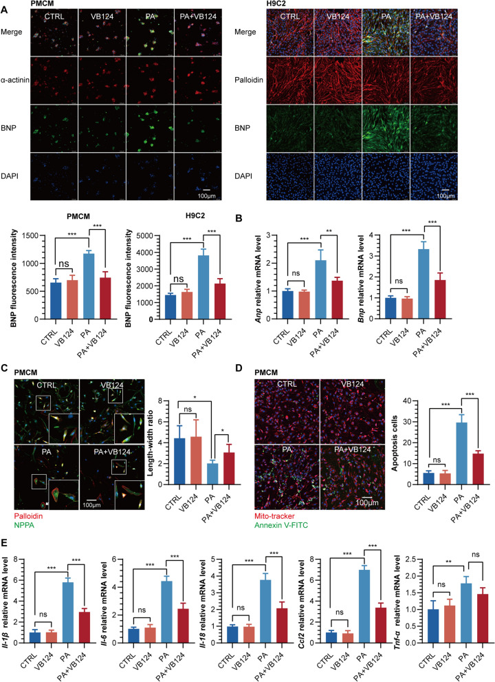
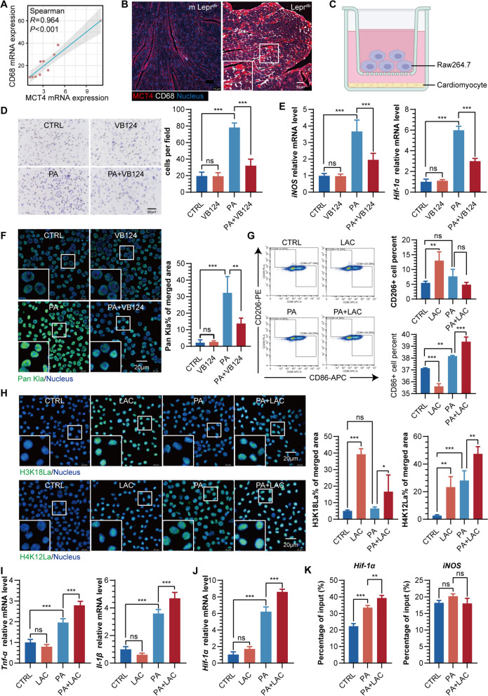
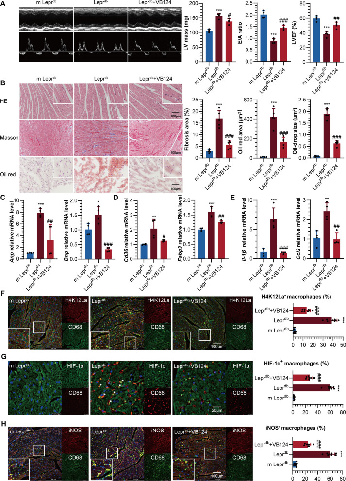
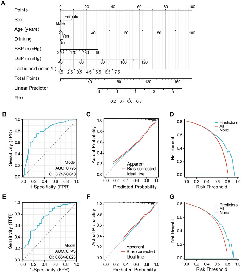
Diabetic cardiomyopathy (DCM) is a major contributor to mortality in diabetic patients, characterized by a multifaceted pathogenesis and limited therapeutic options. While lactate, a byproduct of glycolysis, is known to be significantly elevated in type 2 diabetes, its specific role in DCM remains uncertain. This study reveals an abnormal upregulation of monocarboxylate transporter 4 (MCT4) on the plasma membrane of cardiomyocytes in type 2 diabetes, leading to excessive lactate efflux from these cells. The disruption in lactate transport homeostasis perturbs the intracellular lactate-pyruvate balance in cardiomyocytes, resulting in oxidative stress and inflammatory responses that exacerbate myocardial damage. Additionally, our findings suggest increased lactate efflux augments histone H4K12 lactylation in macrophages, facilitating inflammatory infiltration within the microenvironment. In vivo experiments have demonstrated that inhibiting MCT4 effectively alleviates myocardial oxidative stress and pathological damage, reduces inflammatory macrophage infiltration, and enhances cardiac function in type 2 diabetic mice. Furthermore, a clinical prediction model has been established, demonstrating a notable association between peripheral blood lactate levels and diastolic dysfunction in individuals with type 2 diabetes. This underscores the potential of lactate as a prognostic biomarker for DCM. Ultimately, our findings highlight the pivotal involvement of MCT4 in the dysregulation of cardiac energy metabolism and macrophage-mediated inflammation in type 2 diabetes. These insights offer novel perspectives on the pathogenesis of DCM and pave the way for the development of targeted therapeutic strategies against this debilitating condition.

Keywords: Diabetic cardiomyopathy; Inflammation; Lactate; Lipotoxicity; MCT4.


Conflict of interest statement

The authors declare no competing interests.

Figures

Fig. 1
Fig. 1
A Body weight, blood glucose and triglycerides in m Leprdb and Leprdb mice. B Blood lactic acid in Leprdb vs. m Leprdb mice and T2DM vs. non-T2DM. C Pulsed‐wave Doppler showing diastolic function: peak velocity of the E wave and A wave, E/A ratio in Leprdb vs. m Leprdb mice. D HE and Masson staining of the heart in Leprdb vs. m Leprdb mice. E Volcano plot showing differentially expressed genes (DEGs) of Leprdb vs. m Leprdb mice. F GO and KEGG analysis. G Protein–protein interaction (PPI) hub networks of Slc16a3. H MCT4 immunohistochemical staining of heart tissue in Leprdb vs. m Leprdb mice. I Cardiac Mct1, 3, 4 mRNA expression in Leprdb vs. m Leprdb mice. J Western blot analysis and quantification of MCT4, MCT1, and MCT4:MCT1 ratio in Leprdb vs. m Leprdb mice
Fig. 2
Fig. 2
A Representative diagram and quantification of ROS using DCFH-DA staining in H9C2 cells treated with palmitic acid (PA). B Representative diagram and quantification of ROS in PMCM cells treated with PA. C Quantification of Λψm (mitochondrial membrane potential) in H9C2 cells treated with PA. D Fold change of lactic acid in H9C2 whole cell lysates after time-dependent PA stimulation. E Fold change of lactic acid in H9C2 cell supernatant after time-dependent PA stimulation. F Mct4 mRNA expression in H9C2 cells treated with PA. G Western blot analysis and quantification of MCT4 in H9C2 cells treated with PA. H Fold change in whole-cell abundances of lactic acid in H9C2 treated with PA and/or VB124. I Fold change in cell supernatant abundances of lactic acid in H9C2 treated with PA and/or VB124. J Representative diagram and quantification of ROS in PMCM and H9C2 cells treated with PA and/or VB124. K Representative diagram and quantification of Λψm in H9C2 cells treated with PA and/or VB124. L Fold change of ATP content in H9C2 treated with PA and/or VB124
Fig. 3
Fig. 3
A Representative diagram and quantification of BNP in PMCM and H9C2 cells treated with PA and/or VB124. B Anp and Bnp mRNA expression in H9C2 cells treated with PA and/or VB124. C Representative diagram and quantification of length–width ratio in PMCM treated with PA and/or VB124. D Representative diagram and quantification of Annexin-V positive apoptosis cells in PMCM treated with PA and/or VB124. E mRNA expression of Il-1β, Il-6, Il-18, Ccl2 and Tnf-α in H9C2 cells treated with PA and/or VB124
Fig. 4
Fig. 4
A Spearman correlation analysis between MCT4 and CD68 mRNA expression in the heart of mice. B Representative diagram of CD68 expression around the high expression of MCT4 in the heart of m Leprdb and Leprdb mice. C A diagram to depict the coculture experiment using transwell. RAW264.7 and H9C2 (pre-treated with PA and/or VB124) were cocultured for 24 h for DF. D Representative diagram and quantification of migrated RAW264.7 cells stained with crystal violet. E mRNA expression of iNOS and Hif-1α in coculture RAW264.7. F Representative diagram and quantification of Pan Kla% in coculture RAW264.7. G Flow cytometry analysis of RAW264.7 macrophage subtypes treated with either PA, lactic acid (LAC), or a combination of both. H Representative diagram and quantification of H3K18La and H4K12La in RAW264.7 treated with PA and/or LAC. I mRNA expression of Tnf-α and Il-1β in RAW264.7 treated with PA and/or LAC. J Hif-1α mRNA expression in RAW264.7 treated with PA and/or LAC. K Chromatin IP (ChIP) quantitative real time PCR (ChIP-qRT-PCR) of gene targets for HIF-1α and iNOS at H4K12La in RAW264.7 treated with PA or PA + LAC
Fig. 5
Fig. 5
*Leprdb compared to m Leprdb, #Leprdb + VB124 compared to Leprdb. A Pulsed‐wave Doppler showing: LV mass, E/A ratio and LVEF%. B Representative diagram and quantification of cardiac hypertrophy, fibrosis and oil-drop deposition in mice. C mRNA expression of cardiac Anp and Bnp in mice. D mRNA expression of cardiac Cd36 and Fabp3 in mice. E mRNA expression of cardiac Il-1β and Ccl2 in mice. F Representative diagram and quantification of heart H4K12La positive macrophage (%) in mice. G Representative diagram and quantification of heart HIF-1α positive macrophage (%) in mice. H Representative diagram and quantification of heart iNOS positive macrophage (%) in mice
Fig. 6
Fig. 6
A Nomogram for the prediction of diastolic function in patients with T2DM. B ROC curve of training set, ROC receiver operating characteristic, AUC area under the ROC curve. C Calibration curve for predicting probability of diastolic function in patients with T2DM in training set. D Decision curve analysis in prediction of diastolic function in patients with T2DM in training set. E ROC curve of validation set. F Calibration curve for predicting probability of diastolic function in patients with T2DM in validation set. G Decision curve analysis in prediction of diastolic function in patients with T2DM in the validation set
Fig. 7
Fig. 7
Mechanism of MCT4-dependent lactate transport-mediated diabetic cardiomyopathy

References

    1. Gulsin GS, Athithan L, McCann GP. Diabetic cardiomyopathy: prevalence, determinants and potential treatments. Ther Adv Endocrinol Metab. 2019;10:2042018819834869. doi: 10.1177/2042018819834869. - DOI - PMC - PubMed
    1. Peterson LR, Gropler RJ. Metabolic and molecular imaging of the diabetic cardiomyopathy. Circ Res. 2020;126(11):1628–1645. doi: 10.1161/CIRCRESAHA.120.315899. - DOI - PMC - PubMed
    1. Tan Y, Zhang Z, Zheng C, Wintergerst KA, Keller BB, Cai L. Mechanisms of diabetic cardiomyopathy and potential therapeutic strategies: preclinical and clinical evidence. Nat Rev Cardiol. 2020;17(9):585–607. doi: 10.1038/s41569-020-0339-2. - DOI - PMC - PubMed
    1. Brooks GA, Arevalo JA, Osmond AD, Leija RG, Curl CC, Tovar AP. Lactate in contemporary biology: a phoenix risen. J Physiol. 2022;600(5):1229–1251. doi: 10.1113/JP280955. - DOI - PMC - PubMed
    1. Li X, Yang Y, Zhang B, et al. Lactate metabolism in human health and disease. Signal Transduct Target Ther. 2022;7(1):305. doi: 10.1038/s41392-022-01151-3. - DOI - PMC - PubMed

Publication types