Review
. 2024 Mar 14;24(1):36.
doi: 10.1186/s40644-024-00682-y.

Artificial intelligence-based MRI radiomics and radiogenomics in glioma

Affiliations
Review

Artificial intelligence-based MRI radiomics and radiogenomics in glioma

Haiqing Fan et al. Cancer Imaging. .

Abstract

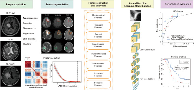
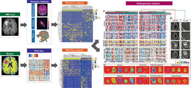
The specific genetic subtypes that gliomas exhibit result in variable clinical courses and the need to involve multidisciplinary teams of neurologists, epileptologists, neurooncologists and neurosurgeons. Currently, the diagnosis of gliomas pivots mainly around the preliminary radiological findings and the subsequent definitive surgical diagnosis (via surgical sampling). Radiomics and radiogenomics present a potential to precisely diagnose and predict survival and treatment responses, via morphological, textural, and functional features derived from MRI data, as well as genomic data. In spite of their advantages, it is still lacking standardized processes of feature extraction and analysis methodology among different research groups, which have made external validations infeasible. Radiomics and radiogenomics can be used to better understand the genomic basis of gliomas, such as tumor spatial heterogeneity, treatment response, molecular classifications and tumor microenvironment immune infiltration. These novel techniques have also been used to predict histological features, grade or even overall survival in gliomas. In this review, workflows of radiomics and radiogenomics are elucidated, with recent research on machine learning or artificial intelligence in glioma.

Keywords: Artificial intelligence; Glioma; MRI; Machine learning; Radiogenomics; Radiomics.


Conflict of interest statement

The authors declare that they have no competing interests.

Figures

Fig. 1
Fig. 1
Pipeline of the general processing steps for radiomic studies. The flowchart presented the major processing steps needed for analysis of radiomic features from MRI in glioma. After skull stripping and artifact removal (bias field, noise, etc.), acquired MRI images are subjected to standardization and segmentation to extract regions of interests (ROIs). Radiomic features are then extracted from the image masks of ROIs via conventional radiomics or deep-learning approaches. After selecting relevant features, advanced statistical analysis is performed to classify and correlate radiomic features, involving machine/deep-learning methods for feature selection, classification, and cross-validation. Finally, endpoints are predicted to evaluate the models, such as patient’s survival, genomics, response to therapy, subsequent location of recurrence, or tumor micro-environment
Fig. 2
Fig. 2
Pipeline of the general processing steps for radiogenomic studies. A typical radiogenomic analysis is usually conducted in four steps: (1) radiomic feature extraction and selection; (2) biopsy and RNA sequencing; (3) radiogenomics analysis and pathway identification; and (4) external validation. First, optimal MRI radiomic features are screened out to predict overall survival. Then, survival-relevant radiomic features are linked with co-expressed gene modules obtained by RNA sequencing. Furthermore, relevant pathways and key genes are identified to be able to annotate prognostic radiomic features. Finally, the reproducibility of prognostic radiomic-annotated pathways and key genes are validated in an external dataset

Similar articles

Cited by

References

    1. Miller KD, Ostrom QT, Kruchko C, Patil N, Tihan T, Cioffi G, Fuchs HE, Waite KA, Jemal A, Siegel RL, et al. Brain and other central nervous system tumor statistics, 2021. CA Cancer J Clin. 2021;71(5):381–406. doi: 10.3322/caac.21693. - DOI - PubMed
    1. Ostrom QT, Cioffi G, Waite K, Kruchko C, Barnholtz-Sloan JS. CBTRUS Statistical Report: primary brain and other Central Nervous System tumors diagnosed in the United States in 2014–2018. Neuro Oncol. 2021;23(12 Suppl 2):iii1–105. doi: 10.1093/neuonc/noab200. - DOI - PMC - PubMed
    1. Tan AC, Ashley DM, Lopez GY, Malinzak M, Friedman HS, Khasraw M. Management of glioblastoma: state of the art and future directions. CA Cancer J Clin. 2020;70(4):299–312. doi: 10.3322/caac.21613. - DOI - PubMed
    1. Aldape K, Zadeh G, Mansouri S, Reifenberger G, von Deimling A. Glioblastoma: pathology, molecular mechanisms and markers. Acta Neuropathol. 2015;129(6):829–48. doi: 10.1007/s00401-015-1432-1. - DOI - PubMed
    1. Kurokawa R, Kurokawa M, Baba A, Ota Y, Pinarbasi E, Camelo-Piragua S, Capizzano AA, Liao E, Srinivasan A, Moritani T. Major Changes in 2021 World Health Organization classification of Central Nervous System tumors. Radiographics. 2022;42(5):1474–93. doi: 10.1148/rg.210236. - DOI - PubMed

LinkOut - more resources